Advertisement

Main menu

  • Home
  • Content
    • Current Issue
    • Accepted Manuscripts
    • Article Preview
    • Past Issue Archive
    • Video Articles
    • AJNR Case Collection
    • Case of the Week Archive
    • Case of the Month Archive
    • Classic Case Archive
  • Special Collections
    • AJNR Awards
    • Low-Field MRI
    • Alzheimer Disease
    • ASNR Foundation Special Collection
    • Photon-Counting CT
    • View All
  • Multimedia
    • AJNR Podcasts
    • AJNR SCANtastic
    • Trainee Corner
    • MRI Safety Corner
    • Imaging Protocols
  • For Authors
    • Submit a Manuscript
    • Submit a Video Article
    • Submit an eLetter to the Editor/Response
    • Manuscript Submission Guidelines
    • Statistical Tips
    • Fast Publishing of Accepted Manuscripts
    • Graphical Abstract Preparation
    • Imaging Protocol Submission
    • Author Policies
  • About Us
    • About AJNR
    • Editorial Board
    • Editorial Board Alumni
  • More
    • Become a Reviewer/Academy of Reviewers
    • Subscribers
    • Permissions
    • Alerts
    • Feedback
    • Advertisers
    • ASNR Home

User menu

  • Alerts
  • Log in

Search

  • Advanced search
American Journal of Neuroradiology

American Journal of Neuroradiology

ASHNR American Society of Functional Neuroradiology ASHNR American Society of Pediatric Neuroradiology ASSR
  • Alerts
  • Log in

Advanced Search

  • Home
  • Content
    • Current Issue
    • Accepted Manuscripts
    • Article Preview
    • Past Issue Archive
    • Video Articles
    • AJNR Case Collection
    • Case of the Week Archive
    • Case of the Month Archive
    • Classic Case Archive
  • Special Collections
    • AJNR Awards
    • Low-Field MRI
    • Alzheimer Disease
    • ASNR Foundation Special Collection
    • Photon-Counting CT
    • View All
  • Multimedia
    • AJNR Podcasts
    • AJNR SCANtastic
    • Trainee Corner
    • MRI Safety Corner
    • Imaging Protocols
  • For Authors
    • Submit a Manuscript
    • Submit a Video Article
    • Submit an eLetter to the Editor/Response
    • Manuscript Submission Guidelines
    • Statistical Tips
    • Fast Publishing of Accepted Manuscripts
    • Graphical Abstract Preparation
    • Imaging Protocol Submission
    • Author Policies
  • About Us
    • About AJNR
    • Editorial Board
    • Editorial Board Alumni
  • More
    • Become a Reviewer/Academy of Reviewers
    • Subscribers
    • Permissions
    • Alerts
    • Feedback
    • Advertisers
    • ASNR Home
  • Follow AJNR on Twitter
  • Visit AJNR on Facebook
  • Follow AJNR on Instagram
  • Join AJNR on LinkedIn
  • RSS Feeds

AJNR Awards, New Junior Editors, and more. Read the latest AJNR updates

Research ArticleSpine Imaging and Spine Image-Guided Interventions

Dural Ectasia in Marfan Syndrome: A Case Control Study

R. Lundby, S. Rand-Hendriksen, J.K. Hald, F.G. Lilleås, A.H. Pripp, S. Skaar, B. Paus, O. Geiran and H.-J. Smith
American Journal of Neuroradiology September 2009, 30 (8) 1534-1540; DOI: https://doi.org/10.3174/ajnr.A1620
R. Lundby
  • Find this author on Google Scholar
  • Find this author on PubMed
  • Search for this author on this site
S. Rand-Hendriksen
  • Find this author on Google Scholar
  • Find this author on PubMed
  • Search for this author on this site
J.K. Hald
  • Find this author on Google Scholar
  • Find this author on PubMed
  • Search for this author on this site
F.G. Lilleås
  • Find this author on Google Scholar
  • Find this author on PubMed
  • Search for this author on this site
A.H. Pripp
  • Find this author on Google Scholar
  • Find this author on PubMed
  • Search for this author on this site
S. Skaar
  • Find this author on Google Scholar
  • Find this author on PubMed
  • Search for this author on this site
B. Paus
  • Find this author on Google Scholar
  • Find this author on PubMed
  • Search for this author on this site
O. Geiran
  • Find this author on Google Scholar
  • Find this author on PubMed
  • Search for this author on this site
H.-J. Smith
  • Find this author on Google Scholar
  • Find this author on PubMed
  • Search for this author on this site
  • Article
  • Figures & Data
  • Info & Metrics
  • Responses
  • References
  • PDF
Loading

Article Figures & Data

Figures

  • Tables
  • Fig 1.
    • Download figure
    • Open in new tab
    • Download powerpoint
    Fig 1.

    Sagittal T2-weighted MR image of a patient with MFS showing a huge anterior meningocele herniating into the pelvis through a large sacral defect (arrows). The urinary bladder (asterisk) is compressed anteriorly.

  • Fig 2.
    • Download figure
    • Open in new tab
    • Download powerpoint
    Fig 2.

    Coronal T2-weighted MR image showing lateral meningoceles/herniations (arrows) of the nerve root sleeves at level S1 in a patient with MFS.

  • Fig 3.
    • Download figure
    • Open in new tab
    • Download powerpoint
    Fig 3.

    A and B, T2-weighted sagittal MR images of a patient with MFS showing craniocaudal and AP measurements of the vertebral bodies (white arrows, A and B) and AP measurements of the dural sac (black arrows, B). Scalloping is present at levels L5, S1, and S2.
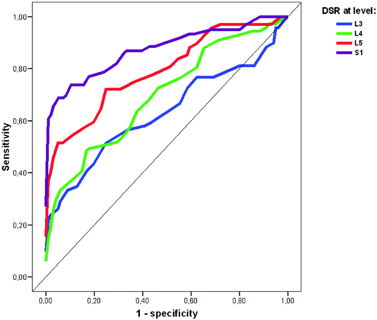
  • Fig 4.
    • Download figure
    • Open in new tab
    • Download powerpoint
    Fig 4.

    ROC curves of DSR at levels L3-S1. Graph shows sensitivity versus (1-specificity) of DSR as a marker of MFS at levels L3-S1 when comparing Ghent-positive patients independent of DE and controls.

Tables

  • Figures
    • View popup
    Table 1:

    Characteristics of the 4 study groups or the number and percentage of patients with the condition*

    CharacteristicsGhent Independent of DE (n = 73)Ghent Dependent on DE (n = 14)Not Fulfilling Ghent (n = 18)Control (n = 101)P Value
    Age (mean ± SD)39.4 (12.2)40.6 (17.1)36.1 (13.4)39.6 (12.9).74
    Females (No.) (%)45 (61.6)11 (78.6)11 (61.6)64 (63.4).68
    DSR L3 (mean ± SD)0.50b,c (0.11), n = 690.53c (0.06)0.43a (0.08), n = 140.45a,b (0.07)<.001
    DSR L4 (mean ± SD0.50b (0.12), n = 720.51b (0.07)0.41a (0.06)0.43a (0.07)<.001
    DSR L5 (mean ± SD)0.57b (0.18), n = 680.65b (0.20)0.39a (0.09)0.42a (0.08)<.001
    DSR S1 (mean ± SD)0.94b (0.69), n = 611.02b (0.77), n = 130.40a (0.12), n = 120.41a (0.13), n = 96<.001
    DSD sacrum > DSD L438 (55.1%), n = 694 (28.6%)1 (5.6%)5 (5.0%)<.001
    Herniation of nerve root sleeves
        Present in ≥1 level (No.) (%)53 (72.6%)10 (71.4%)2 (11.1%)1 (1%)<.001
        Present in level L59 (12.3%)0 (0%)0 (0%)0 (0%)<.001
        Present in level S135 (47.9%)8 (57.1%)2 (11.1%)0 (0%)<.001
        Present in level S248 (65.8%)8 (57.1%)1 (5.6%)1 (1%)<.001
        Present in level S330 (41.1%)5 (35.7%)0 (0%)0 (0%)<.001
        Present in level S48 (11%)1 (7.1%)0 (0%)0 (0%)<.004
    Odds ratio, ≥1 herniation of the nerve root sleeve265 (34–2029)250 (25–2458)12.5 (1–145)ref<.001
    Anterior meningocele27 (37%)2 (14%)00<.001
    Scalloping L1-S1 ≥1 level44 (61.1 %), n = 72 8 (42.9%) 1 (5.6%)4 (4%), n = 100<.001
    Perineural cysts11 (15.1 %) 1 (7.1%) 1 (5.6%)8 (7.9%).387
    Disk herniation beneath T12 5 (7%), n = 69 1 (7%) 2 (11%)6 (6%).890
    Caudal end of dural sac
        L5, L5/S1, S110 (14.5%) 0 (0%) 9 (50%)22 (21.8%)<.001
        S226 (37.7%) 9 (64.3%) 8 (44.4%)68 (67.3%)<.001
        S3, S4, S533 (47.8%), n = 69 5 (35.7%) 1 (5.6%)11 (10.9%)<.001
    • Note:—DE, dural ectasia; DSR, dural sac ratio; DSD, dural sac diameter.

    • * Study groups not containing similar letters (a, b, c, or d) for means were statistically different at the 5% level. Where n is specified in a cell, there were some observations missing and n is the actual number of observations. Missing values are due to incomplete anatomic coverage with CT, extreme scoliosis at MR, or the dural sac ending above the missed level.

    • View popup
    Table 2:

    Prevalence of signs of enlarged dural sac in the 4 groups*

    CriteriaFulfilling Ghent (No.) (%)
    Independent of DE (n = 73)Dependent on DE (n = 14)Suspected MFS, Not Fulfilling Ghent (n = 18)Controls (n = 101)
    1) Anterior meningocele27 (37)2 (1400%0%
    2) DSD sacrum > DSD L438 (52)4 (29)1 (6)5 (5)
    3) Herniations of ≥1 nerve root sleeves53 (73)10 (71)2 (11)1 (1)
    4) DSR L5 > 0.48 (Oosterhof et al6)41 (56)13 (93)1 (6)19 (19)
    5) DSR S1 > 0.57 (Oosterhof et al6)45 (62)11 (85)1 (6)12 (12)
    Presence of ≥1 of above findings65 (89)14 (100)3 (17)24 (24)
    Only DSR L5 > 0.48 and/or S1 > 0.574 (5)4 (29)1 (6)18 (18)
    6) DSR S1 > 0.59 (current study)43 (59)11 (85)1 (6)7 (7)
    Presence of DE replacing criteria 4 and 5 with 663 (86)12 (86)3 (17)9 (9)
    • Note:—MES indicates Marfan Syndrome.

    • * * Expressed as number and percentage.

PreviousNext
Back to top

In this issue

American Journal of Neuroradiology: 30 (8)
American Journal of Neuroradiology
Vol. 30, Issue 8
1 Sep 2009
  • Table of Contents
  • Index by author
Advertisement
Print
Download PDF
Email Article

Thank you for your interest in spreading the word on American Journal of Neuroradiology.

NOTE: We only request your email address so that the person you are recommending the page to knows that you wanted them to see it, and that it is not junk mail. We do not capture any email address.

Enter multiple addresses on separate lines or separate them with commas.
Dural Ectasia in Marfan Syndrome: A Case Control Study
(Your Name) has sent you a message from American Journal of Neuroradiology
(Your Name) thought you would like to see the American Journal of Neuroradiology web site.
CAPTCHA
This question is for testing whether or not you are a human visitor and to prevent automated spam submissions.
Cite this article
R. Lundby, S. Rand-Hendriksen, J.K. Hald, F.G. Lilleås, A.H. Pripp, S. Skaar, B. Paus, O. Geiran, H.-J. Smith
Dural Ectasia in Marfan Syndrome: A Case Control Study
American Journal of Neuroradiology Sep 2009, 30 (8) 1534-1540; DOI: 10.3174/ajnr.A1620

Citation Manager Formats

  • BibTeX
  • Bookends
  • EasyBib
  • EndNote (tagged)
  • EndNote 8 (xml)
  • Medlars
  • Mendeley
  • Papers
  • RefWorks Tagged
  • Ref Manager
  • RIS
  • Zotero
0 Responses
Respond to this article
Share
Bookmark this article
Dural Ectasia in Marfan Syndrome: A Case Control Study
R. Lundby, S. Rand-Hendriksen, J.K. Hald, F.G. Lilleås, A.H. Pripp, S. Skaar, B. Paus, O. Geiran, H.-J. Smith
American Journal of Neuroradiology Sep 2009, 30 (8) 1534-1540; DOI: 10.3174/ajnr.A1620
del.icio.us logo Twitter logo Facebook logo Mendeley logo
  • Tweet Widget
  • Facebook Like
  • Google Plus One
Purchase

Jump to section

  • Article
    • Abstract
    • Materials and Methods
    • Results
    • Discussion
    • Footnotes
    • References
  • Figures & Data
  • Info & Metrics
  • Responses
  • References
  • PDF

Related Articles

  • No related articles found.
  • PubMed
  • Google Scholar

Cited By...

  • No citing articles found.
  • Crossref
  • Google Scholar

This article has not yet been cited by articles in journals that are participating in Crossref Cited-by Linking.

More in this TOC Section

  • Optimization of Photon Counting CT Myelography
  • Characteristics of SIH Type I Culprit Lesions
  • Management Outcomes For VO Spine Biopsy
Show more Spine Imaging and Spine Image-Guided Interventions

Similar Articles

Advertisement

Indexed Content

  • Current Issue
  • Accepted Manuscripts
  • Article Preview
  • Past Issues
  • Editorials
  • Editor's Choice
  • Fellows' Journal Club
  • Letters to the Editor
  • Video Articles

Cases

  • Case Collection
  • Archive - Case of the Week
  • Archive - Case of the Month
  • Archive - Classic Case

More from AJNR

  • Trainee Corner
  • Imaging Protocols
  • MRI Safety Corner
  • Book Reviews

Multimedia

  • AJNR Podcasts
  • AJNR Scantastics

Resources

  • Turnaround Time
  • Submit a Manuscript
  • Submit a Video Article
  • Submit an eLetter to the Editor/Response
  • Manuscript Submission Guidelines
  • Statistical Tips
  • Fast Publishing of Accepted Manuscripts
  • Graphical Abstract Preparation
  • Imaging Protocol Submission
  • Evidence-Based Medicine Level Guide
  • Publishing Checklists
  • Author Policies
  • Become a Reviewer/Academy of Reviewers
  • News and Updates

About Us

  • About AJNR
  • Editorial Board
  • Editorial Board Alumni
  • Alerts
  • Permissions
  • Not an AJNR Subscriber? Join Now
  • Advertise with Us
  • Librarian Resources
  • Feedback
  • Terms and Conditions
  • AJNR Editorial Board Alumni

American Society of Neuroradiology

  • Not an ASNR Member? Join Now

© 2025 by the American Society of Neuroradiology All rights, including for text and data mining, AI training, and similar technologies, are reserved.
Print ISSN: 0195-6108 Online ISSN: 1936-959X

Powered by HighWire