Advertisement

Main menu

  • Home
  • Content
    • Current Issue
    • Accepted Manuscripts
    • Article Preview
    • Past Issue Archive
    • Video Articles
    • AJNR Case Collection
    • Case of the Week Archive
    • Case of the Month Archive
    • Classic Case Archive
  • Special Collections
    • AJNR Awards
    • Low-Field MRI
    • Alzheimer Disease
    • ASNR Foundation Special Collection
    • Photon-Counting CT
    • View All
  • Multimedia
    • AJNR Podcasts
    • AJNR SCANtastic
    • Trainee Corner
    • MRI Safety Corner
    • Imaging Protocols
  • For Authors
    • Submit a Manuscript
    • Submit a Video Article
    • Submit an eLetter to the Editor/Response
    • Manuscript Submission Guidelines
    • Statistical Tips
    • Fast Publishing of Accepted Manuscripts
    • Graphical Abstract Preparation
    • Imaging Protocol Submission
    • Author Policies
  • About Us
    • About AJNR
    • Editorial Board
    • Editorial Board Alumni
  • More
    • Become a Reviewer/Academy of Reviewers
    • Subscribers
    • Permissions
    • Alerts
    • Feedback
    • Advertisers
    • ASNR Home

User menu

  • Alerts
  • Log in

Search

  • Advanced search
American Journal of Neuroradiology

American Journal of Neuroradiology

ASHNR American Society of Functional Neuroradiology ASHNR American Society of Pediatric Neuroradiology ASSR
  • Alerts
  • Log in

Advanced Search

  • Home
  • Content
    • Current Issue
    • Accepted Manuscripts
    • Article Preview
    • Past Issue Archive
    • Video Articles
    • AJNR Case Collection
    • Case of the Week Archive
    • Case of the Month Archive
    • Classic Case Archive
  • Special Collections
    • AJNR Awards
    • Low-Field MRI
    • Alzheimer Disease
    • ASNR Foundation Special Collection
    • Photon-Counting CT
    • View All
  • Multimedia
    • AJNR Podcasts
    • AJNR SCANtastic
    • Trainee Corner
    • MRI Safety Corner
    • Imaging Protocols
  • For Authors
    • Submit a Manuscript
    • Submit a Video Article
    • Submit an eLetter to the Editor/Response
    • Manuscript Submission Guidelines
    • Statistical Tips
    • Fast Publishing of Accepted Manuscripts
    • Graphical Abstract Preparation
    • Imaging Protocol Submission
    • Author Policies
  • About Us
    • About AJNR
    • Editorial Board
    • Editorial Board Alumni
  • More
    • Become a Reviewer/Academy of Reviewers
    • Subscribers
    • Permissions
    • Alerts
    • Feedback
    • Advertisers
    • ASNR Home
  • Follow AJNR on Twitter
  • Visit AJNR on Facebook
  • Follow AJNR on Instagram
  • Join AJNR on LinkedIn
  • RSS Feeds

AJNR Awards, New Junior Editors, and more. Read the latest AJNR updates

Research ArticleNeurointervention

Quantitative Hemodynamic Analysis of Brain Aneurysms at Different Locations

A. Chien, M.A. Castro, S. Tateshima, J. Sayre, J. Cebral and F. Viñuela
American Journal of Neuroradiology September 2009, 30 (8) 1507-1512; DOI: https://doi.org/10.3174/ajnr.A1600
A. Chien
  • Find this author on Google Scholar
  • Find this author on PubMed
  • Search for this author on this site
M.A. Castro
  • Find this author on Google Scholar
  • Find this author on PubMed
  • Search for this author on this site
S. Tateshima
  • Find this author on Google Scholar
  • Find this author on PubMed
  • Search for this author on this site
J. Sayre
  • Find this author on Google Scholar
  • Find this author on PubMed
  • Search for this author on this site
J. Cebral
  • Find this author on Google Scholar
  • Find this author on PubMed
  • Search for this author on this site
F. Viñuela
  • Find this author on Google Scholar
  • Find this author on PubMed
  • Search for this author on this site
  • Article
  • Figures & Data
  • Info & Metrics
  • Responses
  • References
  • PDF
Loading

Article Figures & Data

Figures

  • Tables
  • Fig 1.
    • Download figure
    • Open in new tab
    • Download powerpoint
    Fig 1.

    Schematic representations of regions for hemodynamic data analysis. A, Regions in the arteries of terminal or bifurcation aneurysms. B, Regions in the arteries of sidewall aneurysms. C, Regions in the aneurysm dome. D and E, Application to patient-specific aneurysm models are shown for a bifurcation aneurysm (D) and a sidewall aneurysm (E).
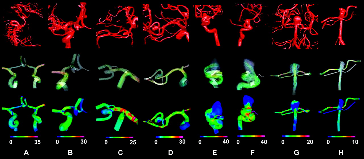
  • Fig 2.
    • Download figure
    • Open in new tab
    • Download powerpoint
    Fig 2.

    Representative hemodynamic results for AcomA (A and B), MCA (C and D ), ICA (E and F ), and BA (G and H ) aneurysms. Top row is the 3DRA images obtained for each aneurysm during the clinical procedure. A diversity of aneurysms and complicated arterial structures can be observed. The middle row (flow pattern) and the bottom row (WSS) are the hemodynamic results obtained from the simulation. Flow pattern and WSS show differences among aneurysms, and each one has a unique pattern in the arteries and the aneurysms. WSS is in pascal units.

  • Fig 3.
    • Download figure
    • Open in new tab
    • Download powerpoint
    Fig 3.

    WSS in aneurysms at each location. Note the average WSS in arteries and aneurysms and the average WSS in the entire aneurysm area.

  • Fig 4.
    • Download figure
    • Open in new tab
    • Download powerpoint
    Fig 4.

    Flow rates in aneurysms at different locations. Note the average flow in arteries and aneurysms and the average flow velocity in the entire aneurysm area.

  • Fig 5.
    • Download figure
    • Open in new tab
    • Download powerpoint
    Fig 5.

    A and B, Validation study comparing the hemodynamic results by using 6 regions (solid) and values collected from the entire dome and parent artery (method of Shojima et al13) (hollow) shows the average WSS in the aneurysms (A ) and the average WSS in the parent arteries (B). Note in cases 5 and 8 that both methods show the same average WSS in the aneurysm.

Tables

  • Figures
  • Summary of aneurysm cases*

    CharacteristicsGroup 1Group 2Group 3Group 4Summary
    Age (year)
        Mean65.264.251.357.359.5
        Range48–8142–7924–7047–7024–81
    Sex (No. of patients)
        Female556319
        Male11035
    Unruptured-ruptured aneurysms (No. of patients)4–24–24–24–216–8
    Total No. of aneurysms666624
    Largest diameter of aneurysm (mm)
        Mean5.26.257.211.07.4
        Range3.6–8.25.1–8.15–86–153.6–15
    Diameter of aneurysm neck (mm)
        Mean3.53.74.47.174.7
        Range2.3–6.53.3–42.7–74–14.62.3–14.6
    Size of aneurysm (No. of patients)
        <7 mm542112
        7–12 mm12429
        13–24 mm00033
    Diameter of parent artery vessel lumen (mm)
        Mean2.12.23.93.42.9
        Range1.3–3.21.6–2.73.4–5.32.7–4.41.3–4.4
    • * Groups 1, 2, 3, and 4 are aneurysms located at the AComA, MCA, ICA, and BA, respectively.

PreviousNext
Back to top

In this issue

American Journal of Neuroradiology: 30 (8)
American Journal of Neuroradiology
Vol. 30, Issue 8
1 Sep 2009
  • Table of Contents
  • Index by author
Advertisement
Print
Download PDF
Email Article

Thank you for your interest in spreading the word on American Journal of Neuroradiology.

NOTE: We only request your email address so that the person you are recommending the page to knows that you wanted them to see it, and that it is not junk mail. We do not capture any email address.

Enter multiple addresses on separate lines or separate them with commas.
Quantitative Hemodynamic Analysis of Brain Aneurysms at Different Locations
(Your Name) has sent you a message from American Journal of Neuroradiology
(Your Name) thought you would like to see the American Journal of Neuroradiology web site.
CAPTCHA
This question is for testing whether or not you are a human visitor and to prevent automated spam submissions.
Cite this article
A. Chien, M.A. Castro, S. Tateshima, J. Sayre, J. Cebral, F. Viñuela
Quantitative Hemodynamic Analysis of Brain Aneurysms at Different Locations
American Journal of Neuroradiology Sep 2009, 30 (8) 1507-1512; DOI: 10.3174/ajnr.A1600

Citation Manager Formats

  • BibTeX
  • Bookends
  • EasyBib
  • EndNote (tagged)
  • EndNote 8 (xml)
  • Medlars
  • Mendeley
  • Papers
  • RefWorks Tagged
  • Ref Manager
  • RIS
  • Zotero
0 Responses
Respond to this article
Share
Bookmark this article
Quantitative Hemodynamic Analysis of Brain Aneurysms at Different Locations
A. Chien, M.A. Castro, S. Tateshima, J. Sayre, J. Cebral, F. Viñuela
American Journal of Neuroradiology Sep 2009, 30 (8) 1507-1512; DOI: 10.3174/ajnr.A1600
del.icio.us logo Twitter logo Facebook logo Mendeley logo
  • Tweet Widget
  • Facebook Like
  • Google Plus One
Purchase

Jump to section

  • Article
    • Abstract
    • Materials and Methods
    • Results
    • Discussion
    • Conclusions
    • Footnotes
    • References
  • Figures & Data
  • Info & Metrics
  • Responses
  • References
  • PDF

Related Articles

  • No related articles found.
  • PubMed
  • Google Scholar

Cited By...

  • A patient-specific intracranial aneurysm model with endothelial lining: a novel in vitro approach to bridge the gap between biology and flow dynamics
  • Nonsphericity Index and Size Ratio Identify Morphologic Differences between Growing and Stable Aneurysms in a Longitudinal Study of 93 Cases
  • Differences in Morphologic and Hemodynamic Characteristics for "PHASES-Based" Intracranial Aneurysm Locations
  • Differences in Hemodynamics and Rupture Rate of Aneurysms at the Bifurcation of the Basilar and Internal Carotid Arteries
  • Morphologic and Hemodynamic Risk Factors in Ruptured Aneurysms Imaged before and after Rupture
  • CFD: Computational Fluid Dynamics or Confounding Factor Dissemination? The Role of Hemodynamics in Intracranial Aneurysm Rupture Risk Assessment
  • Distinct trends of pulsatility found at the necks of ruptured and unruptured aneurysms
  • 3D Cine Phase-Contrast MRI at 3T in Intracranial Aneurysms Compared with Patient-Specific Computational Fluid Dynamics
  • Creation of Bifurcation-Type Elastase-Induced Aneurysms in Rabbits
  • Crossref
  • Google Scholar

This article has not yet been cited by articles in journals that are participating in Crossref Cited-by Linking.

More in this TOC Section

  • Rescue Reentry in Carotid Near-Occlusion
  • Contour Neurovascular System: Five Year Follow Up
  • Effect of SARS-CoV2 on Endovascular Thrombectomy
Show more Neurointervention

Similar Articles

Advertisement

Indexed Content

  • Current Issue
  • Accepted Manuscripts
  • Article Preview
  • Past Issues
  • Editorials
  • Editor's Choice
  • Fellows' Journal Club
  • Letters to the Editor
  • Video Articles

Cases

  • Case Collection
  • Archive - Case of the Week
  • Archive - Case of the Month
  • Archive - Classic Case

More from AJNR

  • Trainee Corner
  • Imaging Protocols
  • MRI Safety Corner
  • Book Reviews

Multimedia

  • AJNR Podcasts
  • AJNR Scantastics

Resources

  • Turnaround Time
  • Submit a Manuscript
  • Submit a Video Article
  • Submit an eLetter to the Editor/Response
  • Manuscript Submission Guidelines
  • Statistical Tips
  • Fast Publishing of Accepted Manuscripts
  • Graphical Abstract Preparation
  • Imaging Protocol Submission
  • Evidence-Based Medicine Level Guide
  • Publishing Checklists
  • Author Policies
  • Become a Reviewer/Academy of Reviewers
  • News and Updates

About Us

  • About AJNR
  • Editorial Board
  • Editorial Board Alumni
  • Alerts
  • Permissions
  • Not an AJNR Subscriber? Join Now
  • Advertise with Us
  • Librarian Resources
  • Feedback
  • Terms and Conditions
  • AJNR Editorial Board Alumni

American Society of Neuroradiology

  • Not an ASNR Member? Join Now

© 2025 by the American Society of Neuroradiology All rights, including for text and data mining, AI training, and similar technologies, are reserved.
Print ISSN: 0195-6108 Online ISSN: 1936-959X

Powered by HighWire