
Article Figures & Data
Figures
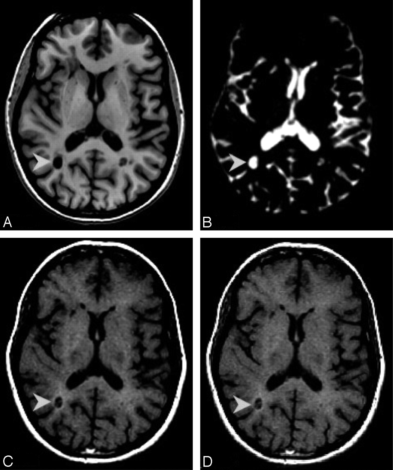
- Fig 1.
A, Axial view of IR-FSPGR image. B, Axial view of 3D-TSI-CSF image. C, Axial view of T1-weighted image acquired at the time of the 3T MR imaging. D, Axial view of T1-weighted image acquired 24 months before images reported in A–C. A–D shows a group-A lesion (arrowheads). Only one of the lesions identified on MR imaging is discussed as an example. It can be seen that the BH in A corresponds to a hyperintense area on the relative TSI volume (B). The BH marked here is defined as CBH because it was present 24 months before the 3T study (D).
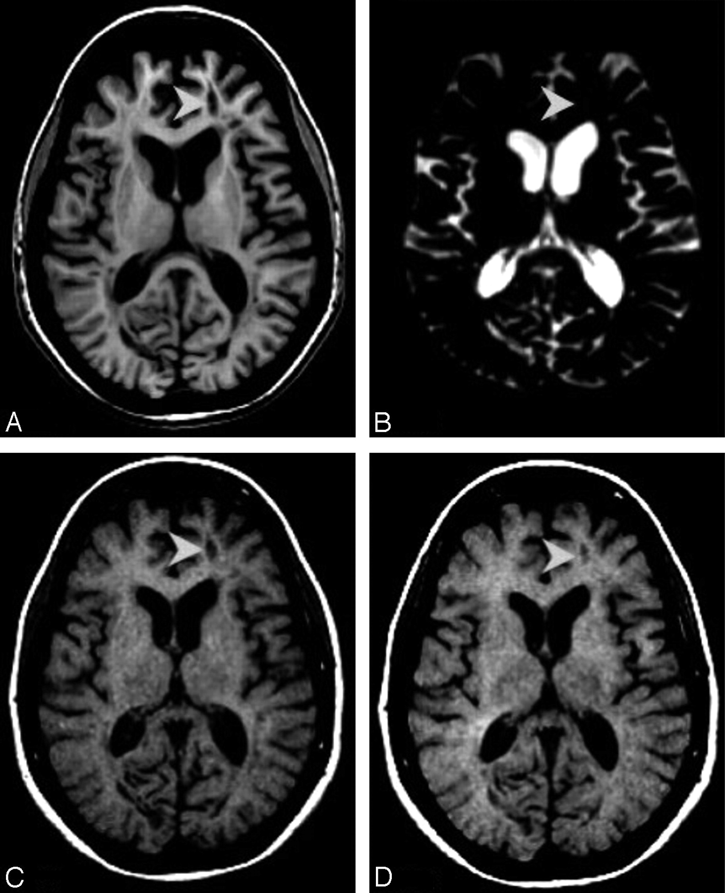
- Fig 2.
A, Axial view of IR-FSPGR image. B, Axial view of 3D-TSI-CSF image. C, Axial view of T1-weighted image acquired at the time of the 3T MR imaging. D, Axial view of T1-weighted image acquired 24 months before images reported in A–C. A–D reports a group-B lesion (arrowheads). Compared with group A, group-B chronic BHs do not have a corresponding hyperintense area on the 3D-TSI-CSF image (B), thought to be present as an area of hypointensity on IR-FSPGR and T1-weighted images (A,C).
- Fig 3.
The figure represents the box plot distribution of mean MTR values of NAWM, the BG, group-B lesions, group-A lesions, and CSF. Although group-B and group-A lesions show a wider distribution, it is possible to note a separation among all observed groups.
- Fig 4.
The white bars represent group-A lesions; the gray bars represent group-B lesions. Patients with SPMS are marked with dotted columns. The graph reports the distribution of the mean MTR of group-A and group-B lesions per patient. The data refer to 15 patients. Of the 18 examined, 1 was excluded from the MTR evaluation because of the poor image quality and 1 because of presenting with no T2W image. Of the remaining 16 patients, all except 1 patient had at least 1 CBH.


Tables
Demographic, clinical, and MR imaging characteristics of patients at the time of 3T MR imaging
Patients with RRMS (n = 9) Patients with SPMS (n = 7) P value* Age (y) 41.3 ± 6.3 (range, 32–53) 50.2 ± 6.0 (range, 41–56) .012 Sex 5 women, 4 men 6 women, 1 man NS Years since MS onset 10.4 ± 6.2 (range, 3–20) 18.4 ± 5.2 (range, 13–28) .016 EDSS 1.9 ± 1.1 (range, 0–4) 6.0 ± 0.6 (range, 5–6.5) <.0001 PASAT 48.7 ± 12.8 (range, 22–60) 31.2 ± 8.1 (range, 21–45) .008 BPF 0.81 ± 0.05 (range, 0.76–0.92) 0.75 ± 0.04 (range, 0.71–0.81) .014 T2-LV (cm3) 11.5 ± 8.8 (range, 0.78–25.5) 12.6 ± 8.4 (range, 4.6–30.1) NS Number of patients with CELs 1† 0 N/A CBHs (number) 48.9 ± 51.5 (range, 0–126) 37.4 ± 19.7 (range, 19–76) NS CBHs (volume in cm3) 2.8 ± 3.3 (range, 0.1–9.6) 3.2 ± 2.7 (range, 0.8–8.4) NS CBHs-LV/T2-LV (%) 16.9 ± 11.7 (range, 3–34) 24.1 ± 10.3 (range, 14–48) NS Undergoing therapies Interferon beta 3 3 Daclizumab 4 − Other treatments − 2 None 2 2 Note:—RRMS indicates relapsing-remitting multiple sclerosis; SPMS, secondary-progressive multiple sclerosis; EDSS, Expanded Disability Status Scale; PASAT, Paced Auditory Serial Addition Test; BPF, brain parenchyma fraction; T2-LV, T2 lesion volume; CELs, contrast-enhancing lesions; CBHs, chronic black holes; N/A, not applicable; NS, not significant.
* P values are significant if P ≤ .05.
† A single patient had 1 CEL in the corpus callosum (see text in the Results section).


