Advertisement

Main menu

  • Home
  • Content
    • Current Issue
    • Accepted Manuscripts
    • Article Preview
    • Past Issue Archive
    • Video Articles
    • AJNR Case Collection
    • Case of the Week Archive
    • Case of the Month Archive
    • Classic Case Archive
  • Special Collections
    • AJNR Awards
    • Low-Field MRI
    • Alzheimer Disease
    • ASNR Foundation Special Collection
    • Photon-Counting CT
    • View All
  • Multimedia
    • AJNR Podcasts
    • AJNR SCANtastic
    • Trainee Corner
    • MRI Safety Corner
    • Imaging Protocols
  • For Authors
    • Submit a Manuscript
    • Submit a Video Article
    • Submit an eLetter to the Editor/Response
    • Manuscript Submission Guidelines
    • Statistical Tips
    • Fast Publishing of Accepted Manuscripts
    • Graphical Abstract Preparation
    • Imaging Protocol Submission
    • Author Policies
  • About Us
    • About AJNR
    • Editorial Board
    • Editorial Board Alumni
  • More
    • Become a Reviewer/Academy of Reviewers
    • Subscribers
    • Permissions
    • Alerts
    • Feedback
    • Advertisers
    • ASNR Home

User menu

  • Alerts
  • Log in

Search

  • Advanced search
American Journal of Neuroradiology

American Journal of Neuroradiology

ASHNR American Society of Functional Neuroradiology ASHNR American Society of Pediatric Neuroradiology ASSR
  • Alerts
  • Log in

Advanced Search

  • Home
  • Content
    • Current Issue
    • Accepted Manuscripts
    • Article Preview
    • Past Issue Archive
    • Video Articles
    • AJNR Case Collection
    • Case of the Week Archive
    • Case of the Month Archive
    • Classic Case Archive
  • Special Collections
    • AJNR Awards
    • Low-Field MRI
    • Alzheimer Disease
    • ASNR Foundation Special Collection
    • Photon-Counting CT
    • View All
  • Multimedia
    • AJNR Podcasts
    • AJNR SCANtastic
    • Trainee Corner
    • MRI Safety Corner
    • Imaging Protocols
  • For Authors
    • Submit a Manuscript
    • Submit a Video Article
    • Submit an eLetter to the Editor/Response
    • Manuscript Submission Guidelines
    • Statistical Tips
    • Fast Publishing of Accepted Manuscripts
    • Graphical Abstract Preparation
    • Imaging Protocol Submission
    • Author Policies
  • About Us
    • About AJNR
    • Editorial Board
    • Editorial Board Alumni
  • More
    • Become a Reviewer/Academy of Reviewers
    • Subscribers
    • Permissions
    • Alerts
    • Feedback
    • Advertisers
    • ASNR Home
  • Follow AJNR on Twitter
  • Visit AJNR on Facebook
  • Follow AJNR on Instagram
  • Join AJNR on LinkedIn
  • RSS Feeds

AJNR Awards, New Junior Editors, and more. Read the latest AJNR updates

Research ArticleNeurointervention

Impact of Intra-Arterial Injection Parameters on Arterial, Capillary, and Venous Time-Concentration Curves in a Canine Model

A.S. Ahmed, Y. Deuerling-Zheng, C.M. Strother, K.A. Pulfer, M. Zellerhoff, T. Redel, K. Royalty, D. Consigny, M.J. Lindstrom and D.B. Niemann
American Journal of Neuroradiology August 2009, 30 (7) 1337-1341; DOI: https://doi.org/10.3174/ajnr.A1586
A.S. Ahmed
  • Find this author on Google Scholar
  • Find this author on PubMed
  • Search for this author on this site
Y. Deuerling-Zheng
  • Find this author on Google Scholar
  • Find this author on PubMed
  • Search for this author on this site
C.M. Strother
  • Find this author on Google Scholar
  • Find this author on PubMed
  • Search for this author on this site
K.A. Pulfer
  • Find this author on Google Scholar
  • Find this author on PubMed
  • Search for this author on this site
M. Zellerhoff
  • Find this author on Google Scholar
  • Find this author on PubMed
  • Search for this author on this site
T. Redel
  • Find this author on Google Scholar
  • Find this author on PubMed
  • Search for this author on this site
K. Royalty
  • Find this author on Google Scholar
  • Find this author on PubMed
  • Search for this author on this site
D. Consigny
  • Find this author on Google Scholar
  • Find this author on PubMed
  • Search for this author on this site
M.J. Lindstrom
  • Find this author on Google Scholar
  • Find this author on PubMed
  • Search for this author on this site
D.B. Niemann
  • Find this author on Google Scholar
  • Find this author on PubMed
  • Search for this author on this site
  • Article
  • Figures & Data
  • Info & Metrics
  • Responses
  • References
  • PDF
Loading

Article Figures & Data

Figures

  • Tables
  • Fig 1.
    • Download figure
    • Open in new tab
    • Download powerpoint
    Fig 1.

    Three views from the 3D reconstruction used to determine time concentration curves for arterial, parenchyma and venous structures (A, B, and C). The corresponding time-concentration curves are shown in D, E, and F.
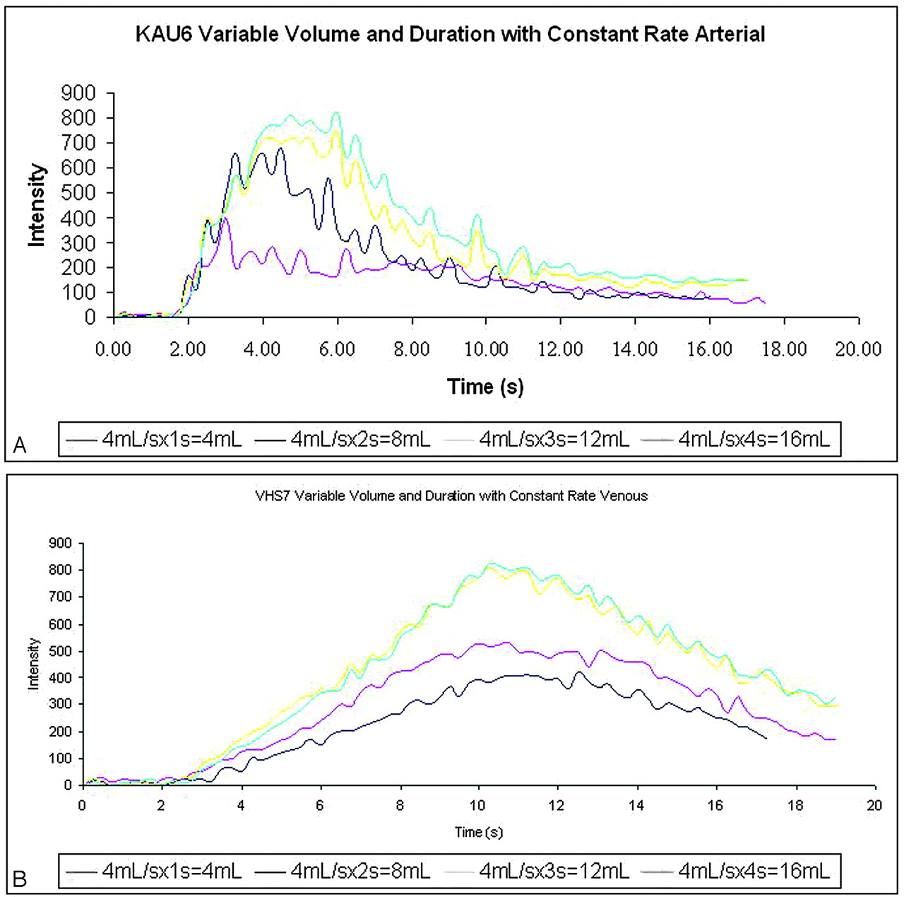
  • Fig 2.
    • Download figure
    • Open in new tab
    • Download powerpoint
    Fig 2.

    Time-concentration curves observed over an artery (A) and vein (B) at 4 different volumes while the rate of injection remained constant.

Tables

  • Figures
    • View popup
    Table 1:

    Injection protocol

    GroupVolume (mL)Rate (mL/s)Duration (s)ConcentrationCatheter Position
    1Constant, 8 mLVariable, 1, 2, 4, 8 mL/sVariable, 1, 2, 4, 8 secondsConstant, 100%Constant, prox ICA
    2Variable, 4, 8, 12, 16 mLConstant, 4 mL/sVariable, 1–4 secondsConstant, 100%Constant, prox ICA
    3Variable, 4, 8, 12, 16 mLVariable, 2, 4, 6, 8 mL/sConstant, 2 secondsConstant, 100%Constant, prox ICA
    4Constant, 8 mLConstant, 4 mL/sConstant, 2 secondsVariable, 25, 50, 75, 100%Constant, prox ICA
    5Constant, 8 mLConstant, 4 mL/sConstant, 2 secondsConstant, 100%Variable, prox, middle, distal ICA
    • Note:—Prox ICA indicates proximal internal carotid artery.

    • View popup
    Table 2:

    P values from the assessment of TTP, peak opacification, and AUC differences when comparing injection protocols: variable rate and volume with constant duration (2 seconds)*

    RateTTP CapillaryTTP ArterialTTP VenousPeak CapillaryPeak ArterialPeak VenousAUC CapillaryAUC ArterialAUC Venous
    Overall.0334.0102.0363.0009.0057.0003.0004.0003<.0001
    2–4 mL/s.9598.4923.4592.1147.0179.0020.1111.0152.0002
    2–6 mL/s.1024.0110.0302.0022.0038.0002.0007.0003<.0001
    2–8 mL/s.0126.0041.0114.0002.0011<.0001<.0001<.0001<.0001
    4–6 mL/s.1117.0404.1165.0503.4158.2065.0171.0512.0584
    4–8 mL/s.0139.0153.0465.0034.1592.0534.0018.0062.0020
    6–8 mL/s.2680.6066.6069.1678.5229.4355.2423.2739.0918
    • Note:—TTP indicates time-to-peak; AUC, area under curve.

    • * P values for paired contrasts were not calculated if no overall significant difference of the group was observed.

    • View popup
    Table 3:

    P values from the assessment of TTP, peak opacification, and AUC differences when comparing injection protocols: variable volume and duration with constant rate*

    TTP CapillaryTTP ArterialTTP VenousPeak CapillaryPeak ArterialPeak VenousAUC CapillaryAUC ArterialAUC Venous
    Overall0.9897<.0001.0161<.0001<.0001<.0001<.0001<.0001<.0001
    4–8 mL.7927.0111.0031<.0001.0014.0033.0016.0015
    4–12 mL.0005.6923<.0001<.0001<.0001<.0001<.0001<.0001
    4–16 mL.0001.9761<.0001<.0001<.0001<.0001<.0001<.0001
    8–12 mL.0003.0046.0004.0027.0003.0004.0001.0002
    8–16 mL<.0001.0104<.0001.0001.0001<.0001<.0001<.0001
    12–16 mL.5841.7144.1945.1677.6919.2334.0495.3619
    • * Pvalues for paired contrasts were not calculated if no overall significant difference of the group was observed.

    • View popup
    Table 4:

    P values from the assessment of TTP, peak opacification, and AUC differences when comparing injection protocols: variable concentration*

    TTP CapillaryTTP ArterialTTP VenousPeak CapillaryPeak ArterialPeak VenousAUC CapillaryAUC ArterialAUC Venous
    Overall.2235<.0001.0072.0432.0040.0035.0789.0009.1901
    100%–25%<.0001.0086.0186.0023.0008.0004
    100%–50%.0977.0132.0122.0015.0038.0064
    100%–75%.2582.9593.1971.1609.1356.6618
    25%–50%.0004.8427.8466.8578.4980.2259
    25%–75%<.0001.0077.2277.0517.0245.0011
    50%–75%.0093.0118.1656.0360.0949.0166
    • * P values for paired contrasts were not calculated if no overall significant difference of the group was observed.

    • View popup
    Table 5:

    P values from the assessment of TTP, peak opacification, and AUC differences when comparing injection protocols: variable rate and duration with constant volume*

    TTP CapillaryTTP ArterialTTP VenousPeak CapillaryPeak ArterialPeak VenousAUC CapillaryAUC ArterialAUC Venous
    Overall.0496.1288.0444.0128.0282.0805.0418.2567.0956
    1–2 mL/s.0483.0075.9195.2043.1035
    1–4 mL/s.0291.0644.0171.0057.2754
    1–8 mL/s.0108.0371.0174.0229.3659
    2–4 mL/s.8036.3116.0137.0854.0109
    2–8 mL/s.4775.4571.0140.2575.0165
    4–8 mL/s.6419.7819.9934.5236.8458
    • * P values for paired contrasts were not calculated if no overall significant difference of the group was observed.

PreviousNext
Back to top

In this issue

American Journal of Neuroradiology: 30 (7)
American Journal of Neuroradiology
Vol. 30, Issue 7
August 2009
  • Table of Contents
  • Index by author
Advertisement
Print
Download PDF
Email Article

Thank you for your interest in spreading the word on American Journal of Neuroradiology.

NOTE: We only request your email address so that the person you are recommending the page to knows that you wanted them to see it, and that it is not junk mail. We do not capture any email address.

Enter multiple addresses on separate lines or separate them with commas.
Impact of Intra-Arterial Injection Parameters on Arterial, Capillary, and Venous Time-Concentration Curves in a Canine Model
(Your Name) has sent you a message from American Journal of Neuroradiology
(Your Name) thought you would like to see the American Journal of Neuroradiology web site.
CAPTCHA
This question is for testing whether or not you are a human visitor and to prevent automated spam submissions.
Cite this article
A.S. Ahmed, Y. Deuerling-Zheng, C.M. Strother, K.A. Pulfer, M. Zellerhoff, T. Redel, K. Royalty, D. Consigny, M.J. Lindstrom, D.B. Niemann
Impact of Intra-Arterial Injection Parameters on Arterial, Capillary, and Venous Time-Concentration Curves in a Canine Model
American Journal of Neuroradiology Aug 2009, 30 (7) 1337-1341; DOI: 10.3174/ajnr.A1586

Citation Manager Formats

  • BibTeX
  • Bookends
  • EasyBib
  • EndNote (tagged)
  • EndNote 8 (xml)
  • Medlars
  • Mendeley
  • Papers
  • RefWorks Tagged
  • Ref Manager
  • RIS
  • Zotero
0 Responses
Respond to this article
Share
Bookmark this article
Impact of Intra-Arterial Injection Parameters on Arterial, Capillary, and Venous Time-Concentration Curves in a Canine Model
A.S. Ahmed, Y. Deuerling-Zheng, C.M. Strother, K.A. Pulfer, M. Zellerhoff, T. Redel, K. Royalty, D. Consigny, M.J. Lindstrom, D.B. Niemann
American Journal of Neuroradiology Aug 2009, 30 (7) 1337-1341; DOI: 10.3174/ajnr.A1586
del.icio.us logo Twitter logo Facebook logo Mendeley logo
  • Tweet Widget
  • Facebook Like
  • Google Plus One
Purchase

Jump to section

  • Article
    • Abstract
    • Materials and Methods
    • Results
    • Discussion
    • Conclusions
    • References
  • Figures & Data
  • Info & Metrics
  • Responses
  • References
  • PDF

Related Articles

  • No related articles found.
  • PubMed
  • Google Scholar

Cited By...

  • Flat Detector CT in the Evaluation of Brain Parenchyma, Intracranial Vasculature, and Cerebral Blood Volume: A Pilot Study in Patients with Acute Symptoms of Cerebral Ischemia
  • Parametric Color Coding of Digital Subtraction Angiography
  • Crossref
  • Google Scholar

This article has not yet been cited by articles in journals that are participating in Crossref Cited-by Linking.

More in this TOC Section

  • Rescue Reentry in Carotid Near-Occlusion
  • Contour Neurovascular System: Five Year Follow Up
  • Effect of SARS-CoV2 on Endovascular Thrombectomy
Show more Neurointervention

Similar Articles

Advertisement

Indexed Content

  • Current Issue
  • Accepted Manuscripts
  • Article Preview
  • Past Issues
  • Editorials
  • Editor's Choice
  • Fellows' Journal Club
  • Letters to the Editor
  • Video Articles

Cases

  • Case Collection
  • Archive - Case of the Week
  • Archive - Case of the Month
  • Archive - Classic Case

More from AJNR

  • Trainee Corner
  • Imaging Protocols
  • MRI Safety Corner
  • Book Reviews

Multimedia

  • AJNR Podcasts
  • AJNR Scantastics

Resources

  • Turnaround Time
  • Submit a Manuscript
  • Submit a Video Article
  • Submit an eLetter to the Editor/Response
  • Manuscript Submission Guidelines
  • Statistical Tips
  • Fast Publishing of Accepted Manuscripts
  • Graphical Abstract Preparation
  • Imaging Protocol Submission
  • Evidence-Based Medicine Level Guide
  • Publishing Checklists
  • Author Policies
  • Become a Reviewer/Academy of Reviewers
  • News and Updates

About Us

  • About AJNR
  • Editorial Board
  • Editorial Board Alumni
  • Alerts
  • Permissions
  • Not an AJNR Subscriber? Join Now
  • Advertise with Us
  • Librarian Resources
  • Feedback
  • Terms and Conditions
  • AJNR Editorial Board Alumni

American Society of Neuroradiology

  • Not an ASNR Member? Join Now

© 2025 by the American Society of Neuroradiology All rights, including for text and data mining, AI training, and similar technologies, are reserved.
Print ISSN: 0195-6108 Online ISSN: 1936-959X

Powered by HighWire