
Article Figures & Data
Figures
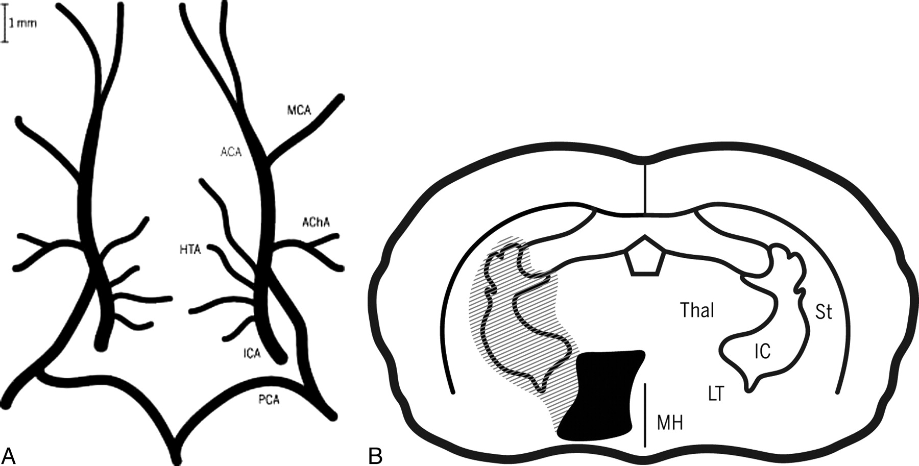
- Fig 1.
Schematic representation of the rat intracranial ICA and the territories of its proximal branches. A, Intracranial arterial supply in the rat with rostral oriented upward. The ACA, MCA, PCA, HTA, and AchoA are labeled.10 B, Axial representation of the rat forebrain 2 to 4 mm caudal to bregma shows the medial thalamus (MH) and lateral thalamus (LT), the thalamus (Thal), internal capsule (IC), and striatum (St). The vascular territories of the HTAs (gray hatching) and AchoA (filled black) are shown.
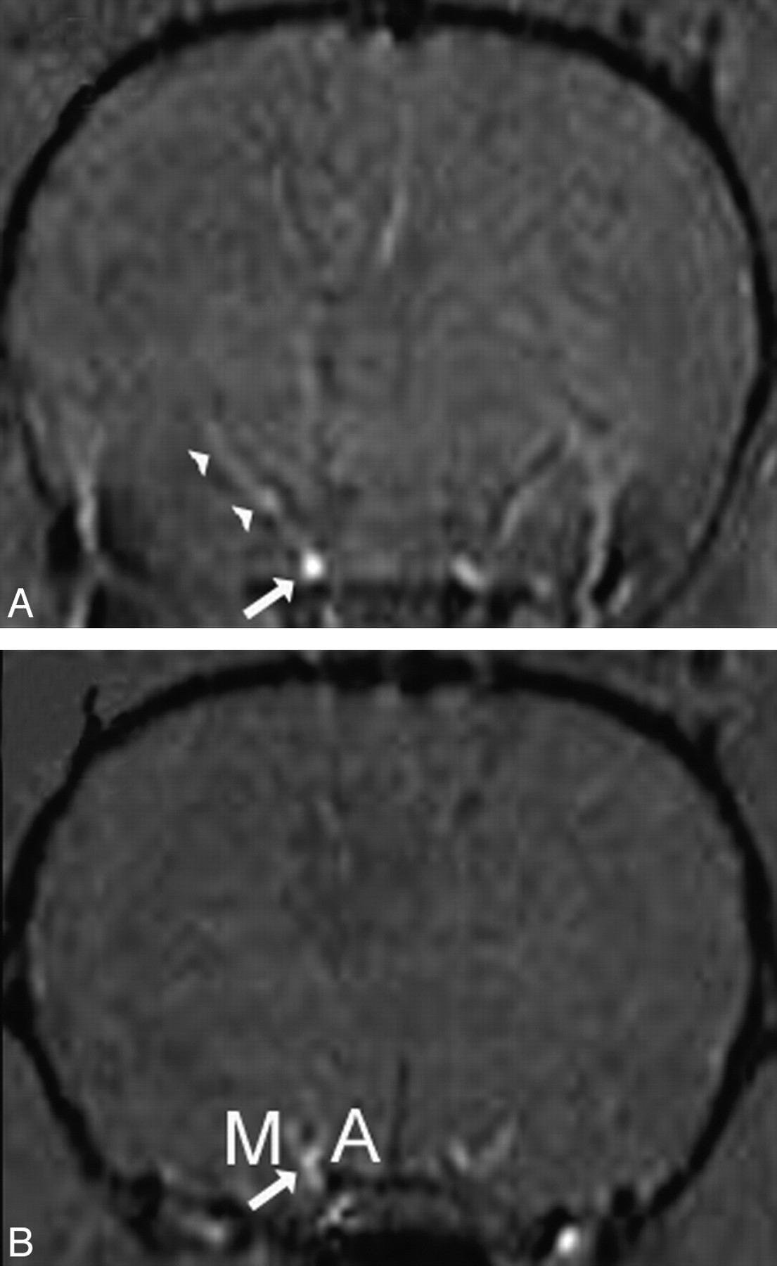
- Fig 2.
3D time-of-flight imaging was used to estimate the ICA diameter. Axial images show the origins of the AchoA (A) and MCA (B). Arrowheads show the course of the AchoA. M and A represent the origins of the MCA and ACA. The HTAs are not visualized.
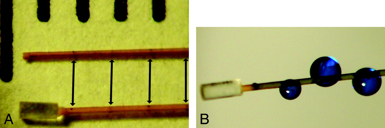
- Fig 3.
Rat brain arterial microcatheters. A, The 169-μm OD polyimide microcatheters have 35-μm perforations at 1, 2, 3, and 4 mm proximal to the tip (arrows). Every other pair of perforations is oriented 90° from the previous. The distal tips of both microcatheters are plugged with epoxy. The bottom microcatheter has a 450-μm bulb glued to its distal tip (μcath2). Markings at the top of the image represent millimeters. B, Methylene blue injected through μcath1 demonstrates side port positions.
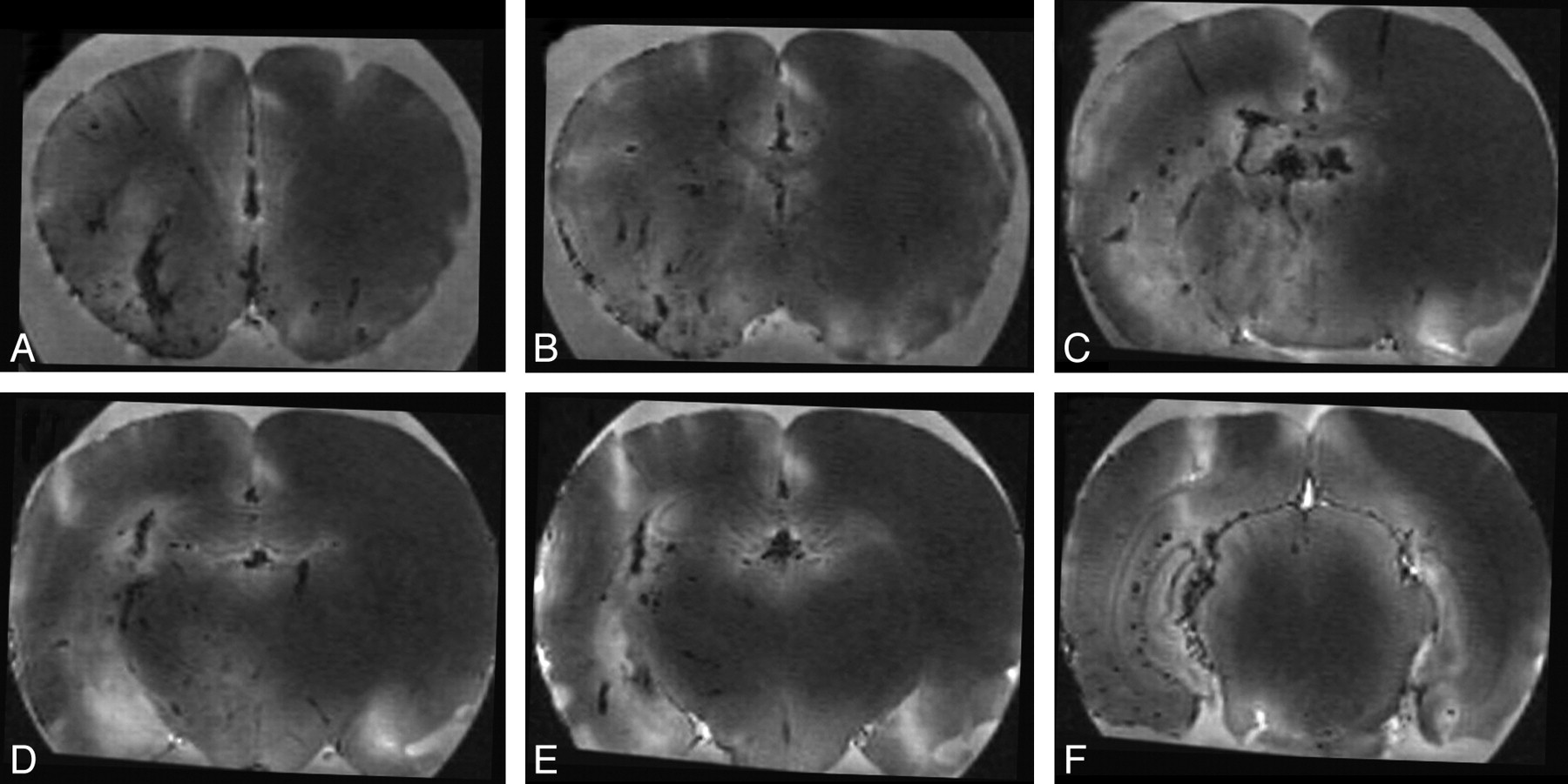
- Fig 4.
Injection of gadolinium into a single cerebral hemisphere demonstrated with ex vivo MR imaging. Thoracotomy was performed and the right atrium sectioned before injection of 300 μL of 1:1 Magnevist:saline for 10 minutes via μcath1. The brain was removed, soaked in formalin for 30 minutes, and imaged with an MPRAGE sequence. Axial images A to F are displayed from rostral to caudal at +1.7, +0.5, −0.9, −1.8, −2.1, and −5.8 mm relative to bregma. Gadolinium-containing vessels were dark because of the concentration effect. Midline vessels (not labeled), irregular vessels seen in long axis within the deep brain, and linear vessels within the cortical mantle are thought to represent veins. Branching vessels arising from the ventral aspect and vessels seen in cross-section are thought to represent arteries (not labeled).
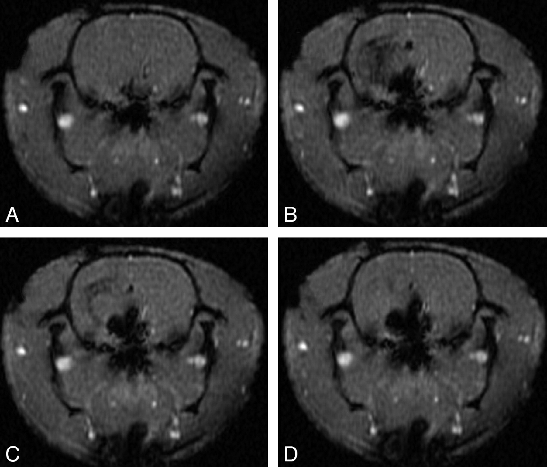
- Fig 5.
Single cerebral hemisphere injection during dynamic MR imaging. Axial T1 GE-MR images acquired 4 to 5 mm caudal to bregma at −1 (A) and 5 (B), 11 (C), and 29 (D) s after the beginning of a 10-s hand-injection of Magnevist:NS via μcath1 (ie, from 0–10 s).
- Fig 6.
Selective injection of gadolinium into the hypothalamus and lateral thalamus. Axial T1 GE-MR images acquired 4 to 5 mm caudal to bregma at 0 (A), 6 (B), 12 (C), and 30 s (D) after the beginning of a 10-s hand-injection of Magnevist:NS via μcath2.
- Fig 7.
Selective injection of 18FDG into the hypothalamus. MicroPET data were acquired 5 minutes after injection of 20 μCi 18FDG in 10-μL NS via μcath2 for 1 minute. Activity is represented by red (270 μCi/mL), yellow (240 μCi/mL), green (195 μCi/mL), and blue (90 μCi/mL). MicroPET images were manually coregistered with 2-mm-thick axial T1-weighted images and coronal and sagittal reformats. Panel A is 1.0 to 0.8 mm rostral to bregma. Panels B through D are 1.0 to 1.2 mm, 3.0 to 3.2 mm, and 5.0 to 5.2 mm caudal to bregma, respectively.


Tables
Experimental protocol*
Animal # μcath Imaging Modality Figure 1 1 Ex vivo MPRAGE 4 2–4 1 In vivo DSC-MR 5 5–7 2 In vivo DSC-MR 6 8 2 In vivo microPET/T1 MR 7 9–12 1 In vivo laser Doppler − 13–16 2 In vivo laser Doppler − Note:—MPRAGE indicates magnetization-prepared rapid acquisition of gradient echo MR imaging; DSC-MR, dynamic susceptibility contrast-enhanced MR imaging.
* In animals #1 to #4 and #9 to #12, μcath1 was placed with its tip 22 to 23 mm distal to the common carotid bifurcation. In animals #5 to #8 and #13 to #16, μcath2 was placed with its tip 18 to 19 mm distal to the common carotid. Gd-DTPA was injected before (#1) or during (#2–#7) image acquisition. In animal #8, 18FDG was injected 5 minutes before imaging was performed. In animals #9 to #16, a μcath was left in place for up to 25 minutes, during which time NS was injected at a rate of 20 μL/min (#9–12) or 10 μL/min (#13–16). Data from animals #9 to #16 are described in the Results section but are not depicted graphically.
In this issue


Jump to section
Related Articles
Cited By...
- No citing articles found.