Advertisement

Main menu

  • Home
  • Content
    • Current Issue
    • Accepted Manuscripts
    • Article Preview
    • Past Issue Archive
    • Video Articles
    • AJNR Case Collection
    • Case of the Week Archive
    • Case of the Month Archive
    • Classic Case Archive
  • Special Collections
    • AJNR Awards
    • Low-Field MRI
    • Alzheimer Disease
    • ASNR Foundation Special Collection
    • Photon-Counting CT
    • View All
  • Multimedia
    • AJNR Podcasts
    • AJNR SCANtastic
    • Trainee Corner
    • MRI Safety Corner
    • Imaging Protocols
  • For Authors
    • Submit a Manuscript
    • Submit a Video Article
    • Submit an eLetter to the Editor/Response
    • Manuscript Submission Guidelines
    • Statistical Tips
    • Fast Publishing of Accepted Manuscripts
    • Graphical Abstract Preparation
    • Imaging Protocol Submission
    • Author Policies
  • About Us
    • About AJNR
    • Editorial Board
    • Editorial Board Alumni
  • More
    • Become a Reviewer/Academy of Reviewers
    • Subscribers
    • Permissions
    • Alerts
    • Feedback
    • Advertisers
    • ASNR Home

User menu

  • Alerts
  • Log in

Search

  • Advanced search
American Journal of Neuroradiology

American Journal of Neuroradiology

ASHNR American Society of Functional Neuroradiology ASHNR American Society of Pediatric Neuroradiology ASSR
  • Alerts
  • Log in

Advanced Search

  • Home
  • Content
    • Current Issue
    • Accepted Manuscripts
    • Article Preview
    • Past Issue Archive
    • Video Articles
    • AJNR Case Collection
    • Case of the Week Archive
    • Case of the Month Archive
    • Classic Case Archive
  • Special Collections
    • AJNR Awards
    • Low-Field MRI
    • Alzheimer Disease
    • ASNR Foundation Special Collection
    • Photon-Counting CT
    • View All
  • Multimedia
    • AJNR Podcasts
    • AJNR SCANtastic
    • Trainee Corner
    • MRI Safety Corner
    • Imaging Protocols
  • For Authors
    • Submit a Manuscript
    • Submit a Video Article
    • Submit an eLetter to the Editor/Response
    • Manuscript Submission Guidelines
    • Statistical Tips
    • Fast Publishing of Accepted Manuscripts
    • Graphical Abstract Preparation
    • Imaging Protocol Submission
    • Author Policies
  • About Us
    • About AJNR
    • Editorial Board
    • Editorial Board Alumni
  • More
    • Become a Reviewer/Academy of Reviewers
    • Subscribers
    • Permissions
    • Alerts
    • Feedback
    • Advertisers
    • ASNR Home
  • Follow AJNR on Twitter
  • Visit AJNR on Facebook
  • Follow AJNR on Instagram
  • Join AJNR on LinkedIn
  • RSS Feeds

AJNR Awards, New Junior Editors, and more. Read the latest AJNR updates

OtherResearch Perspectives
Open Access

Challenges of Using MR Spectroscopy to Detect Neural Progenitor Cells In Vivo

Z. Dong, W. Dreher, D. Leibfritz and B.S. Peterson
American Journal of Neuroradiology June 2009, 30 (6) 1096-1101; DOI: https://doi.org/10.3174/ajnr.A1557
Z. Dong
  • Find this author on Google Scholar
  • Find this author on PubMed
  • Search for this author on this site
W. Dreher
  • Find this author on Google Scholar
  • Find this author on PubMed
  • Search for this author on this site
D. Leibfritz
  • Find this author on Google Scholar
  • Find this author on PubMed
  • Search for this author on this site
B.S. Peterson
  • Find this author on Google Scholar
  • Find this author on PubMed
  • Search for this author on this site
  • Article
  • Figures & Data
  • Info & Metrics
  • Responses
  • References
  • PDF
Loading

Article Figures & Data

Figures

  • Fig 1.
    • Download figure
    • Open in new tab
    • Download powerpoint
    Fig 1.

    Synthesized spectra of proton MR spectroscopy with typical line shapes as seen in vivo and their spectral fitting estimated by using the method of SVD. The same noise was added to the synthesized signals in the time domain. A, An apparent single spectral peak (black) consisting of 3 Lorentzian peaks with zero phase (red, green, and blue) is estimated correctly when the 3 fitted components have identical phases. The complex combination of these components (red) coincides perfectly with the synthesized peak in black, as is true for all the other fittings (B–D). B, A single peak with a Gaussian line shape (black) is decomposed into 4 components, each with differing phases. Unlike the peaks identified in A, where the phases are identical, these components cannot be interpreted as valid peaks even though their combination constitutes a “perfect” fit. C, A single peak with an asymmetric line shape (black), simulating the distortion caused by inhomogeneity of the magnetic field, is decomposed into 5 components (colored), each with differing phases. None of the components can be regarded as a valid peak. D, The synthesized signal (black) consists of 2 components, 1 the same as that in C and the other its duplicate but reduced in amplitude by a factor of 5 and shifted in frequency by 10 Hz. The signal is decomposed into 6 components, each with differing phases (colored), none of which can be interpreted as valid. Freq indicates frequency.
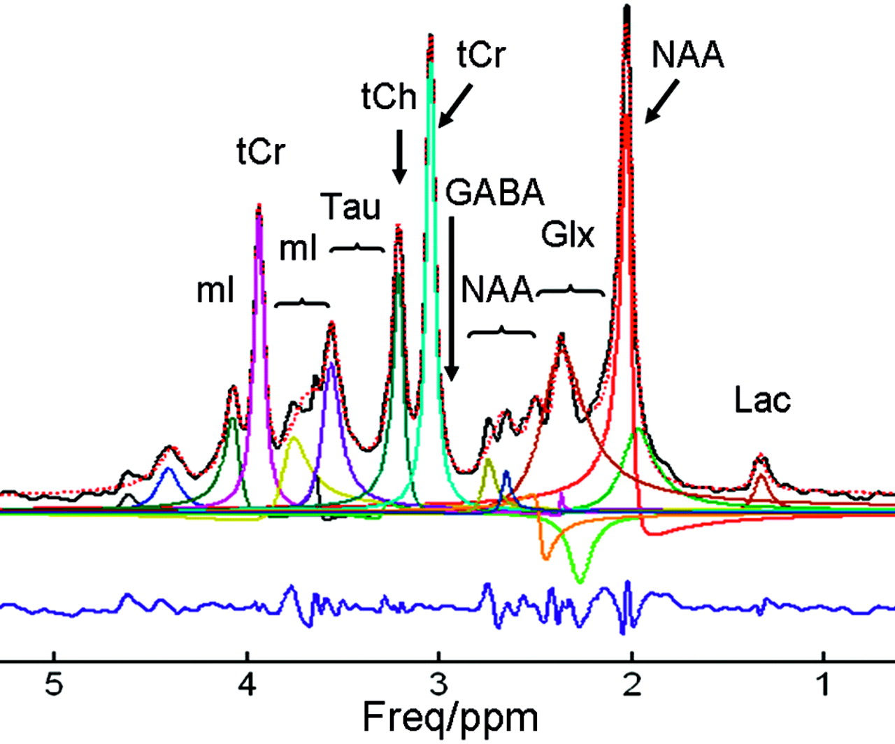
  • Fig 2.
    • Download figure
    • Open in new tab
    • Download powerpoint
    Fig 2.

    Shown are the synthesized in vivo 1H-MR spectroscopy spectrum (solid black), the overall fitting provided by SVD (dashed red), the individual components provided by SVD (solid color), and the residue (blue). The metabolites used in the simulation include N-acetylaspartate (NAA), N-acetyl aspartylglutamate (NAAG), total creatine (tCr), total choline (tCh), GABA, taurine (Tau), lactate (Lac), glutamine/glutamate (Glx), and myo-inositol (mI). The chemical shifts and J-couplings were taken from Govindaraju et al.20 Simulation parameters include the following: system frequency = 200 MHz, spectral width = 4006 Hz, TE = 20 ms, Lorentzian line width = 10 Hz, number of data points = 4096. The first 1024 data points of the FID were used in the SVD procedure. The number of spectral components was 24 (19 were in the range of 0–5 ppm), which was enforced by the user as prior knowledge. The overlapping lines of Cr and PCr, Ch and PCh, NAA and NAAG, and the doublets of Lac cannot be resolved and are instead detected as singlets. The phase error in the NAA singlet at 2.02 ppm is obviously caused by the spurious broad component in its vicinity. Two components with negative phases are detected around 2.3 ppm, clearly indicating that these components are not interpretable and other components in this region are not reliable. Increasing or reducing the number of spectral components as prior knowledge did alter the overall fitting and did not ameliorate the behavior of spectral components. Freq indicates frequency.

PreviousNext
Back to top

In this issue

American Journal of Neuroradiology: 30 (6)
American Journal of Neuroradiology
Vol. 30, Issue 6
June 2009
  • Table of Contents
  • Index by author
Advertisement
Print
Download PDF
Email Article

Thank you for your interest in spreading the word on American Journal of Neuroradiology.

NOTE: We only request your email address so that the person you are recommending the page to knows that you wanted them to see it, and that it is not junk mail. We do not capture any email address.

Enter multiple addresses on separate lines or separate them with commas.
Challenges of Using MR Spectroscopy to Detect Neural Progenitor Cells In Vivo
(Your Name) has sent you a message from American Journal of Neuroradiology
(Your Name) thought you would like to see the American Journal of Neuroradiology web site.
CAPTCHA
This question is for testing whether or not you are a human visitor and to prevent automated spam submissions.
Cite this article
Z. Dong, W. Dreher, D. Leibfritz, B.S. Peterson
Challenges of Using MR Spectroscopy to Detect Neural Progenitor Cells In Vivo
American Journal of Neuroradiology Jun 2009, 30 (6) 1096-1101; DOI: 10.3174/ajnr.A1557

Citation Manager Formats

  • BibTeX
  • Bookends
  • EasyBib
  • EndNote (tagged)
  • EndNote 8 (xml)
  • Medlars
  • Mendeley
  • Papers
  • RefWorks Tagged
  • Ref Manager
  • RIS
  • Zotero
0 Responses
Respond to this article
Share
Bookmark this article
Challenges of Using MR Spectroscopy to Detect Neural Progenitor Cells In Vivo
Z. Dong, W. Dreher, D. Leibfritz, B.S. Peterson
American Journal of Neuroradiology Jun 2009, 30 (6) 1096-1101; DOI: 10.3174/ajnr.A1557
del.icio.us logo Twitter logo Facebook logo Mendeley logo
  • Tweet Widget
  • Facebook Like
  • Google Plus One
Purchase

Jump to section

  • Article
    • Abstract
    • Comments
    • Possible Strategies for Detecting NPCs In Vivo
    • Conclusions
    • Footnotes
    • References
  • Figures & Data
  • Info & Metrics
  • Responses
  • References
  • PDF

Related Articles

  • No related articles found.
  • PubMed
  • Google Scholar

Cited By...

  • ORYX-MRSI: A Fully-Automated Open-Source Software for Three-Dimensional Proton Magnetic Resonance Spectroscopic Imaging Data Analysis
  • Crossref
  • Google Scholar

This article has not yet been cited by articles in journals that are participating in Crossref Cited-by Linking.

More in this TOC Section

  • Hot Topics in Research: Preventive Neuroradiology in Brain Aging and Cognitive Decline
  • Evidence Levels for Neuroradiology Articles: Low Agreement among Raters
  • Imaging Biomarkers in Ischemic Stroke Clinical Trials: An Opportunity for Rigor
Show more Research Perspectives

Similar Articles

Advertisement

Indexed Content

  • Current Issue
  • Accepted Manuscripts
  • Article Preview
  • Past Issues
  • Editorials
  • Editor's Choice
  • Fellows' Journal Club
  • Letters to the Editor
  • Video Articles

Cases

  • Case Collection
  • Archive - Case of the Week
  • Archive - Case of the Month
  • Archive - Classic Case

More from AJNR

  • Trainee Corner
  • Imaging Protocols
  • MRI Safety Corner
  • Book Reviews

Multimedia

  • AJNR Podcasts
  • AJNR Scantastics

Resources

  • Turnaround Time
  • Submit a Manuscript
  • Submit a Video Article
  • Submit an eLetter to the Editor/Response
  • Manuscript Submission Guidelines
  • Statistical Tips
  • Fast Publishing of Accepted Manuscripts
  • Graphical Abstract Preparation
  • Imaging Protocol Submission
  • Evidence-Based Medicine Level Guide
  • Publishing Checklists
  • Author Policies
  • Become a Reviewer/Academy of Reviewers
  • News and Updates

About Us

  • About AJNR
  • Editorial Board
  • Editorial Board Alumni
  • Alerts
  • Permissions
  • Not an AJNR Subscriber? Join Now
  • Advertise with Us
  • Librarian Resources
  • Feedback
  • Terms and Conditions
  • AJNR Editorial Board Alumni

American Society of Neuroradiology

  • Not an ASNR Member? Join Now

© 2025 by the American Society of Neuroradiology All rights, including for text and data mining, AI training, and similar technologies, are reserved.
Print ISSN: 0195-6108 Online ISSN: 1936-959X

Powered by HighWire