Advertisement

Main menu

  • Home
  • Content
    • Current Issue
    • Accepted Manuscripts
    • Article Preview
    • Past Issue Archive
    • Video Articles
    • AJNR Case Collection
    • Case of the Week Archive
    • Case of the Month Archive
    • Classic Case Archive
  • Special Collections
    • AJNR Awards
    • Low-Field MRI
    • Alzheimer Disease
    • ASNR Foundation Special Collection
    • Photon-Counting CT
    • View All
  • Multimedia
    • AJNR Podcasts
    • AJNR SCANtastic
    • Trainee Corner
    • MRI Safety Corner
    • Imaging Protocols
  • For Authors
    • Submit a Manuscript
    • Submit a Video Article
    • Submit an eLetter to the Editor/Response
    • Manuscript Submission Guidelines
    • Statistical Tips
    • Fast Publishing of Accepted Manuscripts
    • Graphical Abstract Preparation
    • Imaging Protocol Submission
    • Author Policies
  • About Us
    • About AJNR
    • Editorial Board
    • Editorial Board Alumni
  • More
    • Become a Reviewer/Academy of Reviewers
    • Subscribers
    • Permissions
    • Alerts
    • Feedback
    • Advertisers
    • ASNR Home

User menu

  • Alerts
  • Log in

Search

  • Advanced search
American Journal of Neuroradiology

American Journal of Neuroradiology

ASHNR American Society of Functional Neuroradiology ASHNR American Society of Pediatric Neuroradiology ASSR
  • Alerts
  • Log in

Advanced Search

  • Home
  • Content
    • Current Issue
    • Accepted Manuscripts
    • Article Preview
    • Past Issue Archive
    • Video Articles
    • AJNR Case Collection
    • Case of the Week Archive
    • Case of the Month Archive
    • Classic Case Archive
  • Special Collections
    • AJNR Awards
    • Low-Field MRI
    • Alzheimer Disease
    • ASNR Foundation Special Collection
    • Photon-Counting CT
    • View All
  • Multimedia
    • AJNR Podcasts
    • AJNR SCANtastic
    • Trainee Corner
    • MRI Safety Corner
    • Imaging Protocols
  • For Authors
    • Submit a Manuscript
    • Submit a Video Article
    • Submit an eLetter to the Editor/Response
    • Manuscript Submission Guidelines
    • Statistical Tips
    • Fast Publishing of Accepted Manuscripts
    • Graphical Abstract Preparation
    • Imaging Protocol Submission
    • Author Policies
  • About Us
    • About AJNR
    • Editorial Board
    • Editorial Board Alumni
  • More
    • Become a Reviewer/Academy of Reviewers
    • Subscribers
    • Permissions
    • Alerts
    • Feedback
    • Advertisers
    • ASNR Home
  • Follow AJNR on Twitter
  • Visit AJNR on Facebook
  • Follow AJNR on Instagram
  • Join AJNR on LinkedIn
  • RSS Feeds

AJNR Awards, New Junior Editors, and more. Read the latest AJNR updates

Research ArticleBrain

Multidetector Row CT Angiography in Spontaneous Lobar Intracerebral Hemorrhage: A Prospective Comparison with Conventional Angiography

D.Y. Yoon, S.K. Chang, C.S. Choi, W.-K. Kim and J.-H. Lee
American Journal of Neuroradiology May 2009, 30 (5) 962-967; DOI: https://doi.org/10.3174/ajnr.A1471
D.Y. Yoon
  • Find this author on Google Scholar
  • Find this author on PubMed
  • Search for this author on this site
S.K. Chang
  • Find this author on Google Scholar
  • Find this author on PubMed
  • Search for this author on this site
C.S. Choi
  • Find this author on Google Scholar
  • Find this author on PubMed
  • Search for this author on this site
W.-K. Kim
  • Find this author on Google Scholar
  • Find this author on PubMed
  • Search for this author on this site
J.-H. Lee
  • Find this author on Google Scholar
  • Find this author on PubMed
  • Search for this author on this site
  • Article
  • Figures & Data
  • Info & Metrics
  • Responses
  • References
  • PDF
Loading

Article Figures & Data

Figures

  • Tables
  • Fig 1.
    • Download figure
    • Open in new tab
    • Download powerpoint
    Fig 1.

    A 46-year-old man with an aneurysm of the distal MCA. A, Unenhanced CT scan demonstrates hemorrhage (arrow) in the right parietal lobe with mild surrounding edema. B and C, Left anterior oblique projection of the volume-rendered image (B) and the coronal multiplanar reformatted image (C) from MDCTA demonstrates a 2.5-mm saccular aneurysm (arrow) at the distal branch of the right MCA with an upward orientation of the sac. Note the relationship of the hematoma (arrowheads) to the aneurysm. D, DSA image of the right ICA, anteroposterior projection, shows the same aneurysm (arrow), which was confirmed at surgical exploration.

  • Fig 2.
    • Download figure
    • Open in new tab
    • Download powerpoint
    Fig 2.

    A 29-year-old man with an arteriovenous malformation. A, Unenhanced CT scan demonstrates hemorrhage (arrow) in the right frontal lobe. B, Volume-rendered MDCTA image, lateral projection, clearly demonstrates the abnormal collections of vessels in the right frontal lobe (arrowhead) and the enlarged draining cortical veins (arrows). C, Corresponding DSA image of the right ICA confirms these findings, demonstrating the nidus (arrowhead) and the early draining veins (arrows).
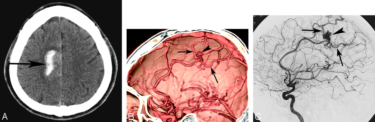
  • Fig 3.
    • Download figure
    • Open in new tab
    • Download powerpoint
    Fig 3.

    A 28-year-old woman with Moyamoya disease. A, Unenhanced CT scan demonstrates pericallosal hematoma (arrow) with extension of hemorrhage into the lateral ventricle. B, Volume-rendered MDCTA image with bone subtraction, anteroposterior projection, demonstrates symmetric occlusion of the bilateral proximal MCAs (arrows). MDCTA image fails to show collateral vessels. C and D, Corresponding DSA images of the right (C) and left (D) carotid arteries, anteroposterior projection, confirm occlusion of the bilateral proximal MCAs (arrows). In addition, reconstitution of the distal MCAs via fine collateral vessels is also noted.

Tables

  • Figures
    • View popup
    Table 1:

    Characteristics of patients and aneurysms

    Patient No.Age (yr)/SexLocationN/D RatioOrientationLobularityBranches from AneurysmSevere Vasospasm*
    DSAMDCTADSAMDCTAMDCTADSADSAMDCTADSAMDCTA
    148/FAcomA<1/3SameAnteriorSameMulti-Same(–)SameBilateral A1, A2; left M2Same
    240/FMCAB<1/31/3–2/3SuperiorSameSingle-Same(–)Same(–)Same
    356/MMCAB1/3–2/3SameSuperiorSameMulti-SameNeckSame(–)Same
    433/FAcomA>2/3SameInferiorSameMulti-Same(–)Same(–)Same
    539/FAcomA<1/3SameSuperiorSameSingle-Same(–)Same(–)Same
    654/MMCAB<1/3SameInferiorSameSingle-SameNeckSame(–)Same
    740/MDistal ICA>2/3SameSuperiorSameMulti-Same(–)Same(–)Same
    851/MMCAB1/3–2/3SameLateralSameSingle-SameDomeSameRight M2Same
    942/FMCAB>2/3SameLateralSameMulti-SameNeckSame(–)Same
    1046/MDistal MCA1/3–2/3SameSuperiorSameSingle-Same(–)Same(–)Same
    1144/MDistal ACA1/3–2/3SameAnteriorSameSingle-Same(–)Same(–)Same
    • Note:—AcomA indicates anterior communicating artery; MCAB, MCA bifurcation; A1, first segment of the ACA; A2, second segment of the ACA; M2, second segment of the MCA; DSA, digital subtraction angiography; ICA, internal carotid artery; MCA, middle cerebral artery; ACA, anterior cerebral artery.

    • * Arterial spasm was categorized as severe when the vessel diameters were reduced by >50%.

    • View popup
    Table 2:

    Characteristics of patients and AVMs

    Patient No.Age (yr)/SexLocationFeeding ArteriesNidusDraining Veins
    DSAMDCTADSAMDCTADSAMDCTA
    129/MFrontal lobeACASameVisibleSameCortical veins, SSSSame
    225/FFrontal lobeACANot visibleVisibleNot visibleDeep veins, ICVSame
    344/FFrontal lobeMCA, ACANot visibleVisibleSameCortical veins, SSSSame
    430/FTemporal lobeMCASameVisibleSameCortical veins, SSSSame
    535/FFrontal lobeACASameVisibleSameCortical veins, SSSSame
    631/MOccipital lobePCASameVisibleSameCortical veins, SSSame
    755/MParietal lobeMCANot visibleVisibleNot visibleCortical veins, SSSNot visible
    • Note:—SSS indicates superior sagittal sinus; ICV, internal cerebral vein; SS, sigmoid sinus; AVM, arteriovenous malformation; PCA, posterior cerebral artery.

    • View popup
    Table 3:

    Characteristics of patients and Moyamoya disease

    Patient No.Age (yr)/SexSite of OcclusionCollateral Vessels
    DSAMDCTADSAMDCTA
    128/FBilateral M1SameBasalNot visible
    231/FBilateral ICASameBasal, leptomeningealNot visible
    347/FBilateral ICASameBasal, leptomeningeal, transduralNot visible
    438/FUnilateral ICASameBasal, leptomeningealNot visible
    • Note:—M1 indicates the first segment of the MCA.

    • View popup
    Table 4:

    Diagnostic performance of MDCTA for the detection of underlying vascular abnormalities causing spontaneous lobar intracerebral hemorrhage in 78 patients

    TP CasesTN CasesFP CasesFN CasesSensitivity (%)Specificity (%)PPV (%)NPV (%)Accuracy (%)
    21560195.510010098.298.7
    • Note:—TP indicates true-positive; TN, true-negative; FP, false-positive; FN, false-negative; PPV, positive predictive value; NPV, negative predictive value; MDCTA, multidetector row CT angiography.

PreviousNext
Back to top

In this issue

American Journal of Neuroradiology: 30 (5)
American Journal of Neuroradiology
Vol. 30, Issue 5
May 2009
  • Table of Contents
  • Index by author
Advertisement
Print
Download PDF
Email Article

Thank you for your interest in spreading the word on American Journal of Neuroradiology.

NOTE: We only request your email address so that the person you are recommending the page to knows that you wanted them to see it, and that it is not junk mail. We do not capture any email address.

Enter multiple addresses on separate lines or separate them with commas.
Multidetector Row CT Angiography in Spontaneous Lobar Intracerebral Hemorrhage: A Prospective Comparison with Conventional Angiography
(Your Name) has sent you a message from American Journal of Neuroradiology
(Your Name) thought you would like to see the American Journal of Neuroradiology web site.
CAPTCHA
This question is for testing whether or not you are a human visitor and to prevent automated spam submissions.
Cite this article
D.Y. Yoon, S.K. Chang, C.S. Choi, W.-K. Kim, J.-H. Lee
Multidetector Row CT Angiography in Spontaneous Lobar Intracerebral Hemorrhage: A Prospective Comparison with Conventional Angiography
American Journal of Neuroradiology May 2009, 30 (5) 962-967; DOI: 10.3174/ajnr.A1471

Citation Manager Formats

  • BibTeX
  • Bookends
  • EasyBib
  • EndNote (tagged)
  • EndNote 8 (xml)
  • Medlars
  • Mendeley
  • Papers
  • RefWorks Tagged
  • Ref Manager
  • RIS
  • Zotero
0 Responses
Respond to this article
Share
Bookmark this article
Multidetector Row CT Angiography in Spontaneous Lobar Intracerebral Hemorrhage: A Prospective Comparison with Conventional Angiography
D.Y. Yoon, S.K. Chang, C.S. Choi, W.-K. Kim, J.-H. Lee
American Journal of Neuroradiology May 2009, 30 (5) 962-967; DOI: 10.3174/ajnr.A1471
del.icio.us logo Twitter logo Facebook logo Mendeley logo
  • Tweet Widget
  • Facebook Like
  • Google Plus One
Purchase

Jump to section

  • Article
    • Abstract
    • Materials and Methods
    • Results
    • Discussion
    • References
  • Figures & Data
  • Info & Metrics
  • Responses
  • References
  • PDF

Related Articles

  • No related articles found.
  • PubMed
  • Google Scholar

Cited By...

  • Guidelines for the Management of Spontaneous Intracerebral Hemorrhage: A Guideline for Healthcare Professionals From the American Heart Association/American Stroke Association
  • Neuroimaging in Intracerebral Hemorrhage
  • Emergency Noninvasive Angiography for Acute Intracerebral Hemorrhage
  • Yield of catheter angiography in patients with intracerebral hemorrhage with and without intraventricular extension
  • Dual-Energy CT in the Evaluation of Intracerebral Hemorrhage of Unknown Origin: Differentiation between Tumor Bleeding and Pure Hemorrhage
  • Frequency of Adequate Contrast Opacification of the Major Intracranial Venous Structures with CT Angiography in the Setting of Intracerebral Hemorrhage: Comparison of 16- and 64-Section CT Angiography Techniques
  • Computed Tomographic Angiography and Venography for Young or Nonhypertensive Patients With Acute Spontaneous Intracerebral Hemorrhage
  • Practical Scoring System for the Identification of Patients with Intracerebral Hemorrhage at Highest Risk of Harboring an Underlying Vascular Etiology: The Secondary Intracerebral Hemorrhage Score
  • Radiological Investigation of Spontaneous Intracerebral Hemorrhage: Systematic Review and Trinational Survey
  • Crossref
  • Google Scholar

This article has not yet been cited by articles in journals that are participating in Crossref Cited-by Linking.

More in this TOC Section

  • Usefulness of Quantitative Susceptibility Mapping for the Diagnosis of Parkinson Disease
  • White Matter Alterations in the Brains of Patients with Active, Remitted, and Cured Cushing Syndrome: A DTI Study
  • Qualitative and Quantitative Analysis of MR Imaging Findings in Patients with Middle Cerebral Artery Stroke Implanted with Mesenchymal Stem Cells
Show more Brain

Similar Articles

Advertisement

Indexed Content

  • Current Issue
  • Accepted Manuscripts
  • Article Preview
  • Past Issues
  • Editorials
  • Editor's Choice
  • Fellows' Journal Club
  • Letters to the Editor
  • Video Articles

Cases

  • Case Collection
  • Archive - Case of the Week
  • Archive - Case of the Month
  • Archive - Classic Case

More from AJNR

  • Trainee Corner
  • Imaging Protocols
  • MRI Safety Corner
  • Book Reviews

Multimedia

  • AJNR Podcasts
  • AJNR Scantastics

Resources

  • Turnaround Time
  • Submit a Manuscript
  • Submit a Video Article
  • Submit an eLetter to the Editor/Response
  • Manuscript Submission Guidelines
  • Statistical Tips
  • Fast Publishing of Accepted Manuscripts
  • Graphical Abstract Preparation
  • Imaging Protocol Submission
  • Evidence-Based Medicine Level Guide
  • Publishing Checklists
  • Author Policies
  • Become a Reviewer/Academy of Reviewers
  • News and Updates

About Us

  • About AJNR
  • Editorial Board
  • Editorial Board Alumni
  • Alerts
  • Permissions
  • Not an AJNR Subscriber? Join Now
  • Advertise with Us
  • Librarian Resources
  • Feedback
  • Terms and Conditions
  • AJNR Editorial Board Alumni

American Society of Neuroradiology

  • Not an ASNR Member? Join Now

© 2025 by the American Society of Neuroradiology All rights, including for text and data mining, AI training, and similar technologies, are reserved.
Print ISSN: 0195-6108 Online ISSN: 1936-959X

Powered by HighWire