Advertisement

Main menu

  • Home
  • Content
    • Current Issue
    • Accepted Manuscripts
    • Article Preview
    • Past Issue Archive
    • Video Articles
    • AJNR Case Collection
    • Case of the Week Archive
    • Case of the Month Archive
    • Classic Case Archive
  • Special Collections
    • AJNR Awards
    • Low-Field MRI
    • Alzheimer Disease
    • ASNR Foundation Special Collection
    • Photon-Counting CT
    • View All
  • Multimedia
    • AJNR Podcasts
    • AJNR SCANtastic
    • Trainee Corner
    • MRI Safety Corner
    • Imaging Protocols
  • For Authors
    • Submit a Manuscript
    • Submit a Video Article
    • Submit an eLetter to the Editor/Response
    • Manuscript Submission Guidelines
    • Statistical Tips
    • Fast Publishing of Accepted Manuscripts
    • Graphical Abstract Preparation
    • Imaging Protocol Submission
    • Author Policies
  • About Us
    • About AJNR
    • Editorial Board
    • Editorial Board Alumni
  • More
    • Become a Reviewer/Academy of Reviewers
    • Subscribers
    • Permissions
    • Alerts
    • Feedback
    • Advertisers
    • ASNR Home

User menu

  • Alerts
  • Log in

Search

  • Advanced search
American Journal of Neuroradiology

American Journal of Neuroradiology

ASHNR American Society of Functional Neuroradiology ASHNR American Society of Pediatric Neuroradiology ASSR
  • Alerts
  • Log in

Advanced Search

  • Home
  • Content
    • Current Issue
    • Accepted Manuscripts
    • Article Preview
    • Past Issue Archive
    • Video Articles
    • AJNR Case Collection
    • Case of the Week Archive
    • Case of the Month Archive
    • Classic Case Archive
  • Special Collections
    • AJNR Awards
    • Low-Field MRI
    • Alzheimer Disease
    • ASNR Foundation Special Collection
    • Photon-Counting CT
    • View All
  • Multimedia
    • AJNR Podcasts
    • AJNR SCANtastic
    • Trainee Corner
    • MRI Safety Corner
    • Imaging Protocols
  • For Authors
    • Submit a Manuscript
    • Submit a Video Article
    • Submit an eLetter to the Editor/Response
    • Manuscript Submission Guidelines
    • Statistical Tips
    • Fast Publishing of Accepted Manuscripts
    • Graphical Abstract Preparation
    • Imaging Protocol Submission
    • Author Policies
  • About Us
    • About AJNR
    • Editorial Board
    • Editorial Board Alumni
  • More
    • Become a Reviewer/Academy of Reviewers
    • Subscribers
    • Permissions
    • Alerts
    • Feedback
    • Advertisers
    • ASNR Home
  • Follow AJNR on Twitter
  • Visit AJNR on Facebook
  • Follow AJNR on Instagram
  • Join AJNR on LinkedIn
  • RSS Feeds

AJNR Awards, New Junior Editors, and more. Read the latest AJNR updates

OtherNeurointervention

Multi-Detector Row CT Angiography with Direct Intra-Arterial Contrast Injection for the Evaluation of Neurovascular Disease: Technique, Applications, and Initial Experience

D. Gandhi, A. Pandey, S.A. Ansari, J.J. Gemmete, B.G. Thompson and S.K. Mukherji
American Journal of Neuroradiology May 2009, 30 (5) 1054-1058; DOI: https://doi.org/10.3174/ajnr.A1438
D. Gandhi
  • Find this author on Google Scholar
  • Find this author on PubMed
  • Search for this author on this site
A. Pandey
  • Find this author on Google Scholar
  • Find this author on PubMed
  • Search for this author on this site
S.A. Ansari
  • Find this author on Google Scholar
  • Find this author on PubMed
  • Search for this author on this site
J.J. Gemmete
  • Find this author on Google Scholar
  • Find this author on PubMed
  • Search for this author on this site
B.G. Thompson Jr
  • Find this author on Google Scholar
  • Find this author on PubMed
  • Search for this author on this site
S.K. Mukherji
  • Find this author on Google Scholar
  • Find this author on PubMed
  • Search for this author on this site
  • Article
  • Figures & Data
  • Supplemental
  • Info & Metrics
  • Responses
  • References
  • PDF
Loading

Article Figures & Data

Figures

  • Fig 1.
    • Download figure
    • Open in new tab
    • Download powerpoint
    Fig 1.

    Patient 7. This patient presented with acute onset of left leg numbness. A, A noncontrast CT scan demonstrates a small bleed in the right parietal lobe. B, An oblique, magnified image of cerebral DSA reveals a tiny pial AVF (black arrow) drained by an early filling cortical vein (white arrows). Given the fairly superficial location of the lesion and recent history of bleed, surgical resection was recommended. C, A source image of IA-CTA reveals the point of connection between the parietal branch of the right anterior cerebral artery (arrow) and adjacent cortical vein. D, 3D volume-rendered images reveal the feeding artery, fistula, and the draining vein (labeled blue). The knowledge of the cross-sectional anatomy helped significantly when planning surgical approach. This lesion could be easily found, confirmed at surgery, and clipped.

  • Fig 2.
    • Download figure
    • Open in new tab
    • Download powerpoint
    Fig 2.

    Patient 10. This patient is a 50-year-old-woman who presented at our emergency department with diffuse SAH. A, Lateral view of the LICA demonstrates a multilobulated, complex aneurysm. B, 3D DSA reveals that the posterior communicating artery is originating from the aneurysmal neck (black arrowhead). Even on 3D imaging, the origin of the anterior choroidal artery (white arrows) cannot be located in relationship to the aneurysmal neck. C, A 3D IA-CTA clearly reveals that the anterior choroidal artery (white arrows) arises from the proximal aneurysmal dome. D, Magnified axial source image reveals the supraclinoid internal carotid artery anteriorly (black arrowheads). The anterior choroidal artery origin from the dome of the aneurysm is again easily identified. This excluded endovascular management, and this aneurysm was surgically clipped.
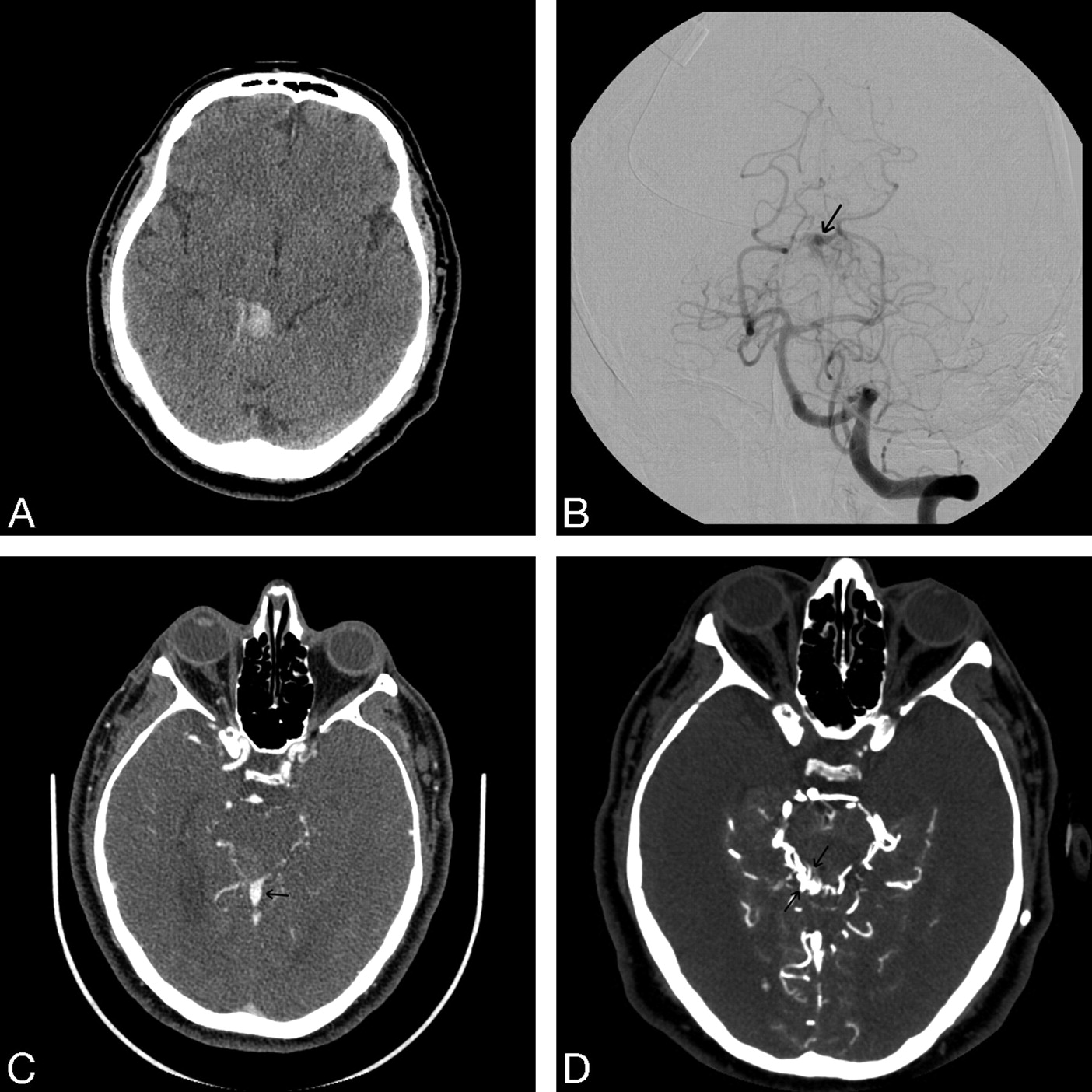
  • Fig 3.
    • Download figure
    • Open in new tab
    • Download powerpoint
    Fig 3.

    Patient 9. This patient is a 56-year-old man who presented with acute-onset headache and sudden neurologic deficits. A, Noncontrast CT scan demonstrates a focal hematoma in the midbrain. B, A high-frame rate DSA image of a left vertebral angiogram reveals small branches of the superior cerebellar artery in the region of the midbrain with an adjacent, early filling superior vermian vein (arrow). However, it could not be inferred with certainty whether this lesion is a fistula or a true AVM. C, A source image of IV-CTA reveals an abnormal, early filling superior vermian vein (arrow), but the nidus of the AVM is very difficult to identify. D, Source image of IA-CTA clearly demonstrates the nidus of a micro-AVM (arrows) in the region of the midbrain tectum as well as multiple tiny arterial feeders. The IA-CTA was considered superior to DSA as well as to IV-CTA in mapping as well as characterization of this tiny AVM. Wider window settings are intentionally used to demonstrate the individual tiny branches supplying this lesion.

PreviousNext
Back to top

In this issue

American Journal of Neuroradiology: 30 (5)
American Journal of Neuroradiology
Vol. 30, Issue 5
May 2009
  • Table of Contents
  • Index by author
Advertisement
Print
Download PDF
Email Article

Thank you for your interest in spreading the word on American Journal of Neuroradiology.

NOTE: We only request your email address so that the person you are recommending the page to knows that you wanted them to see it, and that it is not junk mail. We do not capture any email address.

Enter multiple addresses on separate lines or separate them with commas.
Multi-Detector Row CT Angiography with Direct Intra-Arterial Contrast Injection for the Evaluation of Neurovascular Disease: Technique, Applications, and Initial Experience
(Your Name) has sent you a message from American Journal of Neuroradiology
(Your Name) thought you would like to see the American Journal of Neuroradiology web site.
CAPTCHA
This question is for testing whether or not you are a human visitor and to prevent automated spam submissions.
Cite this article
D. Gandhi, A. Pandey, S.A. Ansari, J.J. Gemmete, B.G. Thompson, S.K. Mukherji
Multi-Detector Row CT Angiography with Direct Intra-Arterial Contrast Injection for the Evaluation of Neurovascular Disease: Technique, Applications, and Initial Experience
American Journal of Neuroradiology May 2009, 30 (5) 1054-1058; DOI: 10.3174/ajnr.A1438

Citation Manager Formats

  • BibTeX
  • Bookends
  • EasyBib
  • EndNote (tagged)
  • EndNote 8 (xml)
  • Medlars
  • Mendeley
  • Papers
  • RefWorks Tagged
  • Ref Manager
  • RIS
  • Zotero
0 Responses
Respond to this article
Share
Bookmark this article
Multi-Detector Row CT Angiography with Direct Intra-Arterial Contrast Injection for the Evaluation of Neurovascular Disease: Technique, Applications, and Initial Experience
D. Gandhi, A. Pandey, S.A. Ansari, J.J. Gemmete, B.G. Thompson, S.K. Mukherji
American Journal of Neuroradiology May 2009, 30 (5) 1054-1058; DOI: 10.3174/ajnr.A1438
del.icio.us logo Twitter logo Facebook logo Mendeley logo
  • Tweet Widget
  • Facebook Like
  • Google Plus One
Purchase

Jump to section

  • Article
    • Abstract
    • Technique
    • Results
    • Discussion
    • Conclusions
    • Footnotes
    • References
  • Figures & Data
  • Supplemental
  • Info & Metrics
  • Responses
  • References
  • PDF

Related Articles

  • PubMed
  • Google Scholar

Cited By...

  • Initial experience with a combined multidetector CT and biplane digital subtraction angiography suite with a single interactive table for the diagnosis and treatment of neurovascular disease
  • Crossref
  • Google Scholar

This article has not yet been cited by articles in journals that are participating in Crossref Cited-by Linking.

More in this TOC Section

  • Rescue Reentry in Carotid Near-Occlusion
  • Contour Neurovascular System: Five Year Follow Up
  • Effect of SARS-CoV2 on Endovascular Thrombectomy
Show more Neurointervention

Similar Articles

Advertisement

Indexed Content

  • Current Issue
  • Accepted Manuscripts
  • Article Preview
  • Past Issues
  • Editorials
  • Editor's Choice
  • Fellows' Journal Club
  • Letters to the Editor
  • Video Articles

Cases

  • Case Collection
  • Archive - Case of the Week
  • Archive - Case of the Month
  • Archive - Classic Case

More from AJNR

  • Trainee Corner
  • Imaging Protocols
  • MRI Safety Corner
  • Book Reviews

Multimedia

  • AJNR Podcasts
  • AJNR Scantastics

Resources

  • Turnaround Time
  • Submit a Manuscript
  • Submit a Video Article
  • Submit an eLetter to the Editor/Response
  • Manuscript Submission Guidelines
  • Statistical Tips
  • Fast Publishing of Accepted Manuscripts
  • Graphical Abstract Preparation
  • Imaging Protocol Submission
  • Evidence-Based Medicine Level Guide
  • Publishing Checklists
  • Author Policies
  • Become a Reviewer/Academy of Reviewers
  • News and Updates

About Us

  • About AJNR
  • Editorial Board
  • Editorial Board Alumni
  • Alerts
  • Permissions
  • Not an AJNR Subscriber? Join Now
  • Advertise with Us
  • Librarian Resources
  • Feedback
  • Terms and Conditions
  • AJNR Editorial Board Alumni

American Society of Neuroradiology

  • Not an ASNR Member? Join Now

© 2025 by the American Society of Neuroradiology All rights, including for text and data mining, AI training, and similar technologies, are reserved.
Print ISSN: 0195-6108 Online ISSN: 1936-959X

Powered by HighWire