
Article Figures & Data
Figures
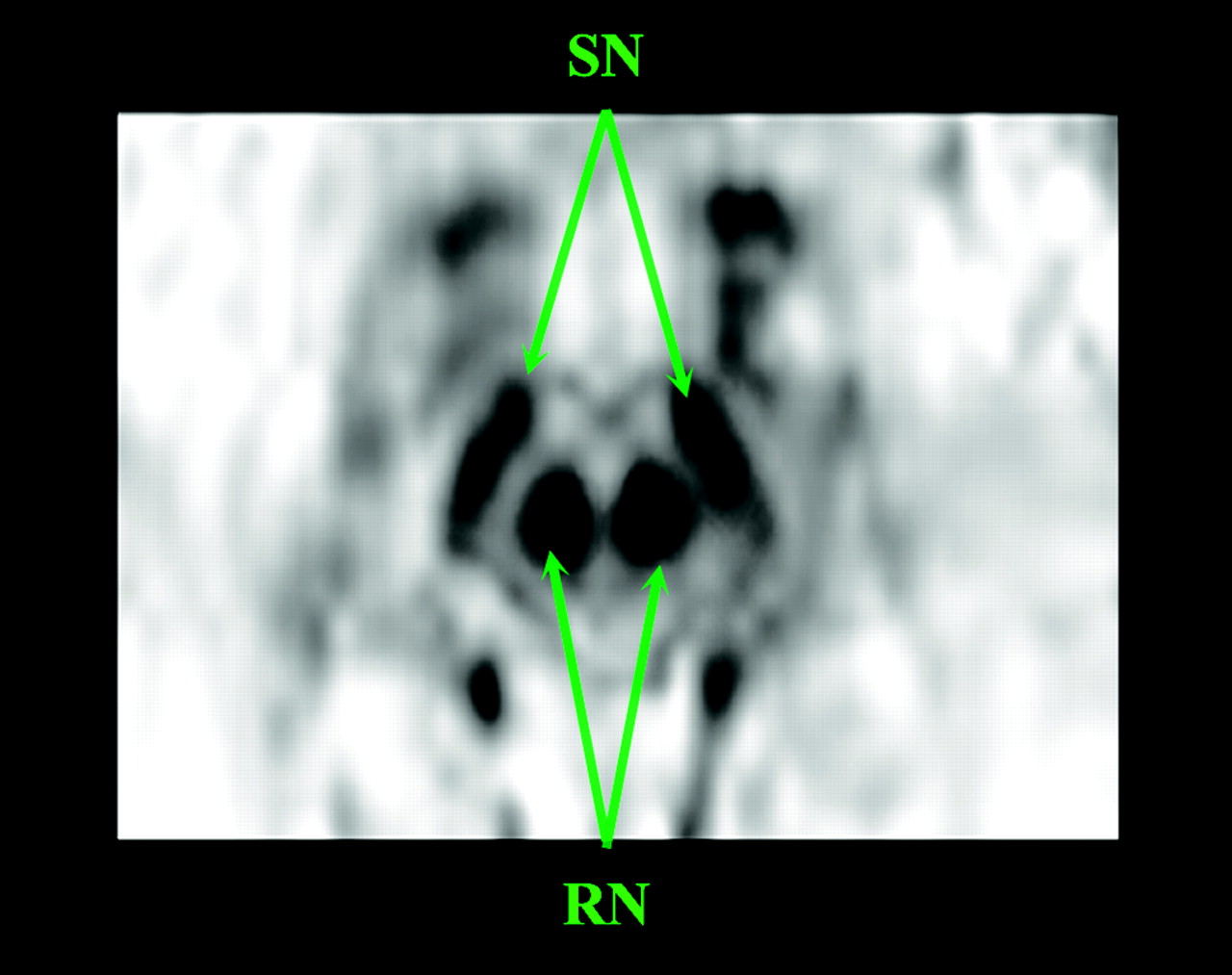
- Fig 1.
Axial T2*-weighted images through the mesencephalon showing the RN and SN on the right and left sides, with a strong signal hypointensity.
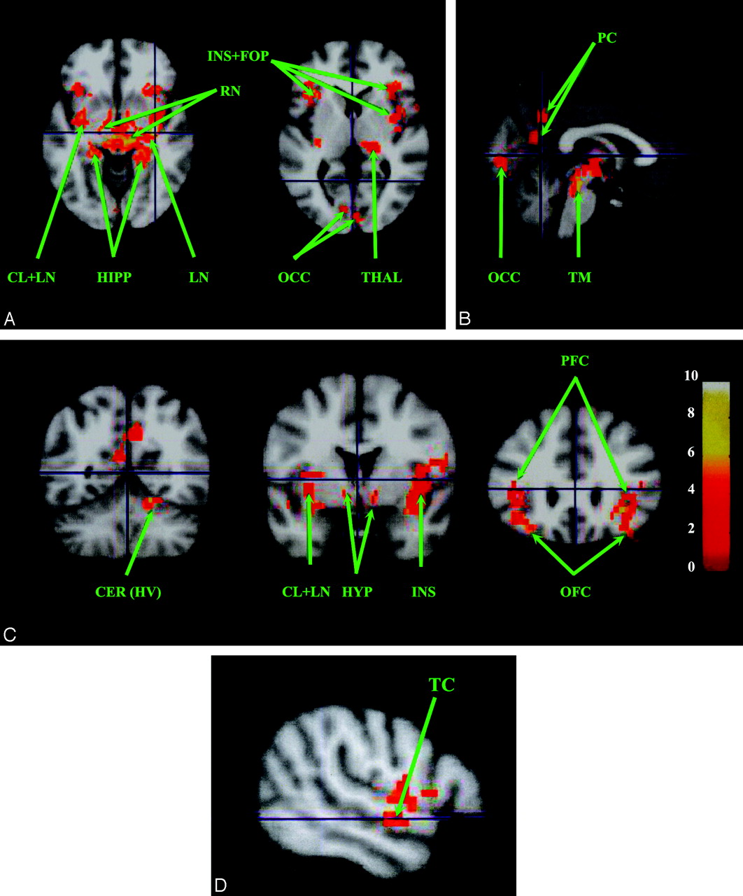
- Fig 2.
Resting-state neural connectivity maps (n = 14; P < .01; and k = 50) showing regions positively correlated with the right RN (A-C) and also found with the left RN. A, Axial sections. B, Sagittal section. C, Coronal sections. D, Sagittal section showing temporal region correlated with the left RN. D, T-score is represented by a color gradient (vertical colored bar). ACC, anterior cingulate cortex; CL, claustrum; CER, cerebellum; FOP, fronto-opercular cortex; HIPP, hippocampus; HYP, hypothalamus; INS, insula; LN, lentiform nucleus (pallidum and putamen); MES, mesencephalon; PFC, prefrontal cortex; RN, red nucleus; SN, substantia nigra; ST, striatum; THAL, thalamus; TC, temporal cortex; TM, tegmentum of the mesencephalon; m, medial; OFC, orbito-frontal cortex; HV, hemisphere of lobule V; OCC, occipital cortex.
- Fig 3.
Resting-state neural connectivity maps (n = 14; P < .01; and k = 50) showing regions positively correlated with the left SN (most of them are also found with the right SN; Table 2). A, Axial sections. B, Coronal sections. C, Sagittal sections. T-score is represented by a color gradient (vertical colored bar).


Tables
Brain Region BA Group-Analysis p <0.01, k = 50 MNI Coordinates (mm) Primary Peak Locations Right Side Left Side x y z x y z Right red nucleus Prefrontal cortex 45 (/47) 35.10 28.08 4.00 –32.76 23.40 –12.00 11 35.10 28.08 –12.00 –36.11 36.02 –13.54 Precuneus 7 m 6.86 –60.12 34.00 –3.86 –60.12 18.46 Occipital cortex 17 7.02 –86.58 –4.00 00.00 –88.92 4.00 36 32.76 –39.78 –16.00 – – – Hippocampus 35 23.40 –42.12 –8.00 –21.06 –35.10 –8.00 Insula 13 35.10 –16.38 –4.00 –36.11 –13.42 11.14 Claustrum 32.16 –17.08 –8.00 –33.08 –2.13 –8.05 Thalamus 15.62 –27.16 0.17 –12.86 –27.16 0.17 Hypothalamus 9.60 –1.52 –8.97 –6.43 –1.52 –8.97 Pallidum 14.04 –7.02 –8.00 –18.72 –16.38 00.00 Mesencephalic tegmentum 3.68 –27.16 –16.28 –3.20 –28.99 –11.79 Cerebellum 16.00 –58.50 –16.28 – – – Left red nucleus Prefrontal cortex 45 (/47) 35.10 28.08 –8.00 –35.10 28.08 –8.00 46 – – – –37.44 39.78 12.00 11 35.10 30.42 –4.00 –32.76 37.44 –12.00 Occipital cortex 37 25.74 –49.14 –12.00 – – – Temporal cortex 20 53.82 –4.68 –8.00 – – – Hippocampus 35 21.06 –35.10 –8.00 –16.38 –35.10 –8.00 Insula 13 44.46 4.68 –4.00 –35.10 14.04 –4.00 Claustrum 30.42 –16.38 –4.00 –28.08 –16.38 00.00 Thalamus 16.38 –21.06 4.00 –9.36 –21.06 4.00 Hypothalamus 9.36 2.34 –8.00 –4.68 –7.02 –12.00 Pallidum 23.10 –14.04 –4.00 –23.10 –16.38 00.00 Mesencephalic tegmentum 9.36 –28.08 –12.00 –7.02 –25.74 –12.00 Cerebellum 20.88 –51.48 –16.52 – – – Note:—BA indicates Brodmann area; MNI, Montreal Neurological Institute.
- Table 2:
Cluster localizations of the substantia nigra group-analysis P < .01, k = 50 MNI coordinates (mm)
BA Brain Region Primary Peak Locations Right Side Left Side x y z x y z Left substantia nigra Prefrontal cortex 11/47 37.44 28.08 –4.00 –32.76 28.08 –8.00 Frontal cortex 6 m 11.70 11.70 48.00 –7.02 2.34 48.00 4 46.80 –7.02 44.00 – – – Orbitofrontal cortex 25 4.68 9.36 –16.00 –2.34 9.36 –13.00 Occipital cortex 19/39 44.46 –79.56 32.00 – – – Temporal cortex 21 53.82 –11.70 –12.00 –51.48 –16.38 –8.00 Hippocampus 35 21.06 –35.10 –12.00 –16.38 –39.78 –12.00 Insula 13 44.46 4.68 –4.00 –30.42 9.36 –4.00 Lentiform nucleus 28.08 11.70 –4.00 –28.08 14.04 00.00 Thalamus 16.38 –16.38 00.00 –11.70 –18.72 4.00 Hypothalamus 7.02 –2.34 –12.00 –2.34 –2.34 –16.00 Mesencephalic tegmentum 4.68 –30.42 –20.00 –4.68 –28.08 –16.00 Pons 00.00 –32.76 –24.00 – – – Right substantia nigra Prefrontal cortex 46 46.80 39.78 20.00 11 (/47) 32.76 25.74 –8.00 –25.74 23.40 –16.00 9 (/6) 51.48 18.72 40.00 – – – 8 4.68 37.44 40.00 –2.34 37.44 44.00 Orbitofrontal cortex 25 7.02 9.36 –12.00 –4.68 9.36 –12.00 Motor cortex 4 51.48 –9.36 40.00 –56.16 –4.68 24.00 Cingulate cortex 24 (/32) 8.27 44.26 20.29 –2.34 37.44 8.00 Hippocampus 35 11.70 –39.78 –12.00 –23.40 –25.74 –12.00 Insula 13 39.78 –4.68 8.00 –41.59 11.70 2.00 Lentiform nucleus 25.74 4.68 –4.00 –25.74 11.70 –8.00 Caudate nucleus 9.60 11.30 2.92 –11.70 16.38 00.00 Claustrum 32.76 14.04 –4.00 –32.76 –11.70 –8.00 Thalamus 15.62 –14.34 4.00 –11.03 –16.17 4.00 Hypothalamus 11.70 –4.68 –8.00 –4.68 –4.68 –8.00 Mesencephalic tegmentum 7.02 –30.42 –20.00 –2.34 –28.08 –20.00 Cerebellum (vermis) 11.70 –56.16 –28.00 –4.68 –60.84 –24.00
In this issue


Jump to section
Related Articles
Cited By...
- Differences in brain activity during sentence repetition in people who stutter: a combined analysis of four fMRI studies
- Midbrain structure volume, estimated myelin and functional connectivity in idiopathic generalised epilepsy
- Hippocampal microstructural and neurobehavioral differences in welders are related to higher R2* in the red nucleus
- Neural Signals in Red Nucleus during Reactive and Proactive Adjustments in Behavior
- Reversible block of cerebellar outflow reveals cortical circuitry for motor coordination
- Valence, Not Utility, Underlies Reward-Driven Prioritization in Human Vision
- Assessment of Iron Deposition in the Brain in Frontotemporal Dementia and Its Correlation with Behavioral Traits
- Metabolic Activity of Red Nucleus and Its Correlation with Cerebral Cortex and Cerebellum: A Study Using a High-Resolution Semiconductor PET System