
Article Figures & Data
Figures
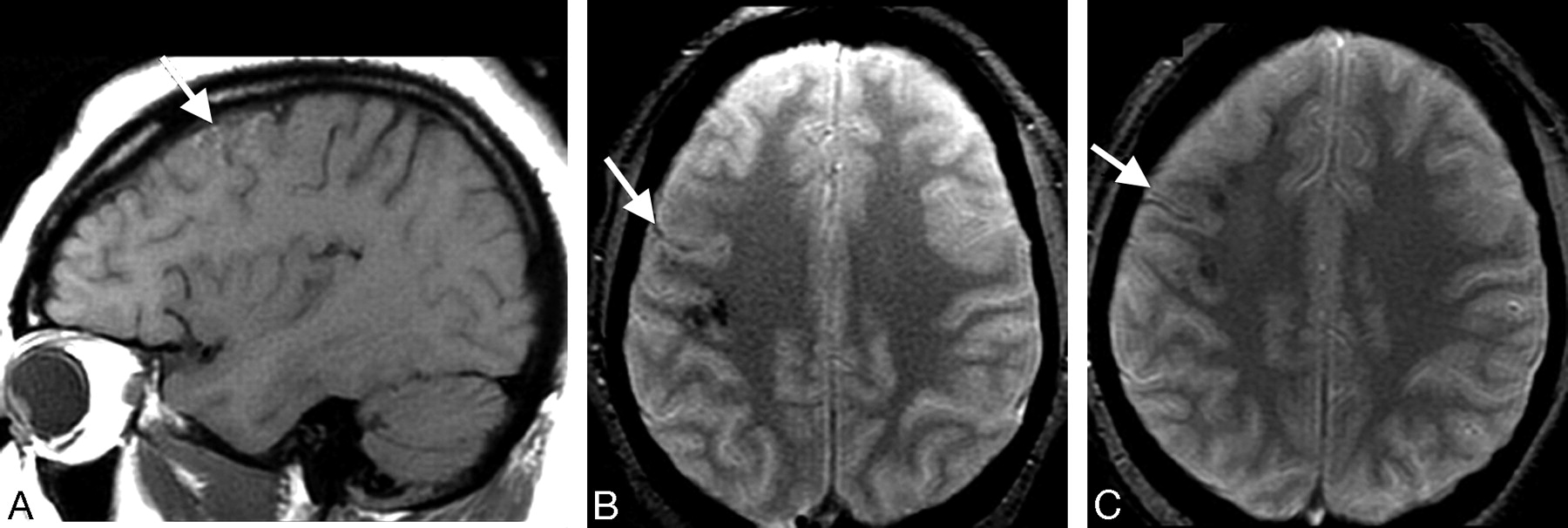
- Fig 1.
Patient 6. A and B, Initial MR imaging at day 7. On T2*GE images, linear hypointensity is seen in the right precentral sulcus, due to MSE at site of the thrombosed veins (small arrow) and hypointense areas corresponding to cortical hemorrhages (long arrows). C, On T1-weighted image, only swollen gyri (arrow) are identified.
- Fig 2.
Patient 6. A, Follow-up at month 3 reveals cortical hyperintensity on the T1-weighted image due to petechial hemorrhages. B, On the T2*GE image (same plane as in Fig 1A), note reduced but still visible MSE at the site of venous thrombosis (arrow), as well as petechial hemorrhages. C, At month 12, T1-weighted imaging findings are normal on T2*GE image (arrow); MSE is still identified.
- Fig 3.
Patient 7. MR imaging at initial examination at day 5. An extra-axial band corresponding to the thrombosed vein of Labbé appears as a marked MSE on T2*GE image (A, arrow), isointense on T1 (B, arrow), hypointense on FLAIR sequence (C, arrow), and as a signal-intensity loss on DWI (D, arrow).
- Fig 4.
Patient 8. Initial MR imaging at day 3. T2*GE shows MSE in the thrombosed cortical vein (A, arrow) and the hematoma, appearing on the T1-weighted image as a mass of mixed signal intensity (B, arrow).
- Fig 5.
Patient 8. A, Follow-up at day 15. MSE is still identified in the thrombosed vein. B, A hyperintense spot is present in the lumen of a cortical vein on the T1-weighted image (arrow), appearing as a flow void on the initial T1-weighted image (Fig 4B, arrow). C−E, On coronal images in the same plane, the aspect of a cord sign is visualized at the surface of the cortex, hyperintense on T1- and T2-weighted images (arrow, C and D) and hypointense on the T2*GE image (E). The underlying hematoma is well identified on T1- and T2*GE images.


In this issue


Jump to section
Related Articles
Cited By...
- A case of isolated cortical venous thrombosis presenting radiographically as a subacute multifocal leukoencephalopathy, and review of literature
- Isolated cortical venous thrombosis after fetal demise
- Current endovascular strategies for cerebral venous thrombosis: report of the SNIS Standards and Guidelines Committee
- Pearls & Oy-sters: Delayed progression of isolated cortical vein thrombosis despite therapeutic INR
- Early Detection and Quantification of Cerebral Venous Thrombosis by Magnetic Resonance Black-Blood Thrombus Imaging
- Teaching NeuroImages: Magnetic resonance susceptibility effect for acute isolated cortical vein thrombosis
- Isolated Cortical Vein Thrombosis: Systematic Review of Case Reports and Case Series
- Vein of Labbe thrombosis by CT and MRI
- Diagnosis and Management of Cerebral Venous Thrombosis: A Statement for Healthcare Professionals From the American Heart Association/American Stroke Association
- Isolated Acute Nontraumatic Cortical Subarachnoid Hemorrhage
- Cerebral Venous Thrombosis: Diagnostic Accuracy of Combined, Dynamic and Static, Contrast-Enhanced 4D MR Venography
- Reply:
- T2* Signal Hyperintensity in Subacute Cerebral Vein Thrombosis
- Diagnosis of cerebral cortical vein thrombosis with T2* weighted magnetic resonance imaging