Advertisement

Main menu

  • Home
  • Content
    • Current Issue
    • Accepted Manuscripts
    • Article Preview
    • Past Issue Archive
    • Video Articles
    • AJNR Case Collection
    • Case of the Week Archive
    • Case of the Month Archive
    • Classic Case Archive
  • Special Collections
    • AJNR Awards
    • Low-Field MRI
    • Alzheimer Disease
    • ASNR Foundation Special Collection
    • Photon-Counting CT
    • View All
  • Multimedia
    • AJNR Podcasts
    • AJNR SCANtastic
    • Trainee Corner
    • MRI Safety Corner
    • Imaging Protocols
  • For Authors
    • Submit a Manuscript
    • Submit a Video Article
    • Submit an eLetter to the Editor/Response
    • Manuscript Submission Guidelines
    • Statistical Tips
    • Fast Publishing of Accepted Manuscripts
    • Graphical Abstract Preparation
    • Imaging Protocol Submission
    • Author Policies
  • About Us
    • About AJNR
    • Editorial Board
    • Editorial Board Alumni
  • More
    • Become a Reviewer/Academy of Reviewers
    • Subscribers
    • Permissions
    • Alerts
    • Feedback
    • Advertisers
    • ASNR Home

User menu

  • Alerts
  • Log in

Search

  • Advanced search
American Journal of Neuroradiology

American Journal of Neuroradiology

ASHNR American Society of Functional Neuroradiology ASHNR American Society of Pediatric Neuroradiology ASSR
  • Alerts
  • Log in

Advanced Search

  • Home
  • Content
    • Current Issue
    • Accepted Manuscripts
    • Article Preview
    • Past Issue Archive
    • Video Articles
    • AJNR Case Collection
    • Case of the Week Archive
    • Case of the Month Archive
    • Classic Case Archive
  • Special Collections
    • AJNR Awards
    • Low-Field MRI
    • Alzheimer Disease
    • ASNR Foundation Special Collection
    • Photon-Counting CT
    • View All
  • Multimedia
    • AJNR Podcasts
    • AJNR SCANtastic
    • Trainee Corner
    • MRI Safety Corner
    • Imaging Protocols
  • For Authors
    • Submit a Manuscript
    • Submit a Video Article
    • Submit an eLetter to the Editor/Response
    • Manuscript Submission Guidelines
    • Statistical Tips
    • Fast Publishing of Accepted Manuscripts
    • Graphical Abstract Preparation
    • Imaging Protocol Submission
    • Author Policies
  • About Us
    • About AJNR
    • Editorial Board
    • Editorial Board Alumni
  • More
    • Become a Reviewer/Academy of Reviewers
    • Subscribers
    • Permissions
    • Alerts
    • Feedback
    • Advertisers
    • ASNR Home
  • Follow AJNR on Twitter
  • Visit AJNR on Facebook
  • Follow AJNR on Instagram
  • Join AJNR on LinkedIn
  • RSS Feeds

AJNR Awards, New Junior Editors, and more. Read the latest AJNR updates

Research ArticlePediatric Neuroimaging
Open Access

Variability of Homotopic and Heterotopic Callosal Connectivity in Partial Agenesis of the Corpus Callosum: A 3T Diffusion Tensor Imaging and Q-Ball Tractography Study

M. Wahl, Z. Strominger, R.J. Jeremy, A.J. Barkovich, M. Wakahiro, E.H. Sherr and P. Mukherjee
American Journal of Neuroradiology February 2009, 30 (2) 282-289; DOI: https://doi.org/10.3174/ajnr.A1361
M. Wahl
  • Find this author on Google Scholar
  • Find this author on PubMed
  • Search for this author on this site
Z. Strominger
  • Find this author on Google Scholar
  • Find this author on PubMed
  • Search for this author on this site
R.J. Jeremy
  • Find this author on Google Scholar
  • Find this author on PubMed
  • Search for this author on this site
A.J. Barkovich
  • Find this author on Google Scholar
  • Find this author on PubMed
  • Search for this author on this site
M. Wakahiro
  • Find this author on Google Scholar
  • Find this author on PubMed
  • Search for this author on this site
E.H. Sherr
  • Find this author on Google Scholar
  • Find this author on PubMed
  • Search for this author on this site
P. Mukherjee
  • Find this author on Google Scholar
  • Find this author on PubMed
  • Search for this author on this site
  • Article
  • Figures & Data
  • Info & Metrics
  • Responses
  • References
  • PDF
Loading

Article Figures & Data

Figures

  • Tables
  • Fig 1.
    • Download figure
    • Open in new tab
    • Download powerpoint
    Fig 1.

    Callosal tract segmentation procedure, shown for a control subject (A−F) and for pAgCC subject 5 (G−L). A region of interest is first placed over the entire callosum, and tracts are colored according to their direction with the standard red-blue-green convention used for DTI color maps (A and G). Individual tracts are then segmented by using 2 additional regions of interest defining lobar regions in each hemisphere. For the control subject, homotopic anterior (B) and posterior (C) frontal, parietal (D), and occipitotemporal (E) tracts are segmented. For the subject with pAgCC, homotopic anterior frontal (H) and occipitotemporal (I) tracts and bilateral heterotopic frontal occipitotemporal (J and K) tracts are isolated. In K, an exclusion region of interest (pink) is used to eliminate homotopic occipitotemporal connections. All segmented tracts are then displayed together (F and L). All 3D tracts are shown projected onto axial sections.

  • Fig 2.
    • Download figure
    • Open in new tab
    • Download powerpoint
    Fig 2.

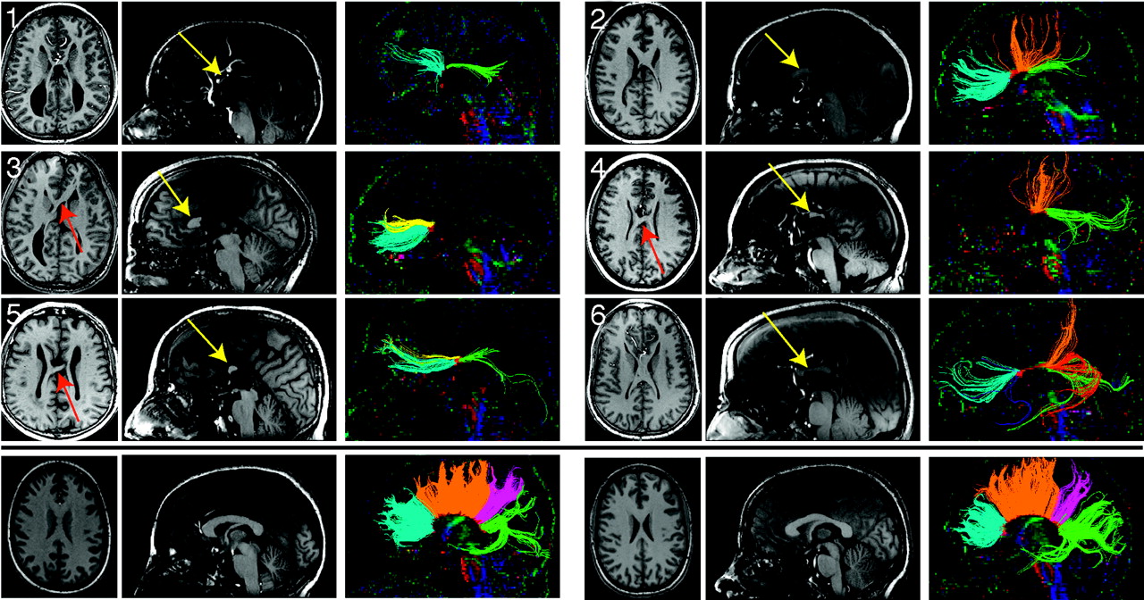
    T1-weighted anatomic images and DTI tractography of 6 subjects with pAgCC (top panels) and 2 representative controls (bottom panel). Axial (left) and midline sagittal (middle) T1 sections are shown for each subject. Callosal fragments are identified with yellow arrows, whereas heterotopic fibers visible on T1-weighted images are denoted by red arrows. Midline sagittal DTI color maps are shown with segmented callosal fibers (right). For subjects with pAgCC, connectivity ranged from anterior frontal connections (subject 3) to only posterior frontal and occipitotemporal connections (subject 4). One individual (subject 5) displayed a discontinuous set of homotopic callosal connections, with anterior frontal and occipitotemporal connectivity without any posterior frontal or parietal connections. Control subjects (not shown) displayed similar callosal morphology and tractography results. Tracts are segmented and colored according to their cortical projections: homotopic anterior frontal, blue; homotopic posterior frontal, orange; homotopic parietal, pink; homotopic occipitotemporal, green; heterotopic left anterior-right posterior, yellow; heterotopic right anterior-left posterior, red.

  • Fig 3.
    • Download figure
    • Open in new tab
    • Download powerpoint
    Fig 3.

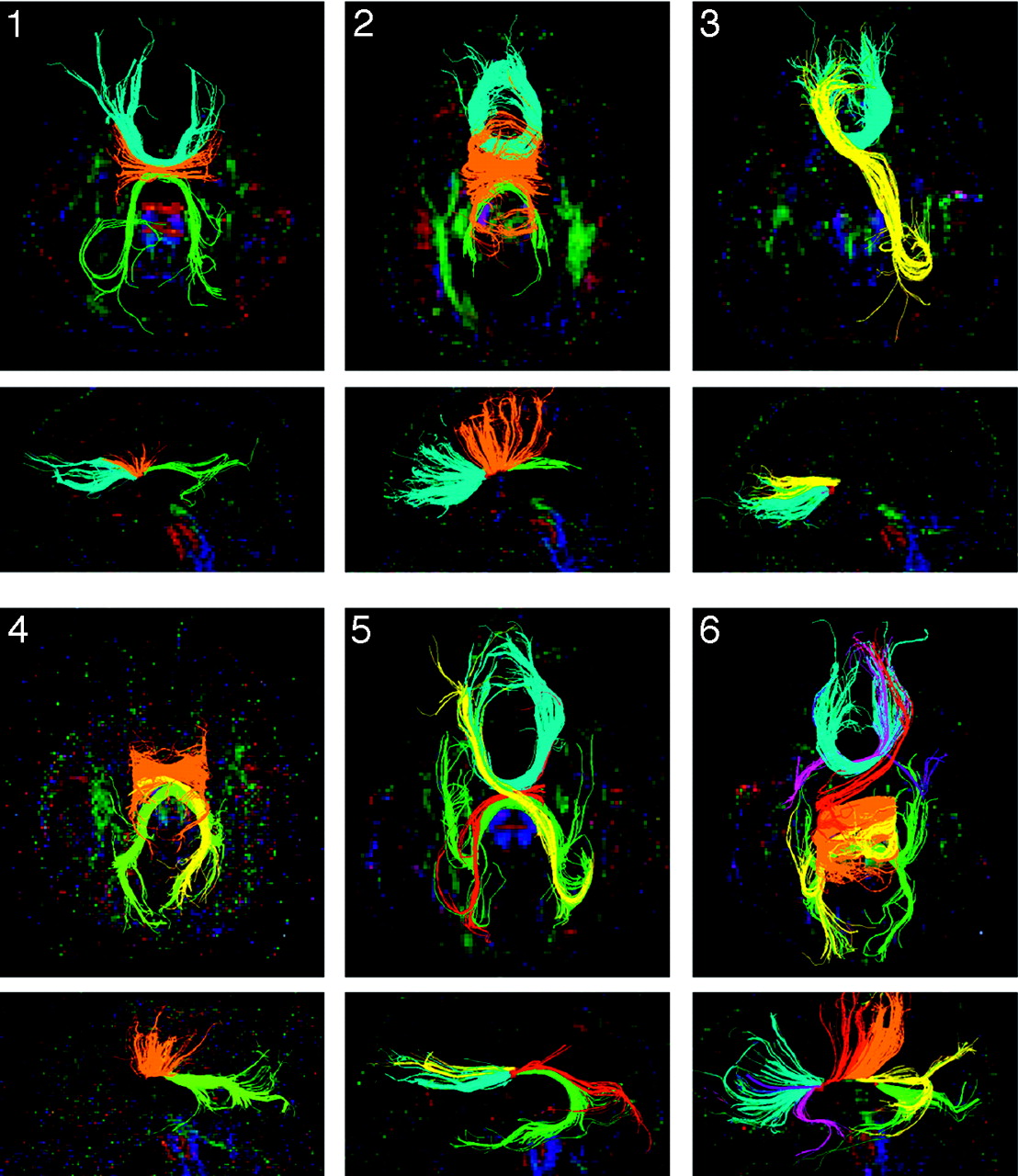
    Q-ball tractography of subjects with pAgCC. All homotopic and heterotopic segmented tracts are shown on both axial (top) and midline sagittal (bottom) projections, with the subject number indicated in the upper left corner of the axial images. Fibers are colored as in Fig 2, with pink and purple fibers for subject 6 representing anterior frontal-temporal heterotopic connections.

  • Fig 4.
    • Download figure
    • Open in new tab
    • Download powerpoint
    Fig 4.

    Q-ball tractography of heterotopic callosal connections. Isolated heterotopic connections are displayed for subjects 3–6. Fibers are shown projected on an axial section (left). A magnified view of the callosal fragment is also shown in the midsagittal plane (right) to demonstrate that all heterotopic connections are well isolated from other homotopic fibers.

  • Fig 5.
    • Download figure
    • Open in new tab
    • Download powerpoint
    Fig 5.

    Comparison of DTI and QBI tractography. For a control subject (top) and a subject with pAgCC (subject 5, bottom), segmented tracts are shown by using DTI tractography performed on a DTI acquisition at b = 1000 s/mm2 (A and D) and on a HARDI acquisition at b = 3000 s/mm2 (B and E). QBI tractography is shown for the same HARDI acquisition at b = 3000 s/mm2 (C and F). QBI tractography yields more extensive fibers, including more lateral frontal fibers and denser temporal fibers in the control subject and an additional heterotopic fiber in the subject with pAgCC (red in F) not recovered by DTI tractography.

Tables

  • Figures
  • Demographic information and summary of imaging findings for 6 subjects with pAgCC

    SubjectAge (yr)SexStructural Malformations*Fractional Fragment PositionPredicted Homotopic Connections†Observed Homotopic Connections‡Observed Heterotopic Connections‡§
    111FNone0.32–0.45PFAF, PF, OTNone
    248FRight lateral periventricular nodular heterotopia0.09–0.44AF, PFAF, PF, OTNone
    336MLeft frontal periventricular nodular heterotopia0.10–0.33AF, PFAFAF(L)-OT(R)
    437FNone0.46–0.70PFPF, OTPF(L)-OT(R)
    570MNone0.38–0.50PFAF, OTAF(L)-OT(R)
    AF(R)-OT(L)
    637MNone0.20–0.83PF, PAAF, PF, OTAF(R)-OT(L)
    AF(L)-OT(R)
    AF(R)-PF(L)
    PF(R)-OT(L)
    • Note:—AF indicates anterior frontal; PF, posterior frontal; PA, parietal; OT, occipitotemporal; L, left; R, right; pAgCC, partial agenesis of the corpus callosum.

    • * Structural malformations and fragment position (as a fraction of normal callosal length) were determined from high-resolution T1-weighted structural images.

    • † Predicted homotopic connectivity is based on fragment position.

    • ‡ Observed homotopic and heterotopic callosal connections are determined from QBI tractography.

    • § For heterotopic connections, the hemisphere of each connected cortical region is also denoted in parentheses.

PreviousNext
Back to top

In this issue

American Journal of Neuroradiology: 30 (2)
American Journal of Neuroradiology
Vol. 30, Issue 2
February 2009
  • Table of Contents
  • Index by author
Advertisement
Print
Download PDF
Email Article

Thank you for your interest in spreading the word on American Journal of Neuroradiology.

NOTE: We only request your email address so that the person you are recommending the page to knows that you wanted them to see it, and that it is not junk mail. We do not capture any email address.

Enter multiple addresses on separate lines or separate them with commas.
Variability of Homotopic and Heterotopic Callosal Connectivity in Partial Agenesis of the Corpus Callosum: A 3T Diffusion Tensor Imaging and Q-Ball Tractography Study
(Your Name) has sent you a message from American Journal of Neuroradiology
(Your Name) thought you would like to see the American Journal of Neuroradiology web site.
CAPTCHA
This question is for testing whether or not you are a human visitor and to prevent automated spam submissions.
Cite this article
M. Wahl, Z. Strominger, R.J. Jeremy, A.J. Barkovich, M. Wakahiro, E.H. Sherr, P. Mukherjee
Variability of Homotopic and Heterotopic Callosal Connectivity in Partial Agenesis of the Corpus Callosum: A 3T Diffusion Tensor Imaging and Q-Ball Tractography Study
American Journal of Neuroradiology Feb 2009, 30 (2) 282-289; DOI: 10.3174/ajnr.A1361

Citation Manager Formats

  • BibTeX
  • Bookends
  • EasyBib
  • EndNote (tagged)
  • EndNote 8 (xml)
  • Medlars
  • Mendeley
  • Papers
  • RefWorks Tagged
  • Ref Manager
  • RIS
  • Zotero
0 Responses
Respond to this article
Share
Bookmark this article
Variability of Homotopic and Heterotopic Callosal Connectivity in Partial Agenesis of the Corpus Callosum: A 3T Diffusion Tensor Imaging and Q-Ball Tractography Study
M. Wahl, Z. Strominger, R.J. Jeremy, A.J. Barkovich, M. Wakahiro, E.H. Sherr, P. Mukherjee
American Journal of Neuroradiology Feb 2009, 30 (2) 282-289; DOI: 10.3174/ajnr.A1361
del.icio.us logo Twitter logo Facebook logo Mendeley logo
  • Tweet Widget
  • Facebook Like
  • Google Plus One
Purchase

Jump to section

  • Article
    • Abstract
    • Materials and Methods
    • Results
    • Discussion
    • Conclusions
    • Acknowledgments
    • Footnotes
    • References
  • Figures & Data
  • Info & Metrics
  • Responses
  • References
  • PDF

Related Articles

  • No related articles found.
  • PubMed
  • Google Scholar

Cited By...

  • Development and plasticity of the corpus callosum
  • Increased cognitive complexity reveals abnormal brain network activity in individuals with corpus callosum dysgenesis
  • Structural Connectivity Analysis in Children with Segmental Callosal Agenesis
  • Structural and functional brain rewiring clarifies preserved interhemispheric transfer in humans born without the corpus callosum
  • Diffusion Imaging of the Congenitally Thickened Corpus Callosum
  • Novel Proximal 14q Deletion: Clinical and Diffusion Tensor Imaging Tractography Findings in a Patient with Lissencephaly, Agenesis of the Corpus Callosum, and Septo-Optic Dysplasia
  • The corpus callosum: white matter or terra incognita
  • Wiring the Brain: The Biology of Neuronal Guidance
  • Human Genetic Disorders of Axon Guidance
  • Partial Development of the Corpus Callosum
  • Reply:
  • Crossref
  • Google Scholar

This article has not yet been cited by articles in journals that are participating in Crossref Cited-by Linking.

More in this TOC Section

  • Frontal Paraventricular Cysts
  • Sodium MRI in Pediatric Brain Tumors
  • FRACTURE MR in Congenital Vertebral Anomalies
Show more Pediatric Neuroimaging

Similar Articles

Advertisement

Indexed Content

  • Current Issue
  • Accepted Manuscripts
  • Article Preview
  • Past Issues
  • Editorials
  • Editor's Choice
  • Fellows' Journal Club
  • Letters to the Editor
  • Video Articles

Cases

  • Case Collection
  • Archive - Case of the Week
  • Archive - Case of the Month
  • Archive - Classic Case

More from AJNR

  • Trainee Corner
  • Imaging Protocols
  • MRI Safety Corner
  • Book Reviews

Multimedia

  • AJNR Podcasts
  • AJNR Scantastics

Resources

  • Turnaround Time
  • Submit a Manuscript
  • Submit a Video Article
  • Submit an eLetter to the Editor/Response
  • Manuscript Submission Guidelines
  • Statistical Tips
  • Fast Publishing of Accepted Manuscripts
  • Graphical Abstract Preparation
  • Imaging Protocol Submission
  • Evidence-Based Medicine Level Guide
  • Publishing Checklists
  • Author Policies
  • Become a Reviewer/Academy of Reviewers
  • News and Updates

About Us

  • About AJNR
  • Editorial Board
  • Editorial Board Alumni
  • Alerts
  • Permissions
  • Not an AJNR Subscriber? Join Now
  • Advertise with Us
  • Librarian Resources
  • Feedback
  • Terms and Conditions
  • AJNR Editorial Board Alumni

American Society of Neuroradiology

  • Not an ASNR Member? Join Now

© 2025 by the American Society of Neuroradiology All rights, including for text and data mining, AI training, and similar technologies, are reserved.
Print ISSN: 0195-6108 Online ISSN: 1936-959X

Powered by HighWire