Advertisement

Main menu

  • Home
  • Content
    • Current Issue
    • Accepted Manuscripts
    • Article Preview
    • Past Issue Archive
    • Video Articles
    • AJNR Case Collection
    • Case of the Week Archive
    • Case of the Month Archive
    • Classic Case Archive
  • Special Collections
    • AJNR Awards
    • Low-Field MRI
    • Alzheimer Disease
    • ASNR Foundation Special Collection
    • Photon-Counting CT
    • View All
  • Multimedia
    • AJNR Podcasts
    • AJNR SCANtastic
    • Trainee Corner
    • MRI Safety Corner
    • Imaging Protocols
  • For Authors
    • Submit a Manuscript
    • Submit a Video Article
    • Submit an eLetter to the Editor/Response
    • Manuscript Submission Guidelines
    • Statistical Tips
    • Fast Publishing of Accepted Manuscripts
    • Graphical Abstract Preparation
    • Imaging Protocol Submission
    • Author Policies
  • About Us
    • About AJNR
    • Editorial Board
    • Editorial Board Alumni
  • More
    • Become a Reviewer/Academy of Reviewers
    • Subscribers
    • Permissions
    • Alerts
    • Feedback
    • Advertisers
    • ASNR Home

User menu

  • Alerts
  • Log in

Search

  • Advanced search
American Journal of Neuroradiology

American Journal of Neuroradiology

ASHNR American Society of Functional Neuroradiology ASHNR American Society of Pediatric Neuroradiology ASSR
  • Alerts
  • Log in

Advanced Search

  • Home
  • Content
    • Current Issue
    • Accepted Manuscripts
    • Article Preview
    • Past Issue Archive
    • Video Articles
    • AJNR Case Collection
    • Case of the Week Archive
    • Case of the Month Archive
    • Classic Case Archive
  • Special Collections
    • AJNR Awards
    • Low-Field MRI
    • Alzheimer Disease
    • ASNR Foundation Special Collection
    • Photon-Counting CT
    • View All
  • Multimedia
    • AJNR Podcasts
    • AJNR SCANtastic
    • Trainee Corner
    • MRI Safety Corner
    • Imaging Protocols
  • For Authors
    • Submit a Manuscript
    • Submit a Video Article
    • Submit an eLetter to the Editor/Response
    • Manuscript Submission Guidelines
    • Statistical Tips
    • Fast Publishing of Accepted Manuscripts
    • Graphical Abstract Preparation
    • Imaging Protocol Submission
    • Author Policies
  • About Us
    • About AJNR
    • Editorial Board
    • Editorial Board Alumni
  • More
    • Become a Reviewer/Academy of Reviewers
    • Subscribers
    • Permissions
    • Alerts
    • Feedback
    • Advertisers
    • ASNR Home
  • Follow AJNR on Twitter
  • Visit AJNR on Facebook
  • Follow AJNR on Instagram
  • Join AJNR on LinkedIn
  • RSS Feeds

AJNR Awards, New Junior Editors, and more. Read the latest AJNR updates

Research ArticleBrain

Mild Hypoxic-Ischemic Injury in the Neonatal Rat Brain: Longitudinal Evaluation of White Matter Using Diffusion Tensor MR Imaging

S. Wang, E.X. Wu, K. Cai, H.-F. Lau, P.-T. Cheung and P.-L. Khong
American Journal of Neuroradiology November 2009, 30 (10) 1907-1913; DOI: https://doi.org/10.3174/ajnr.A1697
S. Wang
  • Find this author on Google Scholar
  • Find this author on PubMed
  • Search for this author on this site
E.X. Wu
  • Find this author on Google Scholar
  • Find this author on PubMed
  • Search for this author on this site
K. Cai
  • Find this author on Google Scholar
  • Find this author on PubMed
  • Search for this author on this site
H.-F. Lau
  • Find this author on Google Scholar
  • Find this author on PubMed
  • Search for this author on this site
P.-T. Cheung
  • Find this author on Google Scholar
  • Find this author on PubMed
  • Search for this author on this site
P.-L. Khong
  • Find this author on Google Scholar
  • Find this author on PubMed
  • Search for this author on this site
  • Article
  • Figures & Data
  • Info & Metrics
  • Responses
  • References
  • PDF
Loading

Article Figures & Data

Figures

  • Tables
  • Fig 1.
    • Download figure
    • Open in new tab
    • Download powerpoint
    Fig 1.

    Regions of interest are manually drawn over the both sides of the EC on an FA map at day 30 post-HI injury (circled area). Then, the regions of interest are placed on the identical sites on the trace, λ//, and λ⊥ maps.
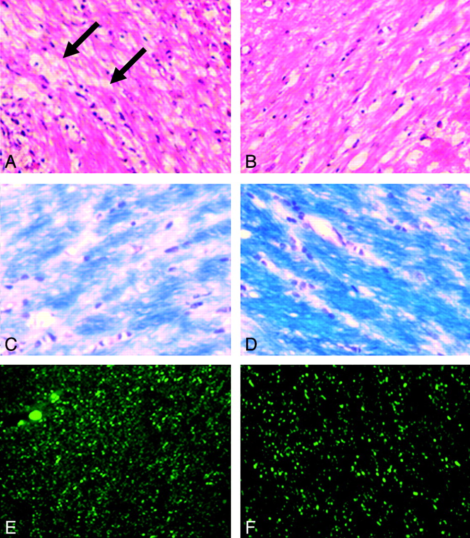
  • Fig 2.
    • Download figure
    • Open in new tab
    • Download powerpoint
    Fig 2.

    Histologic evaluations of mild HI-induced WM damage. A and B, HE stain at day 1 post-HI injury. No necrosis area is found in the injury EC (A). Mild vacuolation changes are found in the injury EC (arrows) compared with the control EC (B). C and D, LFB stain at day 30 post-HI injury. Nerve fibers appear sparse and thinner in the injury EC (C). LFB stain shows decreased stain intensity compared with the control EC (D). E and F, NF stain at day 90 post-HI injury. Distributions and axonal counts are similar in the injury EC (E) and the control EC (F). Scale bar (in F, applicable to A-F) indicates 25 μm.

Tables

  • Figures
    • View popup
    Table 1:

    FA, trace, λ//, and λ⊥ in injury and control ECs from D1 to D90 in a mild-HI neonatal rat model*

    Time PointsInjury ECControl ECRatioP
    FA
        D10.240 ± 0.051a0.262 ± 0.057a0.922 ± 0.091a<.01
        D70.307 ± 0.056b0.321 ± 0.053b0.962 ± 0.132b<.01
        D140.320 ± 0.040b0.337 ± 0.061b0.967 ± 0.138b<.01
        D300.377 ± 0.043c0.392 ± 0.054c0.968 ± 0.147b<.01
        D900.416 ± 0.037d0.430 ± 0.040d0.970 ± 0.086b<.01
    Trace (μm2/ms)
        D12.808 ± 0.204a2.683 ± 0.351a1.059 ± 0.121a<.01
        D72.272 ± 0.019b2.273 ± 0.016b1.001 ± 0.054b.97
        D142.162 ± 0.154b2.145 ± 0.130b1.008 ± 0.033b.08
        D302.284 ± 0.277b2.260 ± 0.125b1.012 ± 0.053b.13
        D902.272 ± 0.317b2.247 ± 0.303b1.011 ± 0.042b.09
    λ// (μm2/ms)
        D11.107 ± 0.1081.125 ± 0.1070.987 ± 0.083.10
        D71.089 ± 0.1091.091 ± 0.0991.000 ± 0.060.86
        D141.082 ± 0.0791.080 ± 0.0791.003 ± 0.039.73
        D301.075 ± 0.0841.073 ± 0.0981.008 ± 0.113.93
        D901.087 ± 0.1591.081 ± 0.1571.009 ± 0.069.56
    λ⊥ (μm2/ms)
        D10.835 ± 0.083a0.769 ± 0.096a1.092 ± 0.079a<.01
        D70.645 ± 0.060b0.616 ± 0.048b1.050 ± 0.100b<.01
        D140.621 ± 0.061b0.594 ± 0.052b1.049 ± 0.093b<.01
        D300.617 ± 0.043b0.591 ± 0.043b1.046 ± 0.073b<.01
        D900.593 ± 0.076b0.570 ± 0.078b1.043 ± 0.060b<.01
    • Note:—FA indicates fractional anisotropy; EC, external capsule; λ//, axial diffusivity; λ⊥, radial diffusivity; Ratio, injury/control diffusion tensor imaging indices of EC; D1-D90, day 1-day 90 post HI; HI, hypoxic-ischemic.

    • * A paired t test was used to evaluate the statistical significance between injury and control ECs. A linear mixed model, followed by a post hoc pair-wise comparison test, was applied to evaluate significant differences among longitudinal time points. Superscript a,b,c,d,e reflect significant differences between time points. Values in the same column without a common superscript indicate a significant difference less than 0.05 between 2 time points. Values are shown as mean ± SD.

    • View popup
    Table 2:

    Histologic evaluations of mild HI-induced WM damage in injury and control ECs from D1 to D90 post-HI*

    Injury ECControl ECRatioP
    LFB staining intensity (optical density)
        D10.055 ± 0.006a0.081 ± 0.008a0.684 ± 0.086a<.01
        D70.098 ± 0.008b0.129 ± 0.017b0.768 ± 0.097b<.01
        D140.113 ± 0.014c0.143 ± 0.015b0.801 ± 0.160b<.01
        D300.143 ± 0.015d0.173 ± 0.012c0.818 ± 0.083b<.01
        D900.177 ± 0.021e0.214 ± 0.026d0.824 ± 0.092b<.01
    Axon count
        D1201 ± 29a234 ± 32a0.859 ± 0.129.074
        D7463 ± 48a503 ± 85a0.920 ± 0.084.254
        D14679 ± 19697 ± 32a0.974 ± 0.025.087
        D30701 ± 132716 ± 89a0.979 ± 0.084.724
        D901059 ± 476b1240 ± 402b0.854 ± 0.025.335
    • Note:—WM indicates white matter; LFB, Luxol fast blue.

    • * A paired t test was used to evaluate the statistical significance between injury and control ECs. One-way analysis of variance followed by a Tukey test was used to evaluate the statistical significance among longitudinal time points. Superscript a,b,c,d,e reflect significant differences between time points. Values in the same column without a common superscript indicate a significant difference less than 0.05 between 2 time points. Values are shown as mean ± SD.

PreviousNext
Back to top

In this issue

American Journal of Neuroradiology: 30 (10)
American Journal of Neuroradiology
Vol. 30, Issue 10
1 Nov 2009
  • Table of Contents
  • Index by author
Advertisement
Print
Download PDF
Email Article

Thank you for your interest in spreading the word on American Journal of Neuroradiology.

NOTE: We only request your email address so that the person you are recommending the page to knows that you wanted them to see it, and that it is not junk mail. We do not capture any email address.

Enter multiple addresses on separate lines or separate them with commas.
Mild Hypoxic-Ischemic Injury in the Neonatal Rat Brain: Longitudinal Evaluation of White Matter Using Diffusion Tensor MR Imaging
(Your Name) has sent you a message from American Journal of Neuroradiology
(Your Name) thought you would like to see the American Journal of Neuroradiology web site.
CAPTCHA
This question is for testing whether or not you are a human visitor and to prevent automated spam submissions.
Cite this article
S. Wang, E.X. Wu, K. Cai, H.-F. Lau, P.-T. Cheung, P.-L. Khong
Mild Hypoxic-Ischemic Injury in the Neonatal Rat Brain: Longitudinal Evaluation of White Matter Using Diffusion Tensor MR Imaging
American Journal of Neuroradiology Nov 2009, 30 (10) 1907-1913; DOI: 10.3174/ajnr.A1697

Citation Manager Formats

  • BibTeX
  • Bookends
  • EasyBib
  • EndNote (tagged)
  • EndNote 8 (xml)
  • Medlars
  • Mendeley
  • Papers
  • RefWorks Tagged
  • Ref Manager
  • RIS
  • Zotero
0 Responses
Respond to this article
Share
Bookmark this article
Mild Hypoxic-Ischemic Injury in the Neonatal Rat Brain: Longitudinal Evaluation of White Matter Using Diffusion Tensor MR Imaging
S. Wang, E.X. Wu, K. Cai, H.-F. Lau, P.-T. Cheung, P.-L. Khong
American Journal of Neuroradiology Nov 2009, 30 (10) 1907-1913; DOI: 10.3174/ajnr.A1697
del.icio.us logo Twitter logo Facebook logo Mendeley logo
  • Tweet Widget
  • Facebook Like
  • Google Plus One
Purchase

Jump to section

  • Article
    • Abstract
    • Materials and Methods
    • Results
    • Discussion
    • Conclusions
    • Acknowledgments
    • Footnotes
    • References
  • Figures & Data
  • Info & Metrics
  • Responses
  • References
  • PDF

Related Articles

  • No related articles found.
  • PubMed
  • Google Scholar

Cited By...

  • An interactive meta-analysis of MRI biomarkers of myelin
  • Can MRI measure myelin? Systematic review, qualitative assessment, and meta-analysis of studies validating microstructural imaging with myelin histology
  • Does Diffusion Tensor Imaging-Based Tractography at 3 Months of Age Contribute to the Prediction of Motor Outcome After Perinatal Arterial Ischemic Stroke?
  • Crossref
  • Google Scholar

This article has not yet been cited by articles in journals that are participating in Crossref Cited-by Linking.

More in this TOC Section

  • Multimodal CT Provides Improved Performance for Lacunar Infarct Detection
  • Optimal MRI Sequence for Identifying Occlusion Location in Acute Stroke: Which Value of Time-Resolved Contrast-Enhanced MRA?
  • Evaluating the Effects of White Matter Multiple Sclerosis Lesions on the Volume Estimation of 6 Brain Tissue Segmentation Methods
Show more Brain

Similar Articles

Advertisement

Indexed Content

  • Current Issue
  • Accepted Manuscripts
  • Article Preview
  • Past Issues
  • Editorials
  • Editor's Choice
  • Fellows' Journal Club
  • Letters to the Editor
  • Video Articles

Cases

  • Case Collection
  • Archive - Case of the Week
  • Archive - Case of the Month
  • Archive - Classic Case

More from AJNR

  • Trainee Corner
  • Imaging Protocols
  • MRI Safety Corner
  • Book Reviews

Multimedia

  • AJNR Podcasts
  • AJNR Scantastics

Resources

  • Turnaround Time
  • Submit a Manuscript
  • Submit a Video Article
  • Submit an eLetter to the Editor/Response
  • Manuscript Submission Guidelines
  • Statistical Tips
  • Fast Publishing of Accepted Manuscripts
  • Graphical Abstract Preparation
  • Imaging Protocol Submission
  • Evidence-Based Medicine Level Guide
  • Publishing Checklists
  • Author Policies
  • Become a Reviewer/Academy of Reviewers
  • News and Updates

About Us

  • About AJNR
  • Editorial Board
  • Editorial Board Alumni
  • Alerts
  • Permissions
  • Not an AJNR Subscriber? Join Now
  • Advertise with Us
  • Librarian Resources
  • Feedback
  • Terms and Conditions
  • AJNR Editorial Board Alumni

American Society of Neuroradiology

  • Not an ASNR Member? Join Now

© 2025 by the American Society of Neuroradiology All rights, including for text and data mining, AI training, and similar technologies, are reserved.
Print ISSN: 0195-6108 Online ISSN: 1936-959X

Powered by HighWire