Advertisement

Main menu

  • Home
  • Content
    • Current Issue
    • Accepted Manuscripts
    • Article Preview
    • Past Issue Archive
    • Video Articles
    • AJNR Case Collection
    • Case of the Week Archive
    • Case of the Month Archive
    • Classic Case Archive
  • Special Collections
    • AJNR Awards
    • Low-Field MRI
    • Alzheimer Disease
    • ASNR Foundation Special Collection
    • Photon-Counting CT
    • View All
  • Multimedia
    • AJNR Podcasts
    • AJNR SCANtastic
    • Trainee Corner
    • MRI Safety Corner
    • Imaging Protocols
  • For Authors
    • Submit a Manuscript
    • Submit a Video Article
    • Submit an eLetter to the Editor/Response
    • Manuscript Submission Guidelines
    • Statistical Tips
    • Fast Publishing of Accepted Manuscripts
    • Graphical Abstract Preparation
    • Imaging Protocol Submission
    • Author Policies
  • About Us
    • About AJNR
    • Editorial Board
    • Editorial Board Alumni
  • More
    • Become a Reviewer/Academy of Reviewers
    • Subscribers
    • Permissions
    • Alerts
    • Feedback
    • Advertisers
    • ASNR Home

User menu

  • Alerts
  • Log in

Search

  • Advanced search
American Journal of Neuroradiology

American Journal of Neuroradiology

ASHNR American Society of Functional Neuroradiology ASHNR American Society of Pediatric Neuroradiology ASSR
  • Alerts
  • Log in

Advanced Search

  • Home
  • Content
    • Current Issue
    • Accepted Manuscripts
    • Article Preview
    • Past Issue Archive
    • Video Articles
    • AJNR Case Collection
    • Case of the Week Archive
    • Case of the Month Archive
    • Classic Case Archive
  • Special Collections
    • AJNR Awards
    • Low-Field MRI
    • Alzheimer Disease
    • ASNR Foundation Special Collection
    • Photon-Counting CT
    • View All
  • Multimedia
    • AJNR Podcasts
    • AJNR SCANtastic
    • Trainee Corner
    • MRI Safety Corner
    • Imaging Protocols
  • For Authors
    • Submit a Manuscript
    • Submit a Video Article
    • Submit an eLetter to the Editor/Response
    • Manuscript Submission Guidelines
    • Statistical Tips
    • Fast Publishing of Accepted Manuscripts
    • Graphical Abstract Preparation
    • Imaging Protocol Submission
    • Author Policies
  • About Us
    • About AJNR
    • Editorial Board
    • Editorial Board Alumni
  • More
    • Become a Reviewer/Academy of Reviewers
    • Subscribers
    • Permissions
    • Alerts
    • Feedback
    • Advertisers
    • ASNR Home
  • Follow AJNR on Twitter
  • Visit AJNR on Facebook
  • Follow AJNR on Instagram
  • Join AJNR on LinkedIn
  • RSS Feeds

AJNR Awards, New Junior Editors, and more. Read the latest AJNR updates

OtherReview Articles

State of the Art: 3T Imaging of the Membranous Labyrinth

J.I. Lane, R.J. Witte, B. Bolster, M.A. Bernstein, K. Johnson and J. Morris
American Journal of Neuroradiology September 2008, 29 (8) 1436-1440; DOI: https://doi.org/10.3174/ajnr.A1036
J.I. Lane
  • Find this author on Google Scholar
  • Find this author on PubMed
  • Search for this author on this site
R.J. Witte
  • Find this author on Google Scholar
  • Find this author on PubMed
  • Search for this author on this site
B. Bolster
  • Find this author on Google Scholar
  • Find this author on PubMed
  • Search for this author on this site
M.A. Bernstein
  • Find this author on Google Scholar
  • Find this author on PubMed
  • Search for this author on this site
K. Johnson
  • Find this author on Google Scholar
  • Find this author on PubMed
  • Search for this author on this site
J. Morris
  • Find this author on Google Scholar
  • Find this author on PubMed
  • Search for this author on this site
  • Article
  • Figures & Data
  • Info & Metrics
  • Responses
  • References
  • PDF
Loading

Article Figures & Data

Figures

  • Fig 1.
    • Download figure
    • Open in new tab
    • Download powerpoint
    Fig 1.

    Schematic representation of the membranous labyrinth. A, magnified cross-section of the cochlea. B, Magnified exposed view of the crista ampullaris of the superior semicircular duct. C, Magnified exposed view of the utricular macula. SV indicates scala vestibuli; SM, scala media; ST, scala tympani.

  • Fig 2.
    • Download figure
    • Open in new tab
    • Download powerpoint
    Fig 2.

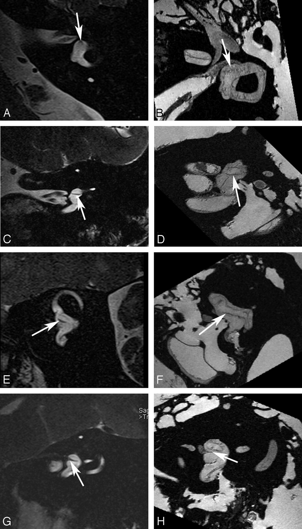
    Utricular macula. Axial (A and B), coronal (C and D), oblique sagittal (Poschl plane) (E and F), and oblique coronal (Stenvers plane) (G and H) images of the utricular macula. In vivo 3T (A, C, E, and G) and ex vivo 9.4T (B, D, F, and H) MR images demonstrate low-signal-intensity focus (arrow) corresponding to the neurosensory epithelium of the utricular macula.

  • Fig 3.
    • Download figure
    • Open in new tab
    • Download powerpoint
    Fig 3.

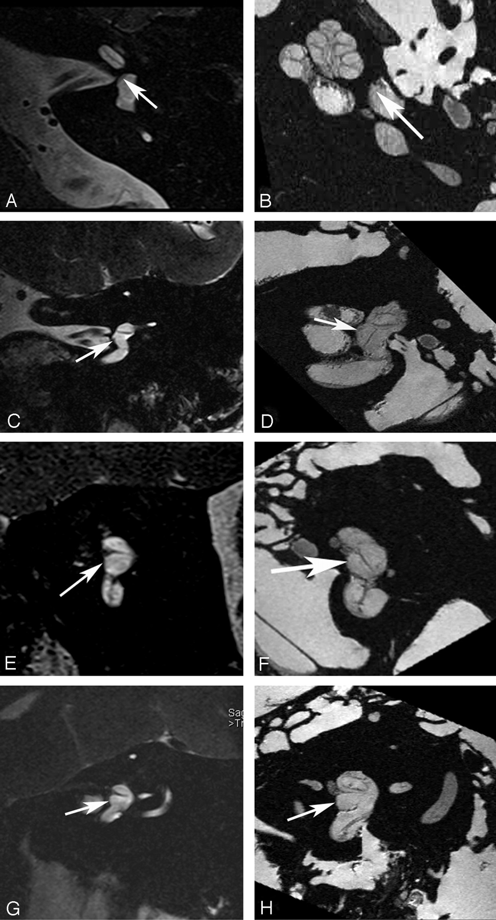
    Saccular macula. Axial (A and B), coronal (C and D), oblique sagittal (Poschl plane) (E and F), and oblique coronal (Stenvers plane) (G and H) images. Ex vivo 9.4T MR images (B, D, F, and H) demonstrate low-signal-intensity curvilinear focus (arrow) corresponding to the neurosensory epithelium of the saccular macula along the medial wall of the vestibule. In vivo 3T MR images (A, C, E, and G) do not differentiate the macula from the medial wall of the vestibule (arrow).

  • Fig 4.
    • Download figure
    • Open in new tab
    • Download powerpoint
    Fig 4.

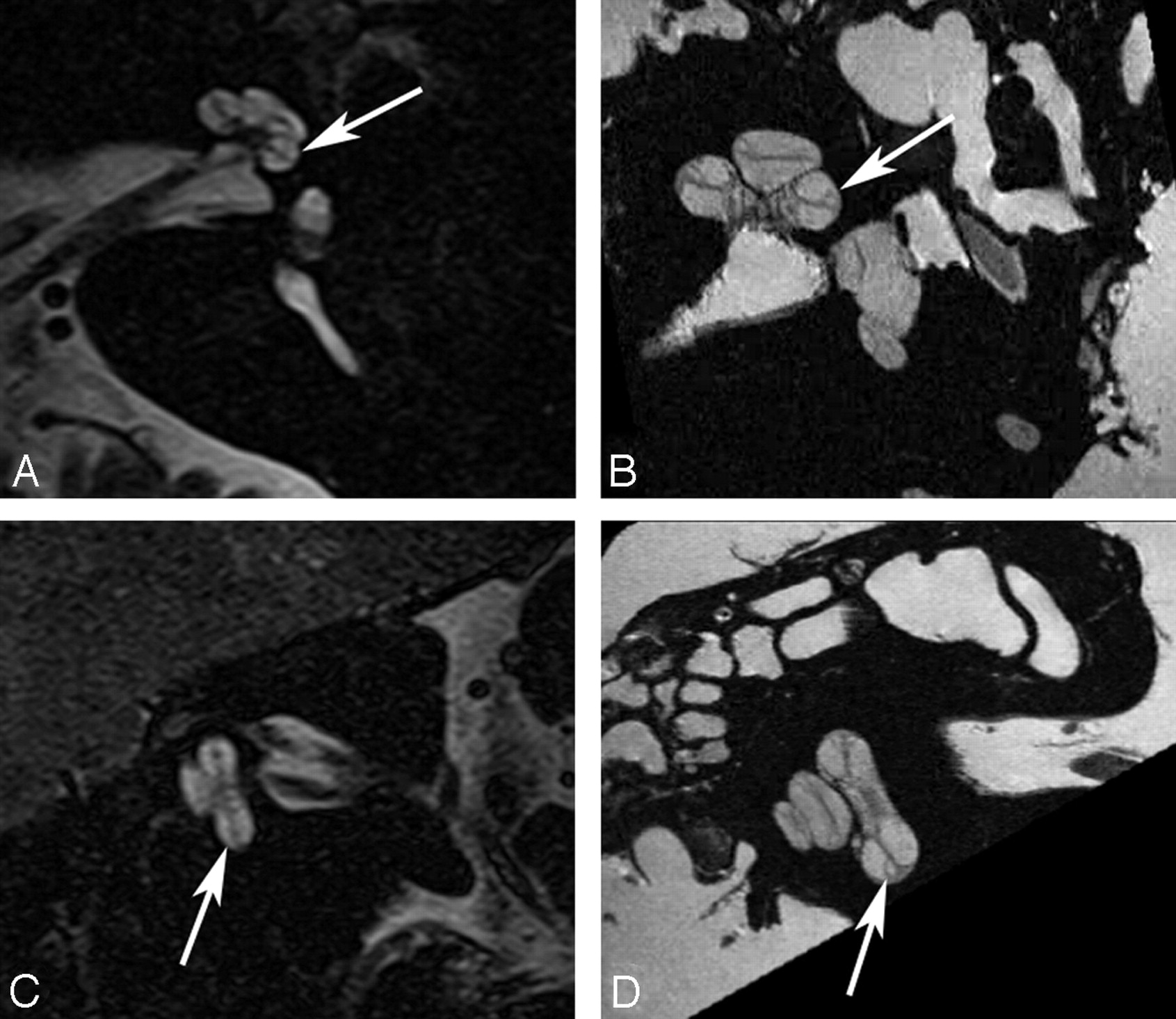
    Crista ampullaris of the superior semicircular duct. A, In vivo 3T oblique sagittal (Poschl plane) image in the plane of the superior canal demonstrates a linear area of low signal intensity (arrow) corresponding to the crista ampullaris of the superior duct. B, Ex vivo 9.4T oblique sagittal image demonstrates similar findings (arrow).

  • Fig 5.
    • Download figure
    • Open in new tab
    • Download powerpoint
    Fig 5.

    Crista ampullaris of the lateral semicircular duct. A, In vivo 3T oblique sagittal (Poschl plane) image in the plane of the superior canal demonstrates a linear area of low signal intensity (arrow) corresponding to the crista ampullaris of the lateral duct. B, Ex vivo 9.4T oblique sagittal image demonstrates similar findings (arrow). C, In vivo 3T coronal image demonstrates focal area of low signal intensity (arrow) in the ampulla of the lateral canal corresponding to the crista. D, Corresponding ex vivo 9.4T image with a similar appearance (arrow).

  • Fig 6.
    • Download figure
    • Open in new tab
    • Download powerpoint
    Fig 6.

    Crista ampullaris of the posterior semicircular duct. A, In vivo 3T oblique sagittal (Poschl plane) image in the plane of the superior canal demonstrates a linear area of low signal intensity (arrow) corresponding to the crista ampullaris of the posterior duct. B, Ex vivo 9.4T oblique sagittal image demonstrates similar findings (arrow).

  • Fig 7.
    • Download figure
    • Open in new tab
    • Download powerpoint
    Fig 7.

    Cochlear duct. B and D, Ex vivo 9.4T MR images in the axial and oblique sagittal (Poschl) plane demonstrate the triangular fluid compartment in the peripheral aspect of the basal turn of the cochlear (arrow), corresponding to the cochlear duct. A and C, corresponding in vivo 3T MR images do not adequately resolve the Reissner membrane and, therefore, do not demarcate the perilymph in the scala vestibuli from the endolymph in the cochlear duct. Approximated location of the duct is indicated (arrow).

PreviousNext
Back to top

In this issue

American Journal of Neuroradiology: 29 (8)
American Journal of Neuroradiology
Vol. 29, Issue 8
September 2008
  • Table of Contents
  • Index by author
Advertisement
Print
Download PDF
Email Article

Thank you for your interest in spreading the word on American Journal of Neuroradiology.

NOTE: We only request your email address so that the person you are recommending the page to knows that you wanted them to see it, and that it is not junk mail. We do not capture any email address.

Enter multiple addresses on separate lines or separate them with commas.
State of the Art: 3T Imaging of the Membranous Labyrinth
(Your Name) has sent you a message from American Journal of Neuroradiology
(Your Name) thought you would like to see the American Journal of Neuroradiology web site.
CAPTCHA
This question is for testing whether or not you are a human visitor and to prevent automated spam submissions.
Cite this article
J.I. Lane, R.J. Witte, B. Bolster, M.A. Bernstein, K. Johnson, J. Morris
State of the Art: 3T Imaging of the Membranous Labyrinth
American Journal of Neuroradiology Sep 2008, 29 (8) 1436-1440; DOI: 10.3174/ajnr.A1036

Citation Manager Formats

  • BibTeX
  • Bookends
  • EasyBib
  • EndNote (tagged)
  • EndNote 8 (xml)
  • Medlars
  • Mendeley
  • Papers
  • RefWorks Tagged
  • Ref Manager
  • RIS
  • Zotero
0 Responses
Respond to this article
Share
Bookmark this article
State of the Art: 3T Imaging of the Membranous Labyrinth
J.I. Lane, R.J. Witte, B. Bolster, M.A. Bernstein, K. Johnson, J. Morris
American Journal of Neuroradiology Sep 2008, 29 (8) 1436-1440; DOI: 10.3174/ajnr.A1036
del.icio.us logo Twitter logo Facebook logo Mendeley logo
  • Tweet Widget
  • Facebook Like
  • Google Plus One
Purchase

Jump to section

  • Article
    • Abstract
    • Imaging Technique
    • Clinical Anatomy
    • Imaging Anatomy
    • Discussion
    • Conclusions
    • References
  • Figures & Data
  • Info & Metrics
  • Responses
  • References
  • PDF

Related Articles

  • No related articles found.
  • PubMed
  • Google Scholar

Cited By...

  • Assessment of the Membranous Labyrinth in Infants Using a Heavily T2-weighted 3D FLAIR Sequence without Contrast Agent Administration
  • Comparison of Enhancement of the Vestibular Perilymph between Variable and Constant Flip Angle-Delayed 3D-FLAIR Sequences in Meniere Disease
  • Crossref
  • Google Scholar

This article has not yet been cited by articles in journals that are participating in Crossref Cited-by Linking.

More in this TOC Section

  • An Atlas of Neonatal Neurovascular Imaging Anatomy as Depicted with Microvascular Imaging: The Intracranial Arteries
  • An Atlas of Neonatal Neurovascular Imaging Anatomy as Depicted with Microvascular Imaging: The Intracranial Veins
  • Clinical Translation of Hyperpolarized 13C Metabolic Probes for Glioma Imaging
Show more Review Articles

Similar Articles

Advertisement

Indexed Content

  • Current Issue
  • Accepted Manuscripts
  • Article Preview
  • Past Issues
  • Editorials
  • Editor's Choice
  • Fellows' Journal Club
  • Letters to the Editor
  • Video Articles

Cases

  • Case Collection
  • Archive - Case of the Week
  • Archive - Case of the Month
  • Archive - Classic Case

More from AJNR

  • Trainee Corner
  • Imaging Protocols
  • MRI Safety Corner
  • Book Reviews

Multimedia

  • AJNR Podcasts
  • AJNR Scantastics

Resources

  • Turnaround Time
  • Submit a Manuscript
  • Submit a Video Article
  • Submit an eLetter to the Editor/Response
  • Manuscript Submission Guidelines
  • Statistical Tips
  • Fast Publishing of Accepted Manuscripts
  • Graphical Abstract Preparation
  • Imaging Protocol Submission
  • Evidence-Based Medicine Level Guide
  • Publishing Checklists
  • Author Policies
  • Become a Reviewer/Academy of Reviewers
  • News and Updates

About Us

  • About AJNR
  • Editorial Board
  • Editorial Board Alumni
  • Alerts
  • Permissions
  • Not an AJNR Subscriber? Join Now
  • Advertise with Us
  • Librarian Resources
  • Feedback
  • Terms and Conditions
  • AJNR Editorial Board Alumni

American Society of Neuroradiology

  • Not an ASNR Member? Join Now

© 2025 by the American Society of Neuroradiology All rights, including for text and data mining, AI training, and similar technologies, are reserved.
Print ISSN: 0195-6108 Online ISSN: 1936-959X

Powered by HighWire