
Article Figures & Data
Figures
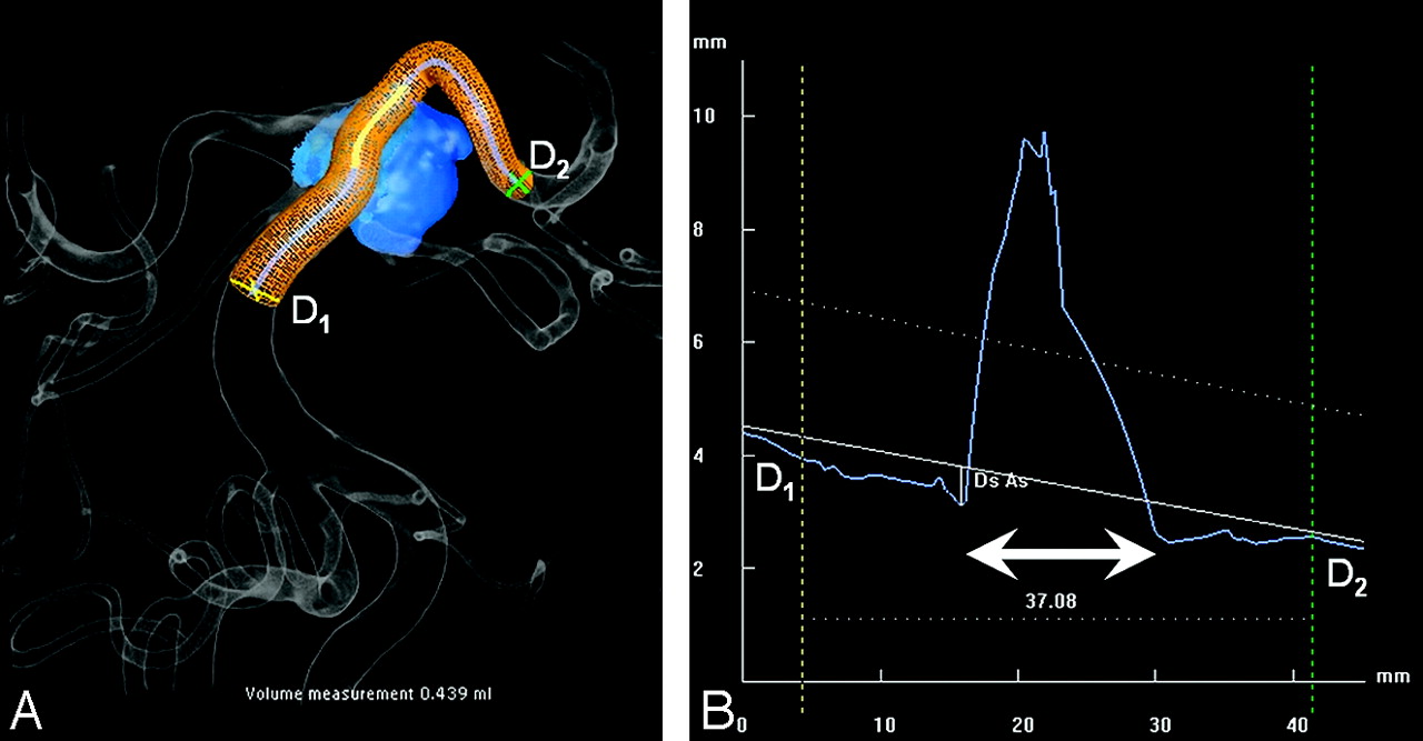
- Fig 1.
Computer simulation before stent placement in a superior cerebellar artery aneurysm (same patient as in Fig 2). A, 3D image after automated aneurysm detection (blue) and stent simulation. B, Corresponding graph indicating vessel diameters in segment lengths. Proximal and distal diameters are indicated by D1 (4.2 mm) and D2 (2.5 mm). The aneurysm neck is indicated by the bidirectional arrow. Use of a 37-mm stent will provide a proximal and distal overlap of 10 mm.
- Fig 2.
A 58-year-old woman with a ruptured superior cerebellar artery aneurysm without a neck. A and B, 2D and 3D vertebral angiograms demonstrate the dysplastic distal basilar segment with a large superior cerebellar artery aneurysm. The superior cerebellar artery arises from base of the aneurysmal sack, and the dysplastic segment extends to the proximal posterior cerebral arteries. C, Result after stent-assisted coiling. Proximal and distal stent markers are indicated by arrows. Flow in the superior cerebellar artery is preserved.
- Fig 3.
A 46-year-old woman with a wide-neck posterior inferior cerebellar artery aneurysm, additional to another ruptured aneurysm. A, 3D angiogram demonstrates the posterior inferior cerebellar artery originating from the base of the aneurysm. B, 3D image after automated aneurysm detection and stent simulation. C, Complete occlusion after stent-assisted coiling with preserved flow in the posterior inferior cerebellar artery.


Tables
Characteristics of 15 patients with 16 aneurysms treated with stent assistance
Patient No. Sex, Age (year) Aneurysm Location Treatment Indication Aneurysm Size Dome-to-Neck Ratio of Aneurysm or Recurrence 1 M, 66 Basilar tip Recurrence 20 0:4 2 M, 60 Basilar tip Recurrence 7 1:5 3 M, 49 Basilar tip Recurrence 16 0:8 4 F, 58 Superior cerebellar artery SAH 12 1:2 F, 58 ICA Additional 11 1:0 5 F, 66 Middle cerebral artery Unruptured 10 1:2 6 F, 52 Ophthalmic artery Recurrence 16 1:4 7 M, 65 Basilar tip Recurrence 13 0:7 8 F, 39 Ophthalmic artery Additional 8 0:8 9 F, 60 Cavernous sinus Recurrence 30 1:3 10 F, 50 Basilar tip Recurrence 10 0:7 11 F, 49 AcomA Unruptured 9 1:1 12 F, 46 PICA Additional 12 1:0 13 M, 51 Extracranial ICA Dissection 7 1:0 14 F, 36 Basilar tip Recurrence 16 1:0 15 F, 56 Basilar tip Recurrence 14 0:8 Note:—SAH indicates subarachnoid hemorrhage; AcomA, anterior communicating artery; PICA, posterior inferior cerebellar artery; ICA, internal carotid artery.
In this issue


Jump to section
Related Articles
Cited By...
- Endovascular treatment of complex intracranial aneurysms using Acandis Acclino stents
- Stent-assisted coil embolization of aneurysms with small parent vessels: safety and efficacy analysis
- Stent-Assisted Coil Embolization of Intracranial Aneurysms: Complications in Acutely Ruptured versus Unruptured Aneurysms
- Clinical and angiographic outcomes after stent-assisted coiling of cerebral aneurysms with Enterprise and Neuroform stents: a comparative analysis of the literature
- Stent-Assisted Coil Embolization of Posterior Communicating Artery Aneurysms
- In-hospital outcomes associated with stent-assisted endovascular treatment of unruptured cerebral aneurysms in the USA
- Stent usage in the treatment of intracranial aneurysms: past, present and future
- Coil Protection Using Small Helical Coils for Wide-Neck Intracranial Aneurysms: A Novel Approach
- Double-barrel entanglement of intracranial Enterprise stents resulting from undetected incomplete stent apposition
- Stenting from the Vertebral Artery to the Posterior Inferior Cerebellar Artery
- Stent-assisted coiling of paraclinoid aneurysms: risks and effectiveness
- Neuroform Stent-Assisted Coiling of Unruptured Intracranial Aneurysms: Short- and Midterm Results from a Single-Center Experience with 68 Patients
- Treatment of Intracranial Aneurysms by Functional Reconstruction of the Parent Artery: The Budapest Experience with the Pipeline Embolization Device
- Late Adverse Events in Coiled Ruptured Aneurysms with Incomplete Occlusion at 6-Month Angiographic Follow-Up
- Advances in Interventional Neuroradiology
- Endovascular Approaches to Acute Stroke, Part 1: Drugs, Devices, and Data
- Dangerous Advances in Measurements from Digital Subtraction Angiography: When Is a Millimeter Not a Millimeter?