
Article Figures & Data
Figures
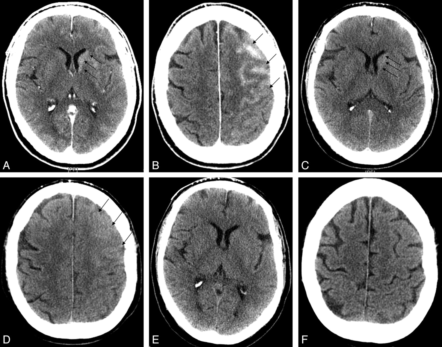
- Fig 1.
A, immediate postembolization noncontrast head CT shows a gyral pattern of enhancement (arrows). B, C, interval resolution of enhancement at 6 hours (B) and 24 hours (C), respectively (arrows).
- Fig 2.
A, B, immediate postembolization noncontrast head CT shows left caudate (A) and gyral (B) enhancement (arrows). C, D, interval improvement of caudate (C) and gyral (D) enhancement at 6 hours (arrows). E, F, resolution of caudate (E) and gyral (F) enhancement at 24 hours.
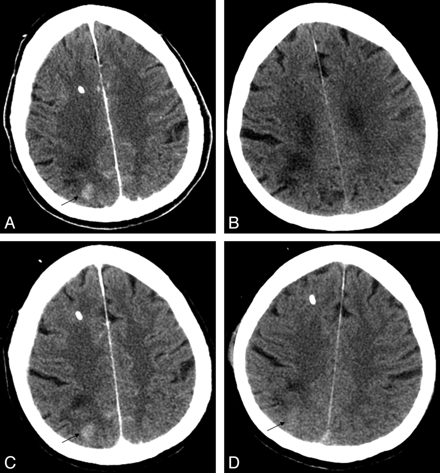
- Fig 3.
A, immediate postembolization noncontrast head CT shows a gyral pattern of enhancement (arrow). B, noncontrast head CT obtained at presentation without gyral subarachnoid blood in the area of postcoiling enhancement. C,D, interval resolution of enhancement at 6 hours (C) and 24 hours (D), respectively (arrows).


Tables
Sex/Age Treated Aneurysm Stent Assist Perioperative Antiplatelets Location of Enhancement HU Postprocedure HU 4–6 Hours HU 20–25 Hours Clinical Change F/42 Unruptured 6 mm left superior hypophyseal No Aspirin* Caudate 48 41 N/A None M/43 Unruptured 6 mm AcomA No Aspirin† clopidogrel† Caudate/Gyral 46/60 43/41 resolved None M/62 Unruptured 8 mm AcomA No Aspirin† abciximab§ Caudate/Gyral 50/67 40/45 resolved None F/65 Unruptured 5 mm left MCA No Aspirin† clopidogrel† abciximab‡ Gyral 59 47 resolved None F/80 SAH, 5 mm basilar tip Yes Aspirin† clopidogrel† abciximab‡§ Gyral 53 52 39 None F/61 Unruptured 22 mm left supraclinoid ICA Yes Aspirin* clopidogrel* Gyral 47 44 resolved None F/68 Unruptured 7 mm AcomA No Aspirin† Gyral 46 42 resolved None Note:—F indicates female; M, male; SAH, subarachnoid hemorrhage; N/A, not available; AcomA, anterior communicating artery; MCA, middle cerebral artery; ICA, internal carotid artery; HU, Hounsfield units.
* Premedication ≥5 days.
† Started day of procedure.
‡ Intra-arterial intraoperative.
§ Intravenous intraoperative and immediately postoperative.
Patient No. CEH Contrast (mL/kg) Contrast (mgI/kg) Procedure Time (min) 1 − 3.33 1065.6 153 2 − 3.18 1017.6 164 3 − 1.70 544.0 160 4 + 7.47 2390.4 102 5 − 5.13 1641.6 237 6 − 2.51 803.2 252 7 − 2.44 780.8 125 8 + 6.77 2166.4 180 9 − 4.07 1302.4 66 10 − 0.86 275.2 116 11 − N/A N/A 199 12 − 4.40 1408.0 97 13 − 3.59 1148.8 160 14 − 4.16 1331.2 139 15 − 4.72 1510.4 356 16 + 6.24 1996.8 243 17 − N/A N/A 127 18 − 7.69 2460.8 41 19 − 4.10 1312.0 225 20 + 5.29 1692.8 188 21 − 6.54 2092.8 300 22 − N/A N/A 260 23 − 1.51 483.2 130 24 − 4.26 1363.2 150 25 − 5.30 1696.0 232 26 − 2.36 755.2 190 27 + 2.56 819.2 310 28 − 0.69 220.8 49 29 + 4.08 1305.6 360 30 + 7.76 2483.2 420 Note:—CEH indicates contrast enhancement hyperdensity; min, minutes; N/A, data not available.
In this issue


Jump to section
Related Articles
Cited By...
- HARMless: Transient Cortical and Sulcal Hyperintensity on Gadolinium-Enhanced FLAIR after Elective Endovascular Coiling of Intracranial Aneurysms
- Subarachnoid Hyperattenuation on Flat Panel Detector-Based Conebeam CT Immediately after Uneventful Coil Embolization of Unruptured Intracranial Aneurysms
- Microcatheter contrast injections during intra-arterial thrombolysis increase intracranial hemorrhage risk
- Intra-Arterial Iodinated Radiographic Contrast Material Injection Administration in a Rat Middle Cerebral Artery Occlusion and Reperfusion Model: Possible Effects on Intracerebral Hemorrhage