
Article Figures & Data
Figures
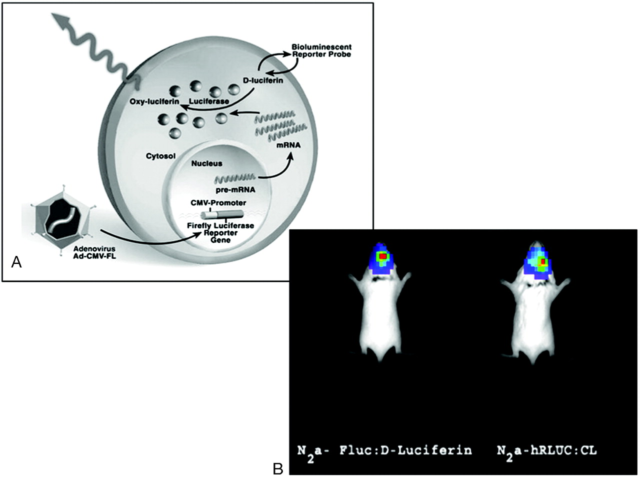
- Fig 1.
Reporter gene imaging. A, Schematic diagram of the principle of reporter gene imaging by using the enzyme firefly luciferase. Once the cell is transduced with a viral vector containing the imaging gene cassette, a promoter of choice drives the transcription of the imaging reporter gene (Fluc). If the promoter leads to transcription of Fluc, then translation of the imaging reporter gene mRNA leads to a protein product (the enzyme firefly luciferase) that can interact with the imaging reporter probe (D-Luciferin). This interaction is a chemiluminescent reaction that catalyzes the transformation of the substrate D-Luciferin into oxyluciferin in a process dependent on ATP, Mg++, and O2, leading to the emission of light, which can be detected by using low-light sensing instruments. Other gene/substrate combinations may be used as well (eg, hRluc and its substrate CL, see text). B, Bioluminescence neuroimaging in mice. Balb/c mice with 105 intracranially injected N2a cells transfected 24 hours previously with CMV-hRluc (right) and CMV-Fluc (left). Intracranial injections were performed immediately before substrate administration. The mice received intraperitoneal injections of the substrates D-Luciferin (1.5 mg) or CL (5μg) respectively. The charge-coupled device camera images were taken approximately 5–7 minutes post-substrate injections. Maximum signal intensity detected in photons per second per square centimeter per steradian is the following: Fluc, 2.1 × 10⁶; hRluc, 9.2 × 10⁴. CMV indicates cytomegalovirus; CMV-FL, adenoviral vector containing an imaging cassette with the CMV promoter driving the transcription of firefly luciferase gene.
- Fig 2.
The Xenogen In Vivo Imaging System (Xenogen Corporation, Hopkinton, Mass) consists of a cooled CCD camera mounted on a light-tight imaging chamber, a cryogenic refrigeration unit, a camera controller, and a computer system for data analysis


Tables
Features of molecular imaging techniques used in reporter gene imaging
Imaging Technique Advantages Disadvantages Bioluminescence optical imaging Very high sensitivity Very low spatial resolution High throughput Only planar imaging, not tomographic Very versatile Surface-weighted images owing to light scatter and absorption Cheap Semiquantitative imaging data Mass amount of probe required (? toxic) Not an established clinical technique MR imaging Very high spatial resolution Low sensitivity Tomographic imaging Mass amount of probe required (? toxic) Widely available established clinical technique PET, SPECT High sensitivity Low spatial resolution Fully quantitative imaging data Probes for using HSV1-tk gene do not cross the blood-brain barrier Tomographic imaging Nanogram amount of probe required (nontoxic and safe) D2R gene normally expressed in basal ganglia interferes with image interpretation when using this reporter system Established clinical techniques Note:—? toxic indicates a question concerning potential toxicity.


