Advertisement

Main menu

  • Home
  • Content
    • Current Issue
    • Accepted Manuscripts
    • Article Preview
    • Past Issue Archive
    • Video Articles
    • AJNR Case Collection
    • Case of the Week Archive
    • Case of the Month Archive
    • Classic Case Archive
  • Special Collections
    • AJNR Awards
    • Low-Field MRI
    • Alzheimer Disease
    • ASNR Foundation Special Collection
    • Photon-Counting CT
    • View All
  • Multimedia
    • AJNR Podcasts
    • AJNR SCANtastic
    • Trainee Corner
    • MRI Safety Corner
    • Imaging Protocols
  • For Authors
    • Submit a Manuscript
    • Submit a Video Article
    • Submit an eLetter to the Editor/Response
    • Manuscript Submission Guidelines
    • Statistical Tips
    • Fast Publishing of Accepted Manuscripts
    • Graphical Abstract Preparation
    • Imaging Protocol Submission
    • Author Policies
  • About Us
    • About AJNR
    • Editorial Board
    • Editorial Board Alumni
  • More
    • Become a Reviewer/Academy of Reviewers
    • Subscribers
    • Permissions
    • Alerts
    • Feedback
    • Advertisers
    • ASNR Home

User menu

  • Alerts
  • Log in

Search

  • Advanced search
American Journal of Neuroradiology

American Journal of Neuroradiology

ASHNR American Society of Functional Neuroradiology ASHNR American Society of Pediatric Neuroradiology ASSR
  • Alerts
  • Log in

Advanced Search

  • Home
  • Content
    • Current Issue
    • Accepted Manuscripts
    • Article Preview
    • Past Issue Archive
    • Video Articles
    • AJNR Case Collection
    • Case of the Week Archive
    • Case of the Month Archive
    • Classic Case Archive
  • Special Collections
    • AJNR Awards
    • Low-Field MRI
    • Alzheimer Disease
    • ASNR Foundation Special Collection
    • Photon-Counting CT
    • View All
  • Multimedia
    • AJNR Podcasts
    • AJNR SCANtastic
    • Trainee Corner
    • MRI Safety Corner
    • Imaging Protocols
  • For Authors
    • Submit a Manuscript
    • Submit a Video Article
    • Submit an eLetter to the Editor/Response
    • Manuscript Submission Guidelines
    • Statistical Tips
    • Fast Publishing of Accepted Manuscripts
    • Graphical Abstract Preparation
    • Imaging Protocol Submission
    • Author Policies
  • About Us
    • About AJNR
    • Editorial Board
    • Editorial Board Alumni
  • More
    • Become a Reviewer/Academy of Reviewers
    • Subscribers
    • Permissions
    • Alerts
    • Feedback
    • Advertisers
    • ASNR Home
  • Follow AJNR on Twitter
  • Visit AJNR on Facebook
  • Follow AJNR on Instagram
  • Join AJNR on LinkedIn
  • RSS Feeds

AJNR Awards, New Junior Editors, and more. Read the latest AJNR updates

Research ArticleBrain

Clinical Characteristics and MR Imaging Features of Nonalcoholic Wernicke Encephalopathy

G.-q. Fei, C. Zhong, L. Jin, J. Wang, Yuhao Zhang, X. Zheng, Yuwen Zhang and Z. Hong
American Journal of Neuroradiology January 2008, 29 (1) 164-169; DOI: https://doi.org/10.3174/ajnr.A0827
G.-q. Fei
  • Find this author on Google Scholar
  • Find this author on PubMed
  • Search for this author on this site
C. Zhong
  • Find this author on Google Scholar
  • Find this author on PubMed
  • Search for this author on this site
L. Jin
  • Find this author on Google Scholar
  • Find this author on PubMed
  • Search for this author on this site
J. Wang
  • Find this author on Google Scholar
  • Find this author on PubMed
  • Search for this author on this site
Yuhao Zhang
  • Find this author on Google Scholar
  • Find this author on PubMed
  • Search for this author on this site
X. Zheng
  • Find this author on Google Scholar
  • Find this author on PubMed
  • Search for this author on this site
Yuwen Zhang
  • Find this author on Google Scholar
  • Find this author on PubMed
  • Search for this author on this site
Z. Hong
  • Find this author on Google Scholar
  • Find this author on PubMed
  • Search for this author on this site
  • Article
  • Figures & Data
  • Info & Metrics
  • Responses
  • References
  • PDF
Loading

Article Figures & Data

Figures

  • Tables
  • Fig 1.
    • Download figure
    • Open in new tab
    • Download powerpoint
    Fig 1.

    Typical MR imaging features of nonalcoholic Wernicke encephalopathy. Axial T1-weighted images (A, F, K) show that no abnormal signal intensity was found. Axial T2-weighted (B, G, L), and FLAIR (C, H, M) images show increased signal intensity symmetric within the medial thalami (B, C), periaqueductal area (G, H), and floor of the fourth ventricle (L, M). DWI (D, I, N) imaging shows only slightly increased signal intensities within the bilateral thalami and periacqueductal area but no abnormal signal intensity in the other brain regions. (The ADC values of sites 1, 2, and 3 in the D image were 620.19 ± 27.39, 513.29 ± 9.60, and 381.92 ± 25.73, respectively; the ADC values of sites 1, 2, and 3 in the I image were 657.00 ± 43.13, 442.23 ± 15.43, and 494.94 ± 12.66, respectively.) Contrasting images (E, J, O) show that enhancement of the mammillary bodies and at the floor of the fourth ventricle by gadolinium contrast medium is found (J). No atrophy of the mammillary bodies (F, G, I, J) and cerebellar vermis (K, L, M, N, O) was found.

  • Fig 2.
    • Download figure
    • Open in new tab
    • Download powerpoint
    Fig 2.

    MR imaging manifestations in patients with drowsiness or without disturbances of consciousness. FLAIR images show that only regional damage surrounding the aqueduct (B, C) and the floor of fourth ventricle (D, enhanced by gadolinium contrast) is observed, and no abnormal signal intensity was found in the bilateral medial thalami (A). Arrowheads indicate cavernous hemangioma in the pons (C, E; E was a CT).
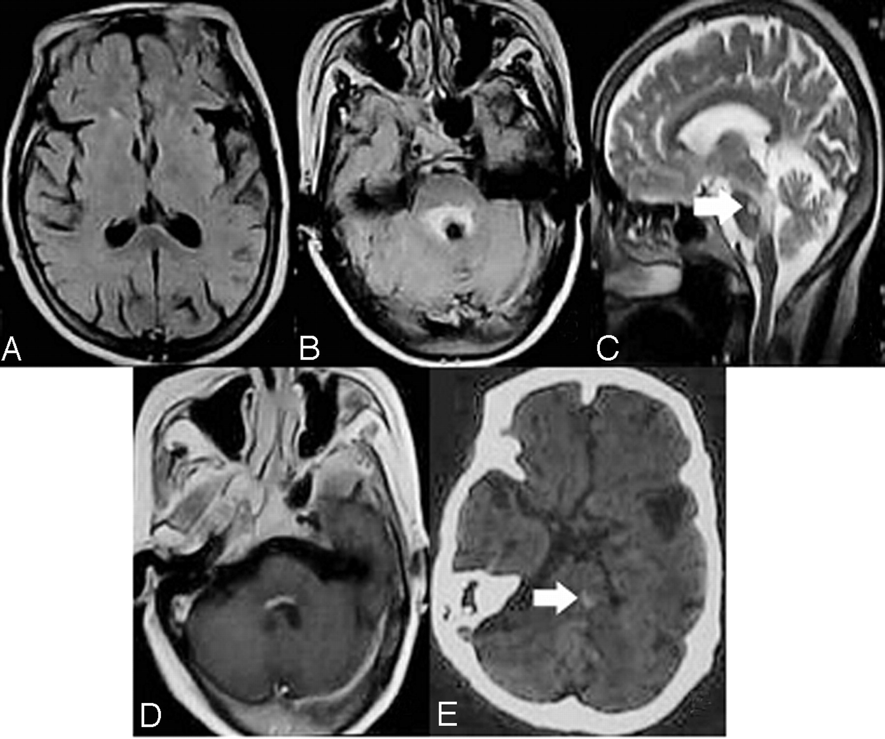
  • Fig 3.
    • Download figure
    • Open in new tab
    • Download powerpoint
    Fig 3.

    MR imaging images demonstrating cortical damage in patients with deep coma.

  • Fig 4.
    • Download figure
    • Open in new tab
    • Download powerpoint
    Fig 4.

    MR imaging follow-up of a patient with mild coma. Axial images show symmetric T2-weighted and FLAIR increased signal intensity in the medial thalami in the acute phase of WE (A, B) and normal signal intensities 1 year after thiamine supplementation (C,D).

  • Fig 5.
    • Download figure
    • Open in new tab
    • Download powerpoint
    Fig 5.

    MR imaging follow-up of a patient with deep coma. The images show progressive atrophy 2 years after the onset of WE.

Tables

  • Figures
    • View popup
    Table 1:

    Predisposing causes of nonalcoholic WE

    CauseAuthor, Year, and Reference
    Bariatric surgerySingh S, 2007 7; Nolli M, 2005 8; Loh Y, 2004 9
    Gastrectomy and gastrojejunostomyWorden RW, 2006 10; Karapanayiotides T, 2006 11
    Therapy with intragastric balloonChaves LC, 2002 13
    ColectomyPagnan L, 1998 12
    Hyperemesis gravidarumChiossi G, 2006 14; Selitsky T, 2006 15; Spruill SC, 2002 16
    Parenteral nutrition, hyperalimentation, prolonged intravenous glucose infusionZhong C, 2005 6; Attard O, 2006 21; Francini-Pesenti F, 2007 22
    Prolonged starvationBasoğlu M, 2006 23; Drenick EJ, 1966 24
    Anorexia nervosaPeters TE, 2007 18
    Terminal tumorYae S, 2005 25
    AIDSButterworth RF, 1991 19
    HemodialysisIhara M, 1999 26
    Chemical therapyD’Aprile P, 2000 17
    Allogenic stem cell transplantationBaek JH, 2005 27
    Wrong formula feedingFattal-Valevski A, 2005 20
    Brain stem diseaseFei G, 2007
    • View popup
    Table 2:

    Clinical manifestations of nonalcoholic WE

    Early-Stage Manifestations
        Non-neurologic specific manifestations
            Fatigue, anorexia, dizziness, nausea, vomiting, weight loss, tachycardia
        Common neurological manifestations
            Learning and memory disturbance, confabulation
            Altered mental state: mental sluggishness, apathy, inattentiveness and inability to concentrate, spatial and personal disorientation, mild or marked confusion
            Ocular abnormalities: nystagmus, diplopia, upward gaze paresis, abducens palsy, conjugate gaze palsies
            Incoordination of gait and trunk ataxia
            Generalized hyporeflexia
            Disturbances of consciousness: drowsiness, lethargy, and mild coma
        Uncommon neurologic manifestations
            Stupor
            Dysarthria
            Hemiparesis, flaccid paraplegia, or quadriplegia
            Choreic dyskinesias
            Pupillary abnormalities: anisocoria, sluggish reactivity
            Optic neuropathy: amaurosis, visual disturbance, papilledema, retinal hemorrhage
            Epileptic seizures
            Hearing loss
            Hallucinations and behavioral disturbances
            Painful paresthesias: burning, prickling, or tingling
    Late-Stage Manifestations
        Hypotension
        Hyperthermia or hypothermia
        Deep coma
PreviousNext
Back to top

In this issue

American Journal of Neuroradiology: 29 (1)
American Journal of Neuroradiology
Vol. 29, Issue 1
January 2008
  • Table of Contents
  • Index by author
Advertisement
Print
Download PDF
Email Article

Thank you for your interest in spreading the word on American Journal of Neuroradiology.

NOTE: We only request your email address so that the person you are recommending the page to knows that you wanted them to see it, and that it is not junk mail. We do not capture any email address.

Enter multiple addresses on separate lines or separate them with commas.
Clinical Characteristics and MR Imaging Features of Nonalcoholic Wernicke Encephalopathy
(Your Name) has sent you a message from American Journal of Neuroradiology
(Your Name) thought you would like to see the American Journal of Neuroradiology web site.
CAPTCHA
This question is for testing whether or not you are a human visitor and to prevent automated spam submissions.
Cite this article
G.-q. Fei, C. Zhong, L. Jin, J. Wang, Yuhao Zhang, X. Zheng, Yuwen Zhang, Z. Hong
Clinical Characteristics and MR Imaging Features of Nonalcoholic Wernicke Encephalopathy
American Journal of Neuroradiology Jan 2008, 29 (1) 164-169; DOI: 10.3174/ajnr.A0827

Citation Manager Formats

  • BibTeX
  • Bookends
  • EasyBib
  • EndNote (tagged)
  • EndNote 8 (xml)
  • Medlars
  • Mendeley
  • Papers
  • RefWorks Tagged
  • Ref Manager
  • RIS
  • Zotero
0 Responses
Respond to this article
Share
Bookmark this article
Clinical Characteristics and MR Imaging Features of Nonalcoholic Wernicke Encephalopathy
G.-q. Fei, C. Zhong, L. Jin, J. Wang, Yuhao Zhang, X. Zheng, Yuwen Zhang, Z. Hong
American Journal of Neuroradiology Jan 2008, 29 (1) 164-169; DOI: 10.3174/ajnr.A0827
del.icio.us logo Twitter logo Facebook logo Mendeley logo
  • Tweet Widget
  • Facebook Like
  • Google Plus One
Purchase

Jump to section

  • Article
    • Abstract
    • Clinical Data
    • Discussion
    • References
  • Figures & Data
  • Info & Metrics
  • Responses
  • References
  • PDF

Related Articles

  • No related articles found.
  • PubMed
  • Google Scholar

Cited By...

  • The Mammillary Bodies: A Review of Causes of Injury in Infants and Children
  • Case of hypoactive delirium precipitated by thiamine deficiency
  • What to see when you are looking at confusion: a review of the neuroimaging of acute encephalopathy
  • Neuroimaging of Rapidly Progressive Dementias, Part 2: Prion, Inflammatory, Neoplastic, and Other Etiologies
  • Spectrum of MR Imaging Findings in Wernicke Encephalopathy: Are Atypical Areas of Involvement Only Present in Nonalcoholic Patients?
  • MR Imaging Findings in Wernicke Encephalopathy: Nonalcoholics May Be Similar to Alcoholics
  • Non-alcoholic Wernicke's encephalopathy: broadening the clinicoradiological spectrum
  • TEACHING NEUROIMAGES: THE FULL-BLOWN NEUROIMAGING OF WERNICKE ENCEPHALOPATHY
  • Teaching NeuroImages: The full-blown neuroimaging of Wernicke encephalopathy
  • MR Imaging Findings in 56 Patients with Wernicke Encephalopathy: Nonalcoholics May Differ from Alcoholics
  • Crossref
  • Google Scholar

This article has not yet been cited by articles in journals that are participating in Crossref Cited-by Linking.

More in this TOC Section

  • Predictors of Reperfusion in Patients with Acute Ischemic Stroke
  • Enhanced Axonal Metabolism during Early Natalizumab Treatment in Relapsing-Remitting Multiple Sclerosis
  • Progression of Microstructural Damage in Spinocerebellar Ataxia Type 2: A Longitudinal DTI Study
Show more Brain

Similar Articles

Advertisement

Indexed Content

  • Current Issue
  • Accepted Manuscripts
  • Article Preview
  • Past Issues
  • Editorials
  • Editor's Choice
  • Fellows' Journal Club
  • Letters to the Editor
  • Video Articles

Cases

  • Case Collection
  • Archive - Case of the Week
  • Archive - Case of the Month
  • Archive - Classic Case

More from AJNR

  • Trainee Corner
  • Imaging Protocols
  • MRI Safety Corner
  • Book Reviews

Multimedia

  • AJNR Podcasts
  • AJNR Scantastics

Resources

  • Turnaround Time
  • Submit a Manuscript
  • Submit a Video Article
  • Submit an eLetter to the Editor/Response
  • Manuscript Submission Guidelines
  • Statistical Tips
  • Fast Publishing of Accepted Manuscripts
  • Graphical Abstract Preparation
  • Imaging Protocol Submission
  • Evidence-Based Medicine Level Guide
  • Publishing Checklists
  • Author Policies
  • Become a Reviewer/Academy of Reviewers
  • News and Updates

About Us

  • About AJNR
  • Editorial Board
  • Editorial Board Alumni
  • Alerts
  • Permissions
  • Not an AJNR Subscriber? Join Now
  • Advertise with Us
  • Librarian Resources
  • Feedback
  • Terms and Conditions
  • AJNR Editorial Board Alumni

American Society of Neuroradiology

  • Not an ASNR Member? Join Now

© 2025 by the American Society of Neuroradiology All rights, including for text and data mining, AI training, and similar technologies, are reserved.
Print ISSN: 0195-6108 Online ISSN: 1936-959X

Powered by HighWire