Advertisement

Main menu

  • Home
  • Content
    • Current Issue
    • Accepted Manuscripts
    • Article Preview
    • Past Issue Archive
    • Video Articles
    • AJNR Case Collection
    • Case of the Week Archive
    • Case of the Month Archive
    • Classic Case Archive
  • Special Collections
    • AJNR Awards
    • Low-Field MRI
    • Alzheimer Disease
    • ASNR Foundation Special Collection
    • Photon-Counting CT
    • View All
  • Multimedia
    • AJNR Podcasts
    • AJNR SCANtastic
    • Trainee Corner
    • MRI Safety Corner
    • Imaging Protocols
  • For Authors
    • Submit a Manuscript
    • Submit a Video Article
    • Submit an eLetter to the Editor/Response
    • Manuscript Submission Guidelines
    • Statistical Tips
    • Fast Publishing of Accepted Manuscripts
    • Graphical Abstract Preparation
    • Imaging Protocol Submission
    • Author Policies
  • About Us
    • About AJNR
    • Editorial Board
    • Editorial Board Alumni
  • More
    • Become a Reviewer/Academy of Reviewers
    • Subscribers
    • Permissions
    • Alerts
    • Feedback
    • Advertisers
    • ASNR Home

User menu

  • Alerts
  • Log in

Search

  • Advanced search
American Journal of Neuroradiology

American Journal of Neuroradiology

ASHNR American Society of Functional Neuroradiology ASHNR American Society of Pediatric Neuroradiology ASSR
  • Alerts
  • Log in

Advanced Search

  • Home
  • Content
    • Current Issue
    • Accepted Manuscripts
    • Article Preview
    • Past Issue Archive
    • Video Articles
    • AJNR Case Collection
    • Case of the Week Archive
    • Case of the Month Archive
    • Classic Case Archive
  • Special Collections
    • AJNR Awards
    • Low-Field MRI
    • Alzheimer Disease
    • ASNR Foundation Special Collection
    • Photon-Counting CT
    • View All
  • Multimedia
    • AJNR Podcasts
    • AJNR SCANtastic
    • Trainee Corner
    • MRI Safety Corner
    • Imaging Protocols
  • For Authors
    • Submit a Manuscript
    • Submit a Video Article
    • Submit an eLetter to the Editor/Response
    • Manuscript Submission Guidelines
    • Statistical Tips
    • Fast Publishing of Accepted Manuscripts
    • Graphical Abstract Preparation
    • Imaging Protocol Submission
    • Author Policies
  • About Us
    • About AJNR
    • Editorial Board
    • Editorial Board Alumni
  • More
    • Become a Reviewer/Academy of Reviewers
    • Subscribers
    • Permissions
    • Alerts
    • Feedback
    • Advertisers
    • ASNR Home
  • Follow AJNR on Twitter
  • Visit AJNR on Facebook
  • Follow AJNR on Instagram
  • Join AJNR on LinkedIn
  • RSS Feeds

AJNR Awards, New Junior Editors, and more. Read the latest AJNR updates

Research ArticleSpine Imaging and Spine Image-Guided Interventions

Symptomatic Spinal Epidural Collections after Lumbar Puncture in Children

B.L. Koch, E.A. Moosbrugger and J.C. Egelhoff
American Journal of Neuroradiology October 2007, 28 (9) 1811-1816; DOI: https://doi.org/10.3174/ajnr.A0634
B.L. Koch
  • Find this author on Google Scholar
  • Find this author on PubMed
  • Search for this author on this site
E.A. Moosbrugger
  • Find this author on Google Scholar
  • Find this author on PubMed
  • Search for this author on this site
J.C. Egelhoff
  • Find this author on Google Scholar
  • Find this author on PubMed
  • Search for this author on this site
  • Article
  • Figures & Data
  • Info & Metrics
  • Responses
  • References
  • PDF
Loading

Article Figures & Data

Figures

  • Tables
  • Fig 1.
    • Download figure
    • Open in new tab
    • Download powerpoint
    Fig 1.

    Sagittal MR images of patient 8 showing thoracolumbar EDC 1 day post-LP. A, Noncontrast sagittal T1-weighted image (TR/TE, 638.3/14) shows low-signal-intensity EDC extending from at least T11-S1. The epidural fat pads are heterogeneous (arrows) secondary to infiltrating fluid. B, Sagittal T2-weighted image (TR/TE, 3260.9/125) shows high-signal-intensity EDC elevating and infiltrating “floating” epidural fat pads (long arrows) and deviating the dura anteriorly (short arrows).

  • Fig 2.
    • Download figure
    • Open in new tab
    • Download powerpoint
    Fig 2.

    Sagittal postcontrast MR images of patient 19 showing very extensive EDC 1 day post-LP. A, Contrast-enhanced T1-weighted image (TR/TE, 500/14) shows high-signal-intensity EDC, which extended from C7-L4 (cervical images not included). B, Contrast-enhanced T2-weighted image (TR/TE, 4000/115) shows very-high-signal-intensity EDC, hyperintense to CSF, with compression of the thecal sac and anterior deviation of the dura (short arrows). Heterogeneous floating fat pads (long arrows) are more obvious on the T2-weighted image.
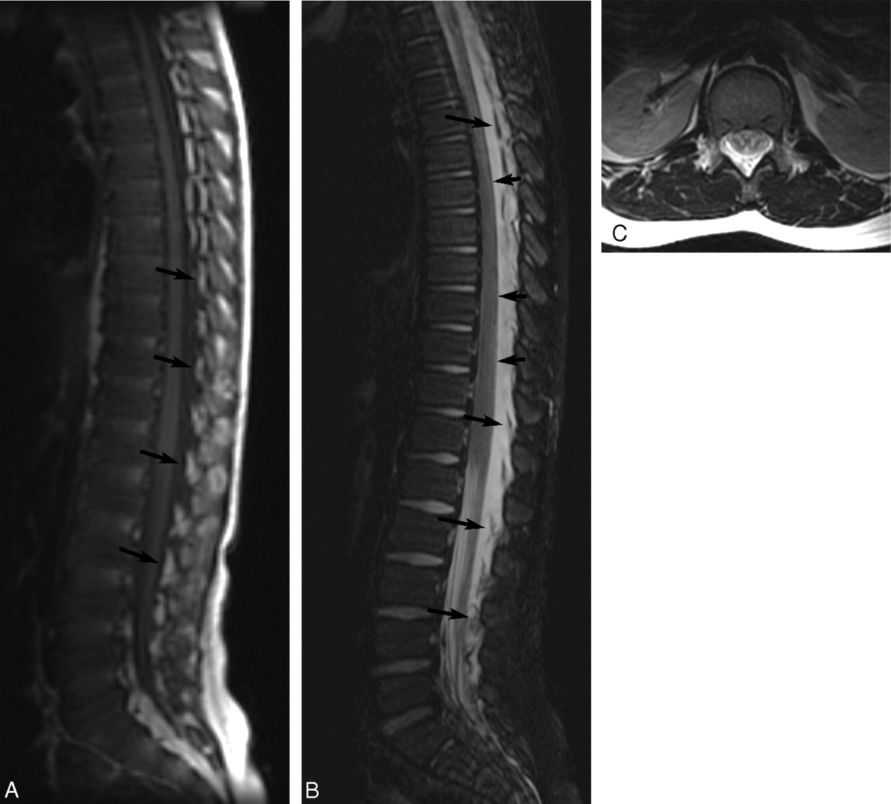
  • Fig 3.
    • Download figure
    • Open in new tab
    • Download powerpoint
    Fig 3.

    MR images of patient 2 showing extensive EDC 1 day post-LP. A, Noncontrast T1-weighted image (TR/TE, 500/9) of the spine shows a low-signal-intensity EDC extending from the upper thoracic level of the spine to the sacral level. Elevated epidural fat pads (arrows) can be seen and appear heterogeneous due to fluid infiltration. B, Noncontrast T2-weighted image (TR/TE, 4000/126) of the spine shows high-signal-intensity EDC. Epidural fat pads (long arrows) are elevated, and the dura is deviated anteriorly (short arrows). C, Axial T2-weighted image (TR/TE, 4000/126) at the level of the conus shows heterogeneous signal intensity in the dorsal epidural space secondary to fluid elevating and infiltrating the epidural fat pads. There is resultant deviation of the dura anteriorly.

  • Fig 4.
    • Download figure
    • Open in new tab
    • Download powerpoint
    Fig 4.

    Sagittal MR images of patient 5 showing initial EDC 4 days post-LP and resolution of EDC 13 days later. A, Noncontrast sagittal T1-weighted image (TR/TE, 500/12) shows hypointense EDC extending from T12-S2. Epidural fat pads (arrows) are shown to be elevated and infiltrated with low-intensity fluid. B, Noncontrast sagittal T2-weighted image (TR/TE, 5000/135) obtained at the same time as A shows hyperintense fluid deviating the dura anteriorly (short arrows) effacing the CSF and elevating the fat pads (long arrows), which are infiltrated with fluid. C and D, Sagittal T1-weighted (C) (TR/TE, 500/12) and sagittal T2-weighted (D) (TR/TE, 5000/133) images obtained 17 days post-LP show resolution of EDC. Epidural fat pads (arrows) are normal in position and homogeneous. The dura is no longer deviated, and the intrathecal CSF is not effaced.

Tables

  • Figures
  • Summary: history and imaging findings

    Patient No.Age/SexTime Between LP and MR ImagingPost-LP Symptoms Leading to Spine MR ImagingLevel of EDCEDC T1 SI Relative to CSFEDC Postcontrast SI Relative to CSFEDC T2 SI Relative to CSF
    12 y 5 months/M1 dayLE pain, decreased LE reflexesT/LLowHigh
    21 y 11 months/M1 dayRefusal to walk, pain with back straightT/L/SLowHigh
    311 y/M1 dayBack and leg pain, right LE numbnessT/LLowHigh
    46 months/F4 daysDecreased LE motionT/LLowHigh
    56 y 11 months/M4 daysLBP, unable to stand up, paraspinal muscle spasmsT/L/SLowHigh
    65 y 8 months/M3 daysBack pain, inability to stand straight/walkAt least T10-L5LowNo CEHigh, minimal stranding
    71 y 5 months/F4 daysAbnormal gaitT/LLowMild dural CEHigh
    88 y 9 months/F1 daySevere LBP, stooped postureAt least T11-S1LowHigh
    911 y 10 months/MUnknownLBPAt least T9-L4LowHigh
    103 y 2 months/F1 dayRefusing to walk, back/knee pain, weak LET/LHighHigh
    111 y 5 months/M2 daysBack pain, refusal to walkAt least T6-L5LowHigh
    121 y/M4 daysIncreased inability to walk, pain on LE movementT/LLowHigh
    134 y 7 months/M1 dayBack/LE pain, thigh/paraspinal muscle spasmT/L/SMildly highHigh
    145 y 5 months/M1 daySevere back/LE pain, refusal to walkAt least T7-S1LowHigh
    1512 y 10 months/F2 daysIncreased paresthesias, weakness in LET/LLowNo CEHigh
    169 y 2 months/M7 daysBack/LE pain, recurring meningeal signsC/T/LLowMild CEHigh
    177 y 8 months/M3 daysHA, LE stiffness, back pain, vomiting/meningeal signsT/LLowNo CEHigh
    182 y 11 months/M1 dayIncreased weakness, back/LE pain, unsteady gaitT/LMinimally highHigh
    191 y 4 months/M2 daysInability to walk/standT/LLowHigh
    207 y 2 months/F1 dayBack painC/T/LHighVery high
    216 y 11 months/M2 daysAtaxia, leg painT/L/SLowMinimal CEHigh
    224 y 7 months/F3 daysRight LE weakness, back painT/LLowMild CEHigh
    2313 y 1 month/F3 daysSevere back/LE painAt least T11-L4LowHigh
    244 y 3 months/M1 dayBilateral hip-to-thigh painLMinimally low, anterior enhancement
    252 y 7 months/M4 daysBack and leg pain, stooped postureT/LLowHigh
    • Note:—SI indicates signal intensity; LE, lower extremity; LBP, lower back pain; HA, headache; T, thoracic; L, lumbar; S, sacral; C, cervical; CE, contrast enhancement.

PreviousNext
Back to top

In this issue

American Journal of Neuroradiology: 28 (9)
American Journal of Neuroradiology
Vol. 28, Issue 9
October 2007
  • Table of Contents
  • Index by author
Advertisement
Print
Download PDF
Email Article

Thank you for your interest in spreading the word on American Journal of Neuroradiology.

NOTE: We only request your email address so that the person you are recommending the page to knows that you wanted them to see it, and that it is not junk mail. We do not capture any email address.

Enter multiple addresses on separate lines or separate them with commas.
Symptomatic Spinal Epidural Collections after Lumbar Puncture in Children
(Your Name) has sent you a message from American Journal of Neuroradiology
(Your Name) thought you would like to see the American Journal of Neuroradiology web site.
CAPTCHA
This question is for testing whether or not you are a human visitor and to prevent automated spam submissions.
Cite this article
B.L. Koch, E.A. Moosbrugger, J.C. Egelhoff
Symptomatic Spinal Epidural Collections after Lumbar Puncture in Children
American Journal of Neuroradiology Oct 2007, 28 (9) 1811-1816; DOI: 10.3174/ajnr.A0634

Citation Manager Formats

  • BibTeX
  • Bookends
  • EasyBib
  • EndNote (tagged)
  • EndNote 8 (xml)
  • Medlars
  • Mendeley
  • Papers
  • RefWorks Tagged
  • Ref Manager
  • RIS
  • Zotero
0 Responses
Respond to this article
Share
Bookmark this article
Symptomatic Spinal Epidural Collections after Lumbar Puncture in Children
B.L. Koch, E.A. Moosbrugger, J.C. Egelhoff
American Journal of Neuroradiology Oct 2007, 28 (9) 1811-1816; DOI: 10.3174/ajnr.A0634
del.icio.us logo Twitter logo Facebook logo Mendeley logo
  • Tweet Widget
  • Facebook Like
  • Google Plus One
Purchase

Jump to section

  • Article
    • Abstract
    • Methods
    • Results
    • Discussion
    • Conclusion
    • References
  • Figures & Data
  • Info & Metrics
  • Responses
  • References
  • PDF

Related Articles

  • No related articles found.
  • PubMed
  • Google Scholar

Cited By...

  • MR Imaging of the Cervical Spine in Nonaccidental Trauma: A Tertiary Institution Experience
  • Crossref
  • Google Scholar

This article has not yet been cited by articles in journals that are participating in Crossref Cited-by Linking.

More in this TOC Section

  • Optimization of Photon Counting CT Myelography
  • Characteristics of SIH Type I Culprit Lesions
  • Management Outcomes For VO Spine Biopsy
Show more Spine Imaging and Spine Image-Guided Interventions

Similar Articles

Advertisement

Indexed Content

  • Current Issue
  • Accepted Manuscripts
  • Article Preview
  • Past Issues
  • Editorials
  • Editor's Choice
  • Fellows' Journal Club
  • Letters to the Editor
  • Video Articles

Cases

  • Case Collection
  • Archive - Case of the Week
  • Archive - Case of the Month
  • Archive - Classic Case

More from AJNR

  • Trainee Corner
  • Imaging Protocols
  • MRI Safety Corner
  • Book Reviews

Multimedia

  • AJNR Podcasts
  • AJNR Scantastics

Resources

  • Turnaround Time
  • Submit a Manuscript
  • Submit a Video Article
  • Submit an eLetter to the Editor/Response
  • Manuscript Submission Guidelines
  • Statistical Tips
  • Fast Publishing of Accepted Manuscripts
  • Graphical Abstract Preparation
  • Imaging Protocol Submission
  • Evidence-Based Medicine Level Guide
  • Publishing Checklists
  • Author Policies
  • Become a Reviewer/Academy of Reviewers
  • News and Updates

About Us

  • About AJNR
  • Editorial Board
  • Editorial Board Alumni
  • Alerts
  • Permissions
  • Not an AJNR Subscriber? Join Now
  • Advertise with Us
  • Librarian Resources
  • Feedback
  • Terms and Conditions
  • AJNR Editorial Board Alumni

American Society of Neuroradiology

  • Not an ASNR Member? Join Now

© 2025 by the American Society of Neuroradiology All rights, including for text and data mining, AI training, and similar technologies, are reserved.
Print ISSN: 0195-6108 Online ISSN: 1936-959X

Powered by HighWire