Advertisement

Main menu

  • Home
  • Content
    • Current Issue
    • Accepted Manuscripts
    • Article Preview
    • Past Issue Archive
    • Video Articles
    • AJNR Case Collection
    • Case of the Week Archive
    • Case of the Month Archive
    • Classic Case Archive
  • Special Collections
    • AJNR Awards
    • Low-Field MRI
    • Alzheimer Disease
    • ASNR Foundation Special Collection
    • Photon-Counting CT
    • View All
  • Multimedia
    • AJNR Podcasts
    • AJNR SCANtastic
    • Trainee Corner
    • MRI Safety Corner
    • Imaging Protocols
  • For Authors
    • Submit a Manuscript
    • Submit a Video Article
    • Submit an eLetter to the Editor/Response
    • Manuscript Submission Guidelines
    • Statistical Tips
    • Fast Publishing of Accepted Manuscripts
    • Graphical Abstract Preparation
    • Imaging Protocol Submission
    • Author Policies
  • About Us
    • About AJNR
    • Editorial Board
    • Editorial Board Alumni
  • More
    • Become a Reviewer/Academy of Reviewers
    • Subscribers
    • Permissions
    • Alerts
    • Feedback
    • Advertisers
    • ASNR Home

User menu

  • Alerts
  • Log in

Search

  • Advanced search
American Journal of Neuroradiology

American Journal of Neuroradiology

ASHNR American Society of Functional Neuroradiology ASHNR American Society of Pediatric Neuroradiology ASSR
  • Alerts
  • Log in

Advanced Search

  • Home
  • Content
    • Current Issue
    • Accepted Manuscripts
    • Article Preview
    • Past Issue Archive
    • Video Articles
    • AJNR Case Collection
    • Case of the Week Archive
    • Case of the Month Archive
    • Classic Case Archive
  • Special Collections
    • AJNR Awards
    • Low-Field MRI
    • Alzheimer Disease
    • ASNR Foundation Special Collection
    • Photon-Counting CT
    • View All
  • Multimedia
    • AJNR Podcasts
    • AJNR SCANtastic
    • Trainee Corner
    • MRI Safety Corner
    • Imaging Protocols
  • For Authors
    • Submit a Manuscript
    • Submit a Video Article
    • Submit an eLetter to the Editor/Response
    • Manuscript Submission Guidelines
    • Statistical Tips
    • Fast Publishing of Accepted Manuscripts
    • Graphical Abstract Preparation
    • Imaging Protocol Submission
    • Author Policies
  • About Us
    • About AJNR
    • Editorial Board
    • Editorial Board Alumni
  • More
    • Become a Reviewer/Academy of Reviewers
    • Subscribers
    • Permissions
    • Alerts
    • Feedback
    • Advertisers
    • ASNR Home
  • Follow AJNR on Twitter
  • Visit AJNR on Facebook
  • Follow AJNR on Instagram
  • Join AJNR on LinkedIn
  • RSS Feeds

AJNR Awards, New Junior Editors, and more. Read the latest AJNR updates

Getting new auth cookie, if you see this message a lot, tell someone!
OtherFunctional

Giant Cystic Widening of Virchow-Robin Spaces: An Anatomofunctional Study

J. Mathias, L. Koessler, H. Brissart, S. Foscolo, E. Schmitt, S. Bracard, M. Braun and S. Kremer
American Journal of Neuroradiology September 2007, 28 (8) 1523-1525; DOI: https://doi.org/10.3174/ajnr.A0622
J. Mathias
  • Find this author on Google Scholar
  • Find this author on PubMed
  • Search for this author on this site
L. Koessler
  • Find this author on Google Scholar
  • Find this author on PubMed
  • Search for this author on this site
H. Brissart
  • Find this author on Google Scholar
  • Find this author on PubMed
  • Search for this author on this site
S. Foscolo
  • Find this author on Google Scholar
  • Find this author on PubMed
  • Search for this author on this site
E. Schmitt
  • Find this author on Google Scholar
  • Find this author on PubMed
  • Search for this author on this site
S. Bracard
  • Find this author on Google Scholar
  • Find this author on PubMed
  • Search for this author on this site
M. Braun
  • Find this author on Google Scholar
  • Find this author on PubMed
  • Search for this author on this site
S. Kremer
  • Find this author on Google Scholar
  • Find this author on PubMed
  • Search for this author on this site
  • Article
  • Figures & Data
  • Info & Metrics
  • Responses
  • References
  • PDF
Loading

Article Figures & Data

Figures

  • Fig 1.
    • Download figure
    • Open in new tab
    • Download powerpoint
    Fig 1.

    Patient 1. Morphologic MR imaging. A and B, respectively, Axial 3D-T1 (TR=13.3 ms, TE=4.2 ms) and axial 3D-T2 (TR=3000 ms, TE=154 ms)-weighted MR images show multiple confluent oval well-defined lesions, isointense to CSF, in the left superior (F1) and inferior (F2) frontal gyri, a portion of the left precentral gyrus, the anterior left cingulate gyrus, and the corpus callosum. These cystic dilations have a mild mass effect, inducing a displacement of the cortical ribbon. C, fMRI (motor task of the right hand) shows a cortical activation in the left motor supplementary area (white curved arrow) in Broadmann areas 4 (white arrowhead) and 6 (white arrow). D, There is also a symmetric cortical activation when the left hand is stimulated. E, The tractography shows a decrease of white matter fibers in the pathologic areas (white arrowhead) compared with the healthy side.
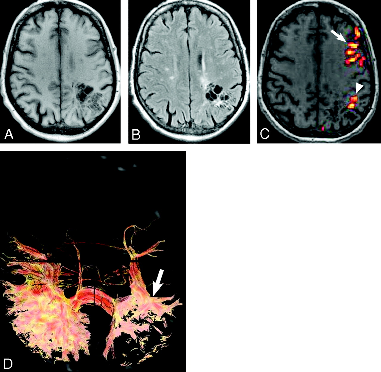
  • Fig 2.
    • Download figure
    • Open in new tab
    • Download powerpoint
    Fig 2.

    Patient 2. Morphologic MR imaging. A and B, respectively, Axial T1 (TR=165 ms, TE=4.2 ms) and axial FLAIR (TR=10 000 ms, TE=155 ms)-weighted MR images show multiple confluent oval well-defined lesions, isointense to CSF, with no mass effect in the left superior and inferior parietal lobules and in the posterior part of the left middle (T2) and inferior (T3) temporal gyri. B, FLAIR-weighted MR image shows a hypersignal interspersed among the cystic dilations, which could correspond to gliosis. C, fMRI (language task) shows a cortical activation in the superior part of the left inferior parietal lobule (Broadmann area 7, arrowhead) in regard to the lesions seen on the morphologic MR imaging. There is also a cortical activation of the left Broadmann area 44 (F3, white arrow), which demonstrates the lateralization of language to the left hemisphere. D, The tractography shows a decrease of white matter fibers in the pathologic areas (white arrow) compared with the healthy side.

PreviousNext
Back to top

In this issue

American Journal of Neuroradiology: 28 (8)
American Journal of Neuroradiology
Vol. 28, Issue 8
September 2007
  • Table of Contents
  • Index by author
Advertisement
Print
Download PDF
Email Article

Thank you for your interest in spreading the word on American Journal of Neuroradiology.

NOTE: We only request your email address so that the person you are recommending the page to knows that you wanted them to see it, and that it is not junk mail. We do not capture any email address.

Enter multiple addresses on separate lines or separate them with commas.
Giant Cystic Widening of Virchow-Robin Spaces: An Anatomofunctional Study
(Your Name) has sent you a message from American Journal of Neuroradiology
(Your Name) thought you would like to see the American Journal of Neuroradiology web site.
CAPTCHA
This question is for testing whether or not you are a human visitor and to prevent automated spam submissions.
Cite this article
J. Mathias, L. Koessler, H. Brissart, S. Foscolo, E. Schmitt, S. Bracard, M. Braun, S. Kremer
Giant Cystic Widening of Virchow-Robin Spaces: An Anatomofunctional Study
American Journal of Neuroradiology Sep 2007, 28 (8) 1523-1525; DOI: 10.3174/ajnr.A0622

Citation Manager Formats

  • BibTeX
  • Bookends
  • EasyBib
  • EndNote (tagged)
  • EndNote 8 (xml)
  • Medlars
  • Mendeley
  • Papers
  • RefWorks Tagged
  • Ref Manager
  • RIS
  • Zotero
0 Responses
Respond to this article
Share
Bookmark this article
Giant Cystic Widening of Virchow-Robin Spaces: An Anatomofunctional Study
J. Mathias, L. Koessler, H. Brissart, S. Foscolo, E. Schmitt, S. Bracard, M. Braun, S. Kremer
American Journal of Neuroradiology Sep 2007, 28 (8) 1523-1525; DOI: 10.3174/ajnr.A0622
del.icio.us logo Twitter logo Facebook logo Mendeley logo
  • Tweet Widget
  • Facebook Like
  • Google Plus One
Purchase

Jump to section

  • Article
    • Abstract
    • Case Reports
    • Discussion
    • References
  • Figures & Data
  • Info & Metrics
  • Responses
  • References
  • PDF

Related Articles

  • No related articles found.
  • PubMed
  • Google Scholar

Cited By...

  • Extreme unilateral widening of Virchow-Robin spaces mimicking stroke
  • Crossref
  • Google Scholar

This article has not yet been cited by articles in journals that are participating in Crossref Cited-by Linking.

More in this TOC Section

  • Kurtosis and Epileptogenic Tubers: A Pilot Study
  • Glutaric Aciduria Type 1: DK vs. Conventional MRI
  • Perifocal Edema in Meningiomas&Brain Connectivity
Show more Functional

Similar Articles

Advertisement

Indexed Content

  • Current Issue
  • Accepted Manuscripts
  • Article Preview
  • Past Issues
  • Editorials
  • Editor's Choice
  • Fellows' Journal Club
  • Letters to the Editor
  • Video Articles

Cases

  • Case Collection
  • Archive - Case of the Week
  • Archive - Case of the Month
  • Archive - Classic Case

More from AJNR

  • Trainee Corner
  • Imaging Protocols
  • MRI Safety Corner
  • Book Reviews

Multimedia

  • AJNR Podcasts
  • AJNR Scantastics

Resources

  • Turnaround Time
  • Submit a Manuscript
  • Submit a Video Article
  • Submit an eLetter to the Editor/Response
  • Manuscript Submission Guidelines
  • Statistical Tips
  • Fast Publishing of Accepted Manuscripts
  • Graphical Abstract Preparation
  • Imaging Protocol Submission
  • Evidence-Based Medicine Level Guide
  • Publishing Checklists
  • Author Policies
  • Become a Reviewer/Academy of Reviewers
  • News and Updates

About Us

  • About AJNR
  • Editorial Board
  • Editorial Board Alumni
  • Alerts
  • Permissions
  • Not an AJNR Subscriber? Join Now
  • Advertise with Us
  • Librarian Resources
  • Feedback
  • Terms and Conditions
  • AJNR Editorial Board Alumni

American Society of Neuroradiology

  • Not an ASNR Member? Join Now

© 2025 by the American Society of Neuroradiology All rights, including for text and data mining, AI training, and similar technologies, are reserved.
Print ISSN: 0195-6108 Online ISSN: 1936-959X

Powered by HighWire