Advertisement

Main menu

  • Home
  • Content
    • Current Issue
    • Accepted Manuscripts
    • Article Preview
    • Past Issue Archive
    • Video Articles
    • AJNR Case Collection
    • Case of the Week Archive
    • Case of the Month Archive
    • Classic Case Archive
  • Special Collections
    • AJNR Awards
    • Low-Field MRI
    • Alzheimer Disease
    • ASNR Foundation Special Collection
    • Photon-Counting CT
    • View All
  • Multimedia
    • AJNR Podcasts
    • AJNR SCANtastic
    • Trainee Corner
    • MRI Safety Corner
    • Imaging Protocols
  • For Authors
    • Submit a Manuscript
    • Submit a Video Article
    • Submit an eLetter to the Editor/Response
    • Manuscript Submission Guidelines
    • Statistical Tips
    • Fast Publishing of Accepted Manuscripts
    • Graphical Abstract Preparation
    • Imaging Protocol Submission
    • Author Policies
  • About Us
    • About AJNR
    • Editorial Board
    • Editorial Board Alumni
  • More
    • Become a Reviewer/Academy of Reviewers
    • Subscribers
    • Permissions
    • Alerts
    • Feedback
    • Advertisers
    • ASNR Home

User menu

  • Alerts
  • Log in

Search

  • Advanced search
American Journal of Neuroradiology

American Journal of Neuroradiology

ASHNR American Society of Functional Neuroradiology ASHNR American Society of Pediatric Neuroradiology ASSR
  • Alerts
  • Log in

Advanced Search

  • Home
  • Content
    • Current Issue
    • Accepted Manuscripts
    • Article Preview
    • Past Issue Archive
    • Video Articles
    • AJNR Case Collection
    • Case of the Week Archive
    • Case of the Month Archive
    • Classic Case Archive
  • Special Collections
    • AJNR Awards
    • Low-Field MRI
    • Alzheimer Disease
    • ASNR Foundation Special Collection
    • Photon-Counting CT
    • View All
  • Multimedia
    • AJNR Podcasts
    • AJNR SCANtastic
    • Trainee Corner
    • MRI Safety Corner
    • Imaging Protocols
  • For Authors
    • Submit a Manuscript
    • Submit a Video Article
    • Submit an eLetter to the Editor/Response
    • Manuscript Submission Guidelines
    • Statistical Tips
    • Fast Publishing of Accepted Manuscripts
    • Graphical Abstract Preparation
    • Imaging Protocol Submission
    • Author Policies
  • About Us
    • About AJNR
    • Editorial Board
    • Editorial Board Alumni
  • More
    • Become a Reviewer/Academy of Reviewers
    • Subscribers
    • Permissions
    • Alerts
    • Feedback
    • Advertisers
    • ASNR Home
  • Follow AJNR on Twitter
  • Visit AJNR on Facebook
  • Follow AJNR on Instagram
  • Join AJNR on LinkedIn
  • RSS Feeds

AJNR Awards, New Junior Editors, and more. Read the latest AJNR updates

Research ArticleFunctional

Incorporating Functional MR Imaging into Diffusion Tensor Tractography in the Preoperative Assessment of the Corticospinal Tract in Patients with Brain Tumors

M. Smits, M.W. Vernooij, P.A. Wielopolski, A.J.P.E. Vincent, G.C. Houston and A. van der Lugt
American Journal of Neuroradiology August 2007, 28 (7) 1354-1361; DOI: https://doi.org/10.3174/ajnr.A0538
M. Smits
  • Find this author on Google Scholar
  • Find this author on PubMed
  • Search for this author on this site
M.W. Vernooij
  • Find this author on Google Scholar
  • Find this author on PubMed
  • Search for this author on this site
P.A. Wielopolski
  • Find this author on Google Scholar
  • Find this author on PubMed
  • Search for this author on this site
A.J.P.E. Vincent
  • Find this author on Google Scholar
  • Find this author on PubMed
  • Search for this author on this site
G.C. Houston
  • Find this author on Google Scholar
  • Find this author on PubMed
  • Search for this author on this site
A. van der Lugt
  • Find this author on Google Scholar
  • Find this author on PubMed
  • Search for this author on this site
  • Article
  • Figures & Data
  • Info & Metrics
  • Responses
  • References
  • PDF
Loading

Article Figures & Data

Figures

  • Tables
  • Fig 1.
    • Download figure
    • Open in new tab
    • Download powerpoint
    Fig 1.

    Orthogonal axial/coronal (A) and axial (B) projection of T1-weighted MR images with fiber tracts depicted in color in the healthy volunteer (violet and yellow, lip fibers; green and red, hand fibers; pale blue and dark blue, foot fibers) The hand and foot fibers of the corticospinal and the lip fibers of the corticobulbar tract can be clearly distinguished and visualized (A and B). The different fMRI activation areas used to choose the seed regions of interest are shown in color (A). The course of the fibers through the corona radiata follows the known somatotopic distribution (B). C, The results are shown from conventional DT tractography (in orange) as well as from fMRI-based fiber tracking with region-of-interest placement in the PMA of the lip of the right hemisphere (in green), projected on a coronal T1-weighted image of the healthy volunteer. fMRI activation (shown in yellow-orange) is visible in the PMA of the lip and supplementary motor area in both images. Clearly, the lip fibers are only visualized by using the fMRI-based fiber tracking approach and not with the conventional fiber tracking approach.

  • Fig 2.
    • Download figure
    • Open in new tab
    • Download powerpoint
    Fig 2.

    Axial T1-weighted MR image of a patient with a brain tumor with fibers of the corticospinal tract projected in color. Results from conventional fiber tracking based on anatomic landmarks (in orange) show dispersion and diminished visualization of the CST fibers in the hemisphere affected by the lesion (arrowheads), compared with the unaffected hemisphere. DT tractography based on seed region-of-interest selection in the PMA of the hand as established with fMRI shows, in blue, the CST fibers of the hand in the affected hemisphere, allowing a clear demarcation of the fibers of interest within the CST.

  • Fig 3.
    • Download figure
    • Open in new tab
    • Download powerpoint
    Fig 3.

    Orthogonal axial/sagittal projection of a T1-weighted MR image with overprojection of the hand PMA and the hand fibers of the CST. In this patient, the left PMA, as shown by fMRI (in orange-yellow), was displaced by tumor, as was the tracked CST (in green).
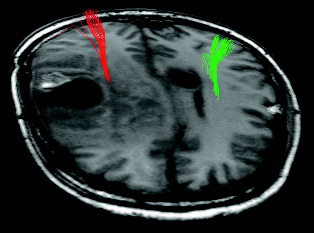
  • Fig 4.
    • Download figure
    • Open in new tab
    • Download powerpoint
    Fig 4.

    Axial T1-weighted MR image with fiber tracts projected in color (red and green for the right and left hemispheres, respectively). In this patient, hand fibers ran through an area of altered signal intensity, due to edema or tumor infiltration.

  • Fig 5.
    • Download figure
    • Open in new tab
    • Download powerpoint
    Fig 5.

    Axial T1-weighted MR images with foot fibers projected in color. Fibers pass through an area of altered signal intensity on T1-weighted images. Varying the FA thresholds for fiber tracking in this patient had a considerable influence on the fibers depicted (A–C, FA thresholds used are shown in each image).

Tables

  • Figures
    • View popup
    Table 1:

    Subject characteristics and fMRI tasks performed by each subject

    SubjectPathologic DiagnosisFunctional Neurologic StatusType of Motor Task
    No./Sex/AgePreoperativePostoperativeFinger TappingFoot TappingLip Pouting
    1/F/27N/AN/AN/AL, RL, RY
    2/F/42Anaplastic oligoastrocytomaSevere left-sided hemiparesisUnchangedL, RY
    3/M/56High-grade oligodendrogliomaMinor left-sided hemiparesisUnchangedL, RB
    4/F/55Anaplastic oligoastrocytomaNo deficitMinor left-sided hemiparesisL, RY
    5/F/48WHO grade II astrocytomaMinor paresis, left handAdditional minor paresis, left legL, RB
    6/F/31Low-grade astrocytomaNo deficitSubtle disturbances of fine motor skills, right handR
    7/M/44Low-grade oligodendrogliomaNo deficitNo deficitL, R
    8/F/42No certain pathologic diagnosis*No deficitNo deficitRB
    9/F/35Low-grade gliomaNo deficitMinor paresis, left handBB
    10/M/59Arteriovenous malformationNo deficitNo deficitBY
    • Note:—F indicates female; M, male; N/A, not applicable (healthy volunteer); B, task performed on both sides simultaneously; R, task performed with right body side; L, task performed with left body side; Y, task performed; WHO, World Health Organization.

    • * Possibly resolving ischemic infarction.

    • View popup
    Table 2:

    Overview of the most important findings by fMRI and DT tractography in each subject

    No.PMA on fMRIFiber Tracts on fMRI-Driven DT Tractography
    1Healthy volunteer: PMAs of hand, foot, and lip on both sides at known anatomic positionHand, foot, and lip fibers on both sides at known anatomic position
    2PMA of hand displaced laterally and caudally by tumorHand fibers on tumor side displaced compared with healthy side, visualization of lip fibers only on healthy side
    3PMA of hand and foot displaced by tumor and in area of decreased signal intensity on T1WIHand and foot fibers displaced on tumor side, run through area of altered signal intensity on T1WI
    4PMA at distance from lesionHand fibers run very close to tumor border, lip fibers visualized only on healthy side
    5PMA of hand and foot in area of altered signal intensity on T1WI, near tumorsHand fibers normal course, foot fibers run through area of altered signal intensity on T1WI, close to tumor
    6PMA of hand displaced cranially and dorsallyHand fibers displaced by tumor
    7PMA not displaced and not related to tumor borderHand fibers not displaced
    8PMA not displaced and not related to tumor borderHand and foot fibers not displaced
    9PMA not displaced and not related to tumor borderHand and foot fibers not displaced
    10PMA not displaced and not related to AVM borderHand fibers not displaced, visualization of lip fibers on both sides
    • Note:—T1WI indicates T1-weighted images; AVM, arteriovenous malformation.

PreviousNext
Back to top

In this issue

American Journal of Neuroradiology: 28 (7)
American Journal of Neuroradiology
Vol. 28, Issue 7
August 2007
  • Table of Contents
  • Index by author
Advertisement
Print
Download PDF
Email Article

Thank you for your interest in spreading the word on American Journal of Neuroradiology.

NOTE: We only request your email address so that the person you are recommending the page to knows that you wanted them to see it, and that it is not junk mail. We do not capture any email address.

Enter multiple addresses on separate lines or separate them with commas.
Incorporating Functional MR Imaging into Diffusion Tensor Tractography in the Preoperative Assessment of the Corticospinal Tract in Patients with Brain Tumors
(Your Name) has sent you a message from American Journal of Neuroradiology
(Your Name) thought you would like to see the American Journal of Neuroradiology web site.
CAPTCHA
This question is for testing whether or not you are a human visitor and to prevent automated spam submissions.
Cite this article
M. Smits, M.W. Vernooij, P.A. Wielopolski, A.J.P.E. Vincent, G.C. Houston, A. van der Lugt
Incorporating Functional MR Imaging into Diffusion Tensor Tractography in the Preoperative Assessment of the Corticospinal Tract in Patients with Brain Tumors
American Journal of Neuroradiology Aug 2007, 28 (7) 1354-1361; DOI: 10.3174/ajnr.A0538

Citation Manager Formats

  • BibTeX
  • Bookends
  • EasyBib
  • EndNote (tagged)
  • EndNote 8 (xml)
  • Medlars
  • Mendeley
  • Papers
  • RefWorks Tagged
  • Ref Manager
  • RIS
  • Zotero
0 Responses
Respond to this article
Share
Bookmark this article
Incorporating Functional MR Imaging into Diffusion Tensor Tractography in the Preoperative Assessment of the Corticospinal Tract in Patients with Brain Tumors
M. Smits, M.W. Vernooij, P.A. Wielopolski, A.J.P.E. Vincent, G.C. Houston, A. van der Lugt
American Journal of Neuroradiology Aug 2007, 28 (7) 1354-1361; DOI: 10.3174/ajnr.A0538
del.icio.us logo Twitter logo Facebook logo Mendeley logo
  • Tweet Widget
  • Facebook Like
  • Google Plus One
Purchase

Jump to section

  • Article
    • Abstract
    • Materials and Methods
    • Results
    • Discussion
    • Conclusion
    • Footnotes
    • References
  • Figures & Data
  • Info & Metrics
  • Responses
  • References
  • PDF

Related Articles

  • No related articles found.
  • PubMed
  • Google Scholar

Cited By...

  • Multimodality Brain Tumor Imaging: MR Imaging, PET, and PET/MR Imaging
  • Tract-Specific and Region of Interest Analysis of Corticospinal Tract Integrity in Subcortical Ischemic Stroke: Reliability and Correlation with Motor Function of Affected Lower Extremity
  • Crossref
  • Google Scholar

This article has not yet been cited by articles in journals that are participating in Crossref Cited-by Linking.

More in this TOC Section

  • Kurtosis and Epileptogenic Tubers: A Pilot Study
  • Glutaric Aciduria Type 1: DK vs. Conventional MRI
  • Brain Iron in Niemann-Pick Type C: 7T Study
Show more Functional

Similar Articles

Advertisement

Indexed Content

  • Current Issue
  • Accepted Manuscripts
  • Article Preview
  • Past Issues
  • Editorials
  • Editor's Choice
  • Fellows' Journal Club
  • Letters to the Editor
  • Video Articles

Cases

  • Case Collection
  • Archive - Case of the Week
  • Archive - Case of the Month
  • Archive - Classic Case

More from AJNR

  • Trainee Corner
  • Imaging Protocols
  • MRI Safety Corner
  • Book Reviews

Multimedia

  • AJNR Podcasts
  • AJNR Scantastics

Resources

  • Turnaround Time
  • Submit a Manuscript
  • Submit a Video Article
  • Submit an eLetter to the Editor/Response
  • Manuscript Submission Guidelines
  • Statistical Tips
  • Fast Publishing of Accepted Manuscripts
  • Graphical Abstract Preparation
  • Imaging Protocol Submission
  • Evidence-Based Medicine Level Guide
  • Publishing Checklists
  • Author Policies
  • Become a Reviewer/Academy of Reviewers
  • News and Updates

About Us

  • About AJNR
  • Editorial Board
  • Editorial Board Alumni
  • Alerts
  • Permissions
  • Not an AJNR Subscriber? Join Now
  • Advertise with Us
  • Librarian Resources
  • Feedback
  • Terms and Conditions
  • AJNR Editorial Board Alumni

American Society of Neuroradiology

  • Not an ASNR Member? Join Now

© 2025 by the American Society of Neuroradiology All rights, including for text and data mining, AI training, and similar technologies, are reserved.
Print ISSN: 0195-6108 Online ISSN: 1936-959X

Powered by HighWire