Advertisement

Main menu

  • Home
  • Content
    • Current Issue
    • Accepted Manuscripts
    • Article Preview
    • Past Issue Archive
    • Video Articles
    • AJNR Case Collection
    • Case of the Week Archive
    • Case of the Month Archive
    • Classic Case Archive
  • Special Collections
    • AJNR Awards
    • Low-Field MRI
    • Alzheimer Disease
    • ASNR Foundation Special Collection
    • Photon-Counting CT
    • View All
  • Multimedia
    • AJNR Podcasts
    • AJNR SCANtastic
    • Trainee Corner
    • MRI Safety Corner
    • Imaging Protocols
  • For Authors
    • Submit a Manuscript
    • Submit a Video Article
    • Submit an eLetter to the Editor/Response
    • Manuscript Submission Guidelines
    • Statistical Tips
    • Fast Publishing of Accepted Manuscripts
    • Graphical Abstract Preparation
    • Imaging Protocol Submission
    • Author Policies
  • About Us
    • About AJNR
    • Editorial Board
    • Editorial Board Alumni
  • More
    • Become a Reviewer/Academy of Reviewers
    • Subscribers
    • Permissions
    • Alerts
    • Feedback
    • Advertisers
    • ASNR Home

User menu

  • Alerts
  • Log in

Search

  • Advanced search
American Journal of Neuroradiology

American Journal of Neuroradiology

ASHNR American Society of Functional Neuroradiology ASHNR American Society of Pediatric Neuroradiology ASSR
  • Alerts
  • Log in

Advanced Search

  • Home
  • Content
    • Current Issue
    • Accepted Manuscripts
    • Article Preview
    • Past Issue Archive
    • Video Articles
    • AJNR Case Collection
    • Case of the Week Archive
    • Case of the Month Archive
    • Classic Case Archive
  • Special Collections
    • AJNR Awards
    • Low-Field MRI
    • Alzheimer Disease
    • ASNR Foundation Special Collection
    • Photon-Counting CT
    • View All
  • Multimedia
    • AJNR Podcasts
    • AJNR SCANtastic
    • Trainee Corner
    • MRI Safety Corner
    • Imaging Protocols
  • For Authors
    • Submit a Manuscript
    • Submit a Video Article
    • Submit an eLetter to the Editor/Response
    • Manuscript Submission Guidelines
    • Statistical Tips
    • Fast Publishing of Accepted Manuscripts
    • Graphical Abstract Preparation
    • Imaging Protocol Submission
    • Author Policies
  • About Us
    • About AJNR
    • Editorial Board
    • Editorial Board Alumni
  • More
    • Become a Reviewer/Academy of Reviewers
    • Subscribers
    • Permissions
    • Alerts
    • Feedback
    • Advertisers
    • ASNR Home
  • Follow AJNR on Twitter
  • Visit AJNR on Facebook
  • Follow AJNR on Instagram
  • Join AJNR on LinkedIn
  • RSS Feeds

AJNR Awards, New Junior Editors, and more. Read the latest AJNR updates

OtherHead and Neck Imaging

Melnick-Needles Syndrome: Report of a Case Associated with Bilateral Hypoplasia of the Cochlea

J.C. Belfield, J.S. Witana and D.J.A. Connolly
American Journal of Neuroradiology June 2007, 28 (6) 1160-1161; DOI: https://doi.org/10.3174/ajnr.A0485
J.C. Belfield
  • Find this author on Google Scholar
  • Find this author on PubMed
  • Search for this author on this site
J.S. Witana
  • Find this author on Google Scholar
  • Find this author on PubMed
  • Search for this author on this site
D.J.A. Connolly
  • Find this author on Google Scholar
  • Find this author on PubMed
  • Search for this author on this site
  • Article
  • Figures & Data
  • Info & Metrics
  • Responses
  • References
  • PDF
Loading

Abstract

SUMMARY: Melnick-Needles Syndrome is a rare disorder associated with many abnormalities. This case report describes a patient with Melnick-Needles Syndrome who was found to have bilateral hypoplasia of the cochlea, a finding not previously described in the literature. The case report describes the syndrome and demonstrates the findings on CT and MR imaging of temporal bones.

Melnick-Needles Syndrome is a rare inherited disorder characterized by skeletal dysplasia, typical facial appearances, and a large cranium. In this report of a female child with Melnick-Needles Syndrome, CT and MR imaging revealed bilateral cochlear hypoplasia.

Case Report

An infant with an unknown syndrome was screened by objective hearing tests as part of a routine protocol at 6 months of age. Auditory brain stem response was performed, which revealed a sensorineural hearing loss.

At 1 year of age, the patient was retested by behavioral audiometry, including distraction testing, and the hearing loss was confirmed. The child's hearing was amplified by using hearing aids from an early age, and her auditory development progressed, though the child displayed general developmental delay. On review at 2 years of age, otoscopic examination and middle ear pressure/compliance measurements (tympanometry) revealed otitis media with effusion and scarring of the tympanic membranes. Hearing tests confirmed that hearing had indeed deteriorated further. On additional review, the right external auditory canal was noted to be narrower due to the presence of an external auditory canal wall osteoma. The patient had no speech because she had a permanent tracheostomy, but there was noticeable change of the child's awareness of speech, environmental sound, nonverbal communication, and social interaction. Adjustment of amplification of the hearing aid seemed to overcome the impairment.

When the patient was reassessed at 5 years of age, her hearing had deteriorated further to levels of severe, bordering on profound, hearing loss. Audiometry showed increasing hearing loss, mostly at low frequencies, and to a lesser degree at high frequencies. The right tympanogram was flat, and otoscopy demonstrated extensive tympanosclerosis (scarring) on the right with bilateral middle ear effusions. At this stage, diagnosis of Melnick-Needles Syndrome was made.

In view of the diagnosis and the progression of the hearing loss, CT and MR imaging of the temporal bones were performed to look for any underlying structural anomalies. CT showed typical calvarial changes of Melnick-Needles Syndrome, as well as fluid filling of the middle ear cavities bilaterally, with an abnormal configuration of the bony ossicles. There was also bilateral hypoplasia of the cochlea, with no evidence of the modiolus, and a large solitary cochlear sac (Fig 1). The internal auditory canal was seen to be of normal width, and the course of the facial nerve canal was found to have an obtuse angle with the anterior genu (Fig 2). The vestibular aqueducts were not enlarged. These findings were confirmed on MR imaging (Fig 3). Hypoplasia of the cochlea has not been previously described in association with the Melnick-Needles Syndrome.

Fig 1.
  • Download figure
  • Open in new tab
  • Download powerpoint
Fig 1.

A, Axial CT image demonstrates bone dysplasia, typical in Melnick-Needles Syndrome. B, Axial CT image through the cochlea demonstrates fluid in the middle ear and lack of a basal turn with no modiolus, consistent with hypoplasia of the cochlea. Prominent eyes can be seen.

Fig 2.
  • Download figure
  • Open in new tab
  • Download powerpoint
Fig 2.

A, Coronal CT reformatted image through the cochlea demonstrates lack of modiolus and a basal turn of the cochlea. B, Axial CT image demonstrates normal vestibular aqueduct and widened angle of the facial nerve at the geniculate ganglion.

Fig 3.
  • Download figure
  • Open in new tab
  • Download powerpoint
Fig 3.

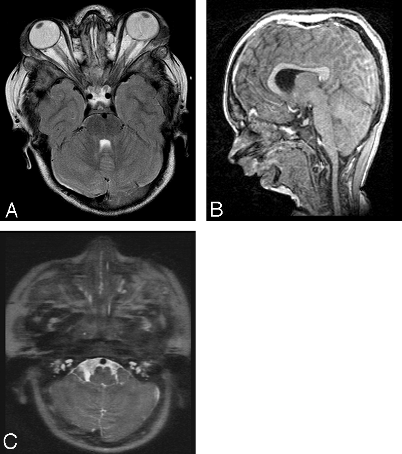
A, Axial T2-weighted MR image shows prominent bulging eyes, as described in Melnick-Needles syndrome. B, Sagittal TI-weighted localizer demonstrates pronounced micrognathia. C, Axial T2-weighted image demonstrates hypoplasia of the cochlea.

Discussion

First described in 1966 by John Melnick and Carl Needles, Melnick-Needles Syndrome is a rare X-linked dominant syndrome characterized by generalized bone dysplasia, a relatively large cranium, and typical facial appearances.1 The face is characterized by prominent supraorbital ridges, prominent eyes, full cheeks, large pinna, marked micrognathia, strabismus, and blue sclerae.2 It is part of the spectrum of X-linked skeletal dysplasias known as otopalatodigital syndrome.3 Melnick-Needles Syndrome is a multisystem disorder, with manifestations including hydronephrosis, lung hypoplasia, malrotation of the bowel, mitral and tricuspid valve prolapse, and short bowed long bones. There is a recognized association with recurrent respiratory tract infections.4 It is more common in females and generally lethal in males, with only 8 surviving male cases before 1994.5

The bony labyrinth of the inner ear comprises the vestibule, the cochlea, and the semicircular canals.6 Axial CT imaging of the cochlea normally demonstrates the central bony modiolus with a notch on the lateral border. Coronal imaging of the normal cochlea demonstrates 2 and a half turns. All of these normal features were absent in the presented patient.

The deafness associated with hypoplasia of the cochlea is usually sensorineural, with some preservation of high-frequency hearing. There may be associated vertigo, rhinorrhea, and recurrent meningitis. It is severe and permanent but can be difficult to assess even after a diagnosis has been made, especially in a young child.7 The case presented is unusual because the deafness was progressive. It is possible that Melnick-Needles Syndrome has progressive sclerotic skull base changes. Deafness has been described in Melnick-Needles Syndrome but is thought to be exceptionally rare.8 No association has been previously reported between Melnick-Needles Syndrome and hypoplasia of the cochlea.

Conclusions

Melnick-Needles Syndrome is a rare disease with multiple manifestations. The high-resolution CT and MR imaging findings of cochlear hypoplasia may represent a further association of the Melnick-Needles Syndrome. The case illustrates the need for early scanning of temporal bones in children with head and neck syndromes who have confirmed hearing loss.

References

  1. ↵
    Greco F, Mauceri L, Finocchiaro M, et al. Melnick-Needles syndrome: report of a case [in Italian]. Pediatr Med Chir 1998;20:149–52
    PubMed
  2. ↵
    Tripi TR. The Melnick-Needles syndrome (osteodysplasia): report of a rare case [in Italian]. Minerva Stomatol 1997;46:687–92
    PubMed
  3. ↵
    Hidalgo-Bravo A, Pompa-Mera EN, Kofman-Alfaro S, et al. A novel filamin A D203Y mutation in a female patient with otopalatodigital type 1 syndrome and extremely skewed X chromosome inactivation. Am J Med Genet A 2005;136:190–93
    PubMed
  4. ↵
    Dmenska H, Olszaniecka M, Gutkowski P. Melnick-Needles syndrome (MNS) and lung involvement: two case reports. Eur Respir J 2002;20:suppl. 38 ,140s
  5. ↵
    Taybi H. Handbook of Syndromes and Metabolic Diseases: Radiologic and Clinical Manifestations. St. Louis: Mosby Yearbook;1999 :875
  6. ↵
    Butler P, Mitchell A, Ellis H, eds. Applied Radiological Anatomy. Cambridge, UK: Cambridge University Press;1999 :89
  7. ↵
    Shah SM, Prabhu SS, Merchant RH. Mondini defect. J Postgrad Med 2001;47:272–73
    PubMed
  8. ↵
    Sellars SL, Beighton PH. Deafness in Osteodysplasty of Melnick and Needles. Arch Otolaryngol 1978;104:225–27
    CrossRefPubMed
  • Received June 1, 2006.
  • Accepted after revision November 8, 2006.
  • Copyright © American Society of Neuroradiology
View Abstract
PreviousNext
Back to top

In this issue

American Journal of Neuroradiology: 28 (6)
American Journal of Neuroradiology
Vol. 28, Issue 6
June 2007
  • Table of Contents
  • Index by author
Advertisement
Print
Download PDF
Email Article

Thank you for your interest in spreading the word on American Journal of Neuroradiology.

NOTE: We only request your email address so that the person you are recommending the page to knows that you wanted them to see it, and that it is not junk mail. We do not capture any email address.

Enter multiple addresses on separate lines or separate them with commas.
Melnick-Needles Syndrome: Report of a Case Associated with Bilateral Hypoplasia of the Cochlea
(Your Name) has sent you a message from American Journal of Neuroradiology
(Your Name) thought you would like to see the American Journal of Neuroradiology web site.
CAPTCHA
This question is for testing whether or not you are a human visitor and to prevent automated spam submissions.
Cite this article
J.C. Belfield, J.S. Witana, D.J.A. Connolly
Melnick-Needles Syndrome: Report of a Case Associated with Bilateral Hypoplasia of the Cochlea
American Journal of Neuroradiology Jun 2007, 28 (6) 1160-1161; DOI: 10.3174/ajnr.A0485

Citation Manager Formats

  • BibTeX
  • Bookends
  • EasyBib
  • EndNote (tagged)
  • EndNote 8 (xml)
  • Medlars
  • Mendeley
  • Papers
  • RefWorks Tagged
  • Ref Manager
  • RIS
  • Zotero
0 Responses
Respond to this article
Share
Bookmark this article
Melnick-Needles Syndrome: Report of a Case Associated with Bilateral Hypoplasia of the Cochlea
J.C. Belfield, J.S. Witana, D.J.A. Connolly
American Journal of Neuroradiology Jun 2007, 28 (6) 1160-1161; DOI: 10.3174/ajnr.A0485
del.icio.us logo Twitter logo Facebook logo Mendeley logo
  • Tweet Widget
  • Facebook Like
  • Google Plus One
Purchase

Jump to section

  • Article
    • Abstract
    • Case Report
    • Discussion
    • Conclusions
    • References
  • Figures & Data
  • Info & Metrics
  • Responses
  • References
  • PDF

Related Articles

  • No related articles found.
  • PubMed
  • Google Scholar

Cited By...

  • No citing articles found.
  • Crossref
  • Google Scholar

This article has not yet been cited by articles in journals that are participating in Crossref Cited-by Linking.

More in this TOC Section

  • MRI of the Parasellar Ligaments
  • ASL Sensitivity for Head and Neck Paraganglioma
  • Post SRS Peritumoral Hyperintense Signal of VSs
Show more Head and Neck Imaging

Similar Articles

Advertisement

Indexed Content

  • Current Issue
  • Accepted Manuscripts
  • Article Preview
  • Past Issues
  • Editorials
  • Editor's Choice
  • Fellows' Journal Club
  • Letters to the Editor
  • Video Articles

Cases

  • Case Collection
  • Archive - Case of the Week
  • Archive - Case of the Month
  • Archive - Classic Case

More from AJNR

  • Trainee Corner
  • Imaging Protocols
  • MRI Safety Corner
  • Book Reviews

Multimedia

  • AJNR Podcasts
  • AJNR Scantastics

Resources

  • Turnaround Time
  • Submit a Manuscript
  • Submit a Video Article
  • Submit an eLetter to the Editor/Response
  • Manuscript Submission Guidelines
  • Statistical Tips
  • Fast Publishing of Accepted Manuscripts
  • Graphical Abstract Preparation
  • Imaging Protocol Submission
  • Evidence-Based Medicine Level Guide
  • Publishing Checklists
  • Author Policies
  • Become a Reviewer/Academy of Reviewers
  • News and Updates

About Us

  • About AJNR
  • Editorial Board
  • Editorial Board Alumni
  • Alerts
  • Permissions
  • Not an AJNR Subscriber? Join Now
  • Advertise with Us
  • Librarian Resources
  • Feedback
  • Terms and Conditions
  • AJNR Editorial Board Alumni

American Society of Neuroradiology

  • Not an ASNR Member? Join Now

© 2025 by the American Society of Neuroradiology All rights, including for text and data mining, AI training, and similar technologies, are reserved.
Print ISSN: 0195-6108 Online ISSN: 1936-959X

Powered by HighWire