
Article Figures & Data
Figures
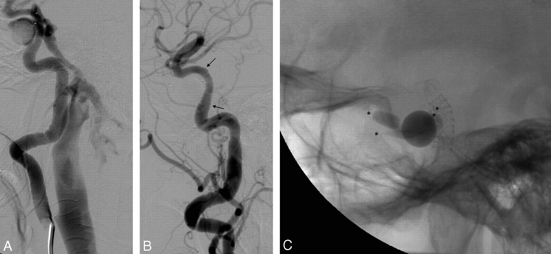
- Fig 1.
Case 3. A and B, lateral right ICA angiograms show the CCF before (A) and after (B) treatment; arrows point to stent extremities.
C, lateral view showing the stent and 4 detachable balloons previously used; 2 balloons partially inflated were used in an attempt to occlude the right CCF, whereas the 2 deflated balloons occluded the left CCF 2 months earlier.
- Fig 2.
Case 8. A, Lateral angiogram of the ICA showing total steal of the flow toward the cavernous sinus; vertebral and contralateral ICA angiographies (not reported) showed steal also from posterior and contralateral circulation.
B, Lateral ICA angiogram at the end of the stent-graft placement procedure showing nearly complete occlusion of the CCF.
C and D, ICA angiogram in lateral projection of day 2 after treatment showing partial reopening of the fistula (C) and the result after angioplasty with a coronary balloon.
E, Lateral ICA angiogram 3 months later showing persistence of the fistula with regularization of intracranial hemispheric circulation (note different diameter of distal ICA and posterior communicating artery between D and E).
- Fig 3.
Case 5. Lateral angiograms show the postprocedural occlusion of the CCF (A) and the intimal hyperplasia causing a 30% reduction of the ICA lumen at the 6-month follow-up (B), with a possible improvement at the 1-year follow-up (C). Arrows point to the stent extremities. The marker of the previously released balloon is visible anterior to the ascending segment of the carotid siphon.


Tables
Treatment of CCFs with covered stents and 6-month and 1-year follow-up
Case 1 Case 2 Case 3 Case 4 Case 5 Case 6 Case 7 Case 8 Sex/age M/31y M/23y F/71y F/45y F/15y M/31y M/29y M/35y Indication Ocular symptoms Ocular symptoms Ocular symptoms Ocular symptoms Ocular symptoms and venous drainage in the subarachnoid cerebral veins Large dilation of the basilar venous plexus with compression of the midbrain requiring urgent treatment Ocular symptoms Recent worsening of ocular symptoms Time between trauma and treatment 2 months 1 month 7 months 1 month 1 month 1 day 11 days 20 years Location of the fistula* 3 5 4 5 5 3,4,5 5 5 U.P. 3 2 2 3 1 1 1 1 Stent size 4/26 mm 4/19 mm 4/16 mm 4.5/19 mm 4/19 mm 4/19 and 4/16 mm 4.5/26 mm 4.5/26 mm Outcome Complete occlusion of the CCF without complications Complete occlusion of the CCF without complications. On day 2 angiography, a residual fistula was treated with a 5-mm angioplasty balloon Complete occlusion of the CCF without complications Complete occlusion of the CCF without complications Complete occlusion of the CCF without complications Complete occlusion of the CCF without complications related to the procedure Near-complete occlusion of the CCF without complications Near-complete occlusion of the CCF without complications. On day 2 angiography, a residual fistula was treated with a 5-mm angioplasty balloon 6-month follow-up Asymptomatic ICA occlusion Good ICA patency Good ICA patency Good ICA patency Intimal hyperplasia (30% stenosis) Not available for follow-up because of death from multiorgan failure (2 months) Persistence of the CCF that required transvenous coil occlusion of the cavernous sinus (3 months) Moderate persistence of the CCF with regression of the ocular symptoms 1-year follow-up Stable result Stable result Stable result Stable result Stable result NA NA NA * According to Debrun ICA segmentation.44
Note:—y indicates years; U.P., unsuccessful procedures; ICA, internal carotid artery; AV, arteriovenous; NA, not available.
In this issue


Jump to section
Related Articles
Cited By...
- The use of PK Papyrus covered coronary stent for carotid reconstruction: an initial institutional experience
- Use of coronary stent grafts for the treatment of high-flow carotid cavernous fistula
- Recurrence risk factors in detachable balloon embolization of traumatic direct carotid cavernous fistulas in 188 patients
- Combined use of Onyx and coils for transarterial balloon-assisted embolization of traumatic carotid-cavernous fistulas: a report of 16 cases with 17 fistulas
- Use of Onyx for Transarterial Balloon-Assisted Embolization of Traumatic Carotid Cavernous Fistulas: A Report of 23 Cases
- Placement of Covered Stents for the Treatment of Direct Carotid Cavernous Fistulas