Advertisement

Main menu

  • Home
  • Content
    • Current Issue
    • Accepted Manuscripts
    • Article Preview
    • Past Issue Archive
    • Video Articles
    • AJNR Case Collection
    • Case of the Week Archive
    • Case of the Month Archive
    • Classic Case Archive
  • Special Collections
    • AJNR Awards
    • Low-Field MRI
    • Alzheimer Disease
    • ASNR Foundation Special Collection
    • Photon-Counting CT
    • View All
  • Multimedia
    • AJNR Podcasts
    • AJNR SCANtastic
    • Trainee Corner
    • MRI Safety Corner
    • Imaging Protocols
  • For Authors
    • Submit a Manuscript
    • Submit a Video Article
    • Submit an eLetter to the Editor/Response
    • Manuscript Submission Guidelines
    • Statistical Tips
    • Fast Publishing of Accepted Manuscripts
    • Graphical Abstract Preparation
    • Imaging Protocol Submission
    • Author Policies
  • About Us
    • About AJNR
    • Editorial Board
    • Editorial Board Alumni
  • More
    • Become a Reviewer/Academy of Reviewers
    • Subscribers
    • Permissions
    • Alerts
    • Feedback
    • Advertisers
    • ASNR Home

User menu

  • Alerts
  • Log in

Search

  • Advanced search
American Journal of Neuroradiology

American Journal of Neuroradiology

ASHNR American Society of Functional Neuroradiology ASHNR American Society of Pediatric Neuroradiology ASSR
  • Alerts
  • Log in

Advanced Search

  • Home
  • Content
    • Current Issue
    • Accepted Manuscripts
    • Article Preview
    • Past Issue Archive
    • Video Articles
    • AJNR Case Collection
    • Case of the Week Archive
    • Case of the Month Archive
    • Classic Case Archive
  • Special Collections
    • AJNR Awards
    • Low-Field MRI
    • Alzheimer Disease
    • ASNR Foundation Special Collection
    • Photon-Counting CT
    • View All
  • Multimedia
    • AJNR Podcasts
    • AJNR SCANtastic
    • Trainee Corner
    • MRI Safety Corner
    • Imaging Protocols
  • For Authors
    • Submit a Manuscript
    • Submit a Video Article
    • Submit an eLetter to the Editor/Response
    • Manuscript Submission Guidelines
    • Statistical Tips
    • Fast Publishing of Accepted Manuscripts
    • Graphical Abstract Preparation
    • Imaging Protocol Submission
    • Author Policies
  • About Us
    • About AJNR
    • Editorial Board
    • Editorial Board Alumni
  • More
    • Become a Reviewer/Academy of Reviewers
    • Subscribers
    • Permissions
    • Alerts
    • Feedback
    • Advertisers
    • ASNR Home
  • Follow AJNR on Twitter
  • Visit AJNR on Facebook
  • Follow AJNR on Instagram
  • Join AJNR on LinkedIn
  • RSS Feeds

AJNR Awards, New Junior Editors, and more. Read the latest AJNR updates

Research ArticleBrain

Reproducibility of Three Whole-Brain N-Acetylaspartate Decline Cohorts in Relapsing-Remitting Multiple Sclerosis

O. Gonen, T.A. Oberndorfer, M. Inglese, J.S. Babb, J. Herbert and R.I. Grossman
American Journal of Neuroradiology February 2007, 28 (2) 267-271;
O. Gonen
  • Find this author on Google Scholar
  • Find this author on PubMed
  • Search for this author on this site
T.A. Oberndorfer
  • Find this author on Google Scholar
  • Find this author on PubMed
  • Search for this author on this site
M. Inglese
  • Find this author on Google Scholar
  • Find this author on PubMed
  • Search for this author on this site
J.S. Babb
  • Find this author on Google Scholar
  • Find this author on PubMed
  • Search for this author on this site
J. Herbert
  • Find this author on Google Scholar
  • Find this author on PubMed
  • Search for this author on this site
R.I. Grossman
  • Find this author on Google Scholar
  • Find this author on PubMed
  • Search for this author on this site
  • Article
  • Figures & Data
  • Info & Metrics
  • Responses
  • References
  • PDF
Loading

Article Figures & Data

Figures

  • Tables
  • Fig 1.
    • Download figure
    • Open in new tab
    • Download powerpoint
    Fig 1.

    Dot-plot distribution of individual patients as a function of average rate of WBNAA decline per year of CD disease duration (R1), as defined by Equations (1) and (5a). The vertical dotted lines at R1 = 0 and 1.7 mmol/L/y partition the cohort into stable (○), moderate (□), and rapid (▿) according to criteria defined in the text.23

  • Fig 2.
    • Download figure
    • Open in new tab
    • Download powerpoint
    Fig 2.

    Same as Fig 1, except that the average individual rates of WBNAA decline per year, R2, were estimated for disease duration from FS, ΔY2, as defined by Equations (2) and (5b). The vertical dotted lines at R1 = 0 and 1.7 mmol/L/y partition the cohort into stable (○), moderate (□), and rapid (▿) decline rates according to criteria defined in the text.23
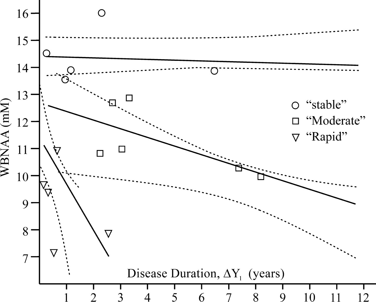
  • Fig 3.
    • Download figure
    • Open in new tab
    • Download powerpoint
    Fig 3.

    Individual WBNAA levels as a function of ΔY1 (from clinical diagnosis) of Equation (1) (labeled according to Fig 1). Solid lines are the regressions calculated for each subgroup. The dashed lines are the ±95% confidence intervals calculated for the subgroups of the previous cohort.23 Note how well the current regressions fit into the previous cohort’s confidence intervals.23

  • Fig 4.
    • Download figure
    • Open in new tab
    • Download powerpoint
    Fig 4.

    Individual WBNAA levels as a function of ΔY2 (from fist symptom) of Equation (2) (labeled according to Fig 2). Solid lines are regressions for each subgroup. The dashed lines are the ±95% confidence intervals calculated for the subgroups of the previous cohort.23 Note how well the regression lines for the current patients fit into the previous cohort’s confidence intervals.23

Tables

  • Figures
    • View popup
    Table 1:

    Cohort details

    Patient No./Age (y)/SexEDSSΔY1ΔY2WBNAA Concentration (mM)Group*Medications
    1/27.6/F2.50.532.787.14RAvonex
    2/28.1/F10.261.6814.52SCopaxone
    3/29.8/F03.323.8312.87MAvonex
    4/31.3/F1.58.198.949.96MAvonex
    5/33.1/M12.73.212.69MAvonex
    6/34.5/F1.56.476.4713.87SAvonex
    7/34.7/F20.153.629.65R/M†Copaxone
    8/37.2/F32.552.727.85RAvonex
    9/37.9/F30.320.329.37RN/A
    10/38.4/M1.50.9518.2913.55SAvonex
    11/41.5/F20.651.0710.91RAvonex
    12/42.8/F11.161.2413.9SAvonex
    13/44.3/F27.377.5310.28MN/A
    14/45.1/M03.0511.0610.98MN/A
    15/45.7/M2.52.242.2410.82MAvonex
    16/54.9/F62.34.5516.01SBestaseron
    Average2.12.644.9711.39 ± 2.5
    • Note:—EDSS indicates Expanded Disability Status Scale; Δ Y1, disease duration since clinically definite diagnosis (years); Δ Y2, disease duration from first clinical event (years); WBNAA, whole-brain N-acetylaspartate. The average age of patients studied was 37.9 ± 7.7.

    • * Subgroup notations are S, stable; M, moderate; R, rapid.

    • † Assignment changed from rapid (based on Δ Y1) to moderate when estimated from Δ Y2 disease duration.

    • View popup
    Table 2:

    Comparison between present and previous cohorts

    PresentPrevious
    CD decline rateaStable−0.0310.02
    Moderate−0.32−0.34
    Rapid−1.71−3.39
    CD group sizeStable5 (31%)10 (20%)
    Moderate7 (44%)27 (55%)
    Rapid4 (25%)12 (25%)
    EDSSbStable2.0 (1.0–6.0)2.0 (1.5–3.5)
    Moderate1.0 (0.0–2.5)2.0 (0.0–6.0)
    Rapid2.5 (2.0–3.0)2.0 (0.0–3.5)
    FS decline rateaStable−0.0570.02
    Moderate−0.20−0.13
    Rapid−1.38−2.24
    FS group sizeStable5 (31%)10 (20%)
    Moderate6 (38%)32 (65%)
    Rapid5 (32%)7 (14%)
    EDSSbStable2.0 (1.0–6.0)2.0 (1.5–3.5)
    Moderate1.5 (0.0–2.5)2.0 (0.0–6.0)
    Rapid2.5 (2.0–3.0)2.0 (0.0–3.5)
    • Note:—Group rates of WBNAA decline

    • a mM/year;

    • b EDSS score (range). Abbreviations: CD–clinical diagnosis, EDSS–Expanded Disability Status Score, FS–first symptom.

PreviousNext
Back to top

In this issue

American Journal of Neuroradiology: 28 (2)
American Journal of Neuroradiology
Vol. 28, Issue 2
February 2007
  • Table of Contents
  • Index by author
Advertisement
Print
Download PDF
Email Article

Thank you for your interest in spreading the word on American Journal of Neuroradiology.

NOTE: We only request your email address so that the person you are recommending the page to knows that you wanted them to see it, and that it is not junk mail. We do not capture any email address.

Enter multiple addresses on separate lines or separate them with commas.
Reproducibility of Three Whole-Brain N-Acetylaspartate Decline Cohorts in Relapsing-Remitting Multiple Sclerosis
(Your Name) has sent you a message from American Journal of Neuroradiology
(Your Name) thought you would like to see the American Journal of Neuroradiology web site.
CAPTCHA
This question is for testing whether or not you are a human visitor and to prevent automated spam submissions.
Cite this article
O. Gonen, T.A. Oberndorfer, M. Inglese, J.S. Babb, J. Herbert, R.I. Grossman
Reproducibility of Three Whole-Brain N-Acetylaspartate Decline Cohorts in Relapsing-Remitting Multiple Sclerosis
American Journal of Neuroradiology Feb 2007, 28 (2) 267-271;

Citation Manager Formats

  • BibTeX
  • Bookends
  • EasyBib
  • EndNote (tagged)
  • EndNote 8 (xml)
  • Medlars
  • Mendeley
  • Papers
  • RefWorks Tagged
  • Ref Manager
  • RIS
  • Zotero
0 Responses
Respond to this article
Share
Bookmark this article
Reproducibility of Three Whole-Brain N-Acetylaspartate Decline Cohorts in Relapsing-Remitting Multiple Sclerosis
O. Gonen, T.A. Oberndorfer, M. Inglese, J.S. Babb, J. Herbert, R.I. Grossman
American Journal of Neuroradiology Feb 2007, 28 (2) 267-271;
del.icio.us logo Twitter logo Facebook logo Mendeley logo
  • Tweet Widget
  • Facebook Like
  • Google Plus One

Jump to section

  • Article
    • Abstract
    • Methods
    • Results
    • Discussion
    • Conclusion
    • Footnotes
    • References
  • Figures & Data
  • Info & Metrics
  • Responses
  • References
  • PDF

Related Articles

  • No related articles found.
  • PubMed
  • Google Scholar

Cited By...

  • Global N-Acetylaspartate Declines Even in Benign Multiple Sclerosis
  • Crossref
  • Google Scholar

This article has not yet been cited by articles in journals that are participating in Crossref Cited-by Linking.

More in this TOC Section

  • Evaluating the Effects of White Matter Multiple Sclerosis Lesions on the Volume Estimation of 6 Brain Tissue Segmentation Methods
  • Quiet PROPELLER MRI Techniques Match the Quality of Conventional PROPELLER Brain Imaging Techniques
  • Predictors of Reperfusion in Patients with Acute Ischemic Stroke
Show more Brain

Similar Articles

Advertisement

Indexed Content

  • Current Issue
  • Accepted Manuscripts
  • Article Preview
  • Past Issues
  • Editorials
  • Editor's Choice
  • Fellows' Journal Club
  • Letters to the Editor
  • Video Articles

Cases

  • Case Collection
  • Archive - Case of the Week
  • Archive - Case of the Month
  • Archive - Classic Case

More from AJNR

  • Trainee Corner
  • Imaging Protocols
  • MRI Safety Corner
  • Book Reviews

Multimedia

  • AJNR Podcasts
  • AJNR Scantastics

Resources

  • Turnaround Time
  • Submit a Manuscript
  • Submit a Video Article
  • Submit an eLetter to the Editor/Response
  • Manuscript Submission Guidelines
  • Statistical Tips
  • Fast Publishing of Accepted Manuscripts
  • Graphical Abstract Preparation
  • Imaging Protocol Submission
  • Evidence-Based Medicine Level Guide
  • Publishing Checklists
  • Author Policies
  • Become a Reviewer/Academy of Reviewers
  • News and Updates

About Us

  • About AJNR
  • Editorial Board
  • Editorial Board Alumni
  • Alerts
  • Permissions
  • Not an AJNR Subscriber? Join Now
  • Advertise with Us
  • Librarian Resources
  • Feedback
  • Terms and Conditions
  • AJNR Editorial Board Alumni

American Society of Neuroradiology

  • Not an ASNR Member? Join Now

© 2025 by the American Society of Neuroradiology All rights, including for text and data mining, AI training, and similar technologies, are reserved.
Print ISSN: 0195-6108 Online ISSN: 1936-959X

Powered by HighWire