Advertisement

Main menu

  • Home
  • Content
    • Current Issue
    • Accepted Manuscripts
    • Article Preview
    • Past Issue Archive
    • Video Articles
    • AJNR Case Collection
    • Case of the Week Archive
    • Case of the Month Archive
    • Classic Case Archive
  • Special Collections
    • AJNR Awards
    • Low-Field MRI
    • Alzheimer Disease
    • ASNR Foundation Special Collection
    • Photon-Counting CT
    • View All
  • Multimedia
    • AJNR Podcasts
    • AJNR SCANtastic
    • Trainee Corner
    • MRI Safety Corner
    • Imaging Protocols
  • For Authors
    • Submit a Manuscript
    • Submit a Video Article
    • Submit an eLetter to the Editor/Response
    • Manuscript Submission Guidelines
    • Statistical Tips
    • Fast Publishing of Accepted Manuscripts
    • Graphical Abstract Preparation
    • Imaging Protocol Submission
    • Author Policies
  • About Us
    • About AJNR
    • Editorial Board
    • Editorial Board Alumni
  • More
    • Become a Reviewer/Academy of Reviewers
    • Subscribers
    • Permissions
    • Alerts
    • Feedback
    • Advertisers
    • ASNR Home

User menu

  • Alerts
  • Log in

Search

  • Advanced search
American Journal of Neuroradiology

American Journal of Neuroradiology

ASHNR American Society of Functional Neuroradiology ASHNR American Society of Pediatric Neuroradiology ASSR
  • Alerts
  • Log in

Advanced Search

  • Home
  • Content
    • Current Issue
    • Accepted Manuscripts
    • Article Preview
    • Past Issue Archive
    • Video Articles
    • AJNR Case Collection
    • Case of the Week Archive
    • Case of the Month Archive
    • Classic Case Archive
  • Special Collections
    • AJNR Awards
    • Low-Field MRI
    • Alzheimer Disease
    • ASNR Foundation Special Collection
    • Photon-Counting CT
    • View All
  • Multimedia
    • AJNR Podcasts
    • AJNR SCANtastic
    • Trainee Corner
    • MRI Safety Corner
    • Imaging Protocols
  • For Authors
    • Submit a Manuscript
    • Submit a Video Article
    • Submit an eLetter to the Editor/Response
    • Manuscript Submission Guidelines
    • Statistical Tips
    • Fast Publishing of Accepted Manuscripts
    • Graphical Abstract Preparation
    • Imaging Protocol Submission
    • Author Policies
  • About Us
    • About AJNR
    • Editorial Board
    • Editorial Board Alumni
  • More
    • Become a Reviewer/Academy of Reviewers
    • Subscribers
    • Permissions
    • Alerts
    • Feedback
    • Advertisers
    • ASNR Home
  • Follow AJNR on Twitter
  • Visit AJNR on Facebook
  • Follow AJNR on Instagram
  • Join AJNR on LinkedIn
  • RSS Feeds

AJNR Awards, New Junior Editors, and more. Read the latest AJNR updates

Getting new auth cookie, if you see this message a lot, tell someone!
Research ArticleBrain

Alberta Stroke Program Early CT Scoring of CT Perfusion in Early Stroke Visualization and Assessment

R.I. Aviv, J. Mandelcorn, S. Chakraborty, D. Gladstone, S. Malham, G. Tomlinson, A.J. Fox and S. Symons
American Journal of Neuroradiology November 2007, 28 (10) 1975-1980; DOI: https://doi.org/10.3174/ajnr.A0689
R.I. Aviv
  • Find this author on Google Scholar
  • Find this author on PubMed
  • Search for this author on this site
J. Mandelcorn
  • Find this author on Google Scholar
  • Find this author on PubMed
  • Search for this author on this site
S. Chakraborty
  • Find this author on Google Scholar
  • Find this author on PubMed
  • Search for this author on this site
D. Gladstone
  • Find this author on Google Scholar
  • Find this author on PubMed
  • Search for this author on this site
S. Malham
  • Find this author on Google Scholar
  • Find this author on PubMed
  • Search for this author on this site
G. Tomlinson
  • Find this author on Google Scholar
  • Find this author on PubMed
  • Search for this author on this site
A.J. Fox
  • Find this author on Google Scholar
  • Find this author on PubMed
  • Search for this author on this site
S. Symons
  • Find this author on Google Scholar
  • Find this author on PubMed
  • Search for this author on this site
  • Article
  • Figures & Data
  • Info & Metrics
  • Responses
  • References
  • PDF
Loading

Article Figures & Data

Figures

  • Tables
  • Fig 1.
    • Download figure
    • Open in new tab
    • Download powerpoint
    Fig 1.

    A 75-year-old man, within 2 hours of right-sided stroke and presentation of NIHSS 20. A, NCCT demonstrates subtle loss of the left posterior putamen, internal capsule, and posterior insular cortex (white arrowhead) (ASPECTS 7). B, Cerebral blood flow. C, Cerebral blood volume. D, Mean transit time. Cerebral blood volume demonstrates an abnormality confined to the posterior putamen and internal capsule (ASPECTS 8), with larger cerebral blood flow and mean-transit-time abnormalities corresponding to the left middle cerebral artery M1 segment occlusion (not shown). E, Follow-up NCCT at day 6 shows an indistinct posterior putamen confirmed on diffusion-weighted MR imaging (F). The patient recovered by 18 points with a final NIHSS of 2.
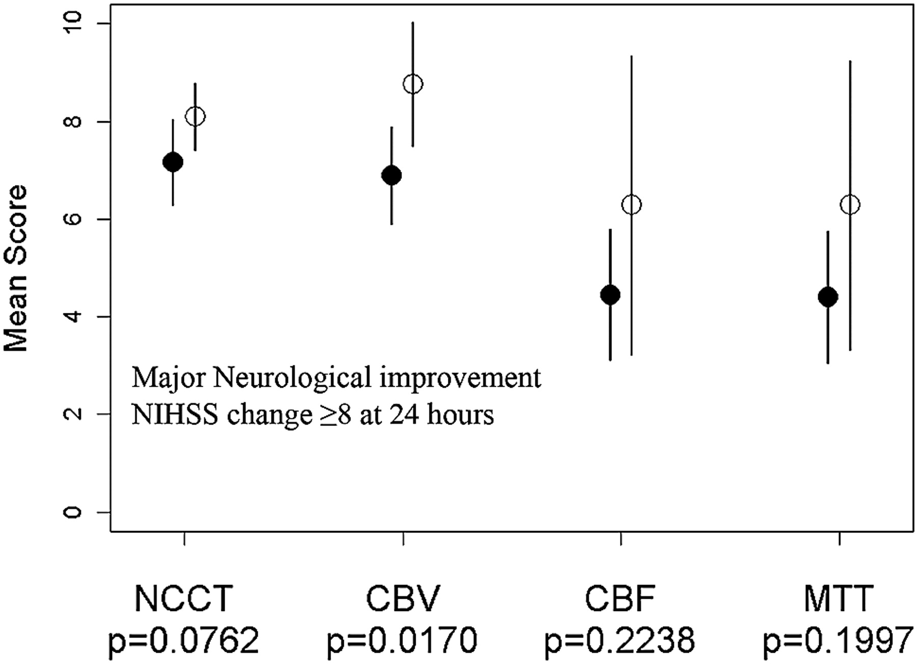
  • Fig 2.
    • Download figure
    • Open in new tab
    • Download powerpoint
    Fig 2.

    A plot of mean baseline ASPECTS NCCT, cerebral blood volume (CBV), cerebral blood flow (CBF), and mean transit time (MTT) against major neurologic improvement, demonstrating that only CBV is predictive of 24-hour NIHSS change.

  • Fig 3.
    • Download figure
    • Open in new tab
    • Download powerpoint
    Fig 3.

    A plot of mean baseline ASPECTS NCCT, cerebral blood volume (CBV), cerebral blood flow (CBF), and mean transit time (MTT). Results are dichotomized for clinical outcome by using 2-sample t tests. Favorable outcome was defined as mRS ≤2. Comparison of mean clinical outcomes was based on dichotomized mRS (0–2 versus 3–6), by using unequal variance 2-sample t tests.

Tables

  • Figures
    • View popup
    Table 1:

    Mean ASPECTS values and 95% CIs for differences in ASPECTS at baseline for NCCT and CTP compared with final NCCT ASPECTS

    NC-CTCBVCBFMTTFinal NC-CT ASPECTS
    MNI (n = 7)a
        Mean8.595.16666759
        NCCT−2.3–1.4NS0.06–3.8S0.11–3.88S−2.1–1.7NS
        CBV0.5–4.3S0.5–4.4S−1.6–2.2NS
        CBF−1.8–1.9NS−4.0 to −0.3S
        MTT−4.0 to −0.3S
    No MNI (n = 29)
        Mean6.86.54.04.05.4
        NCCT−0.4–1.1NS1.7–4.0S**S 1.7–4.0S**0.6–2.2S
        CBV1.3–3.6S**S 1.4–3.6S**0.07–2.3S
        CBF−1.3–1.4NS−2.6–0.05NS
        MTT−2.7–0.01NS
    • Note:—MNI indicates major neurologic improvement; CBV, cerebral blood volume; CBF, cerebral blood flow; MTT, mean transit time; NS, nonsignificant; S, significant (level of significance determined by paired t test, P < .05, or S**, P < .01).

    • a In patients with MNI, mean CBV and NCCT were not significantly different from mean final NCCT ASPECTS, whereas in the absence of MNI, CBF, and MTT most closely approximated final NCCT ASPECTS.

    • View popup
    Table 2:

    Performance measures of NCCT, CTP, final ASPECTS, and 24-hour NIHSS change for clinical outcome

    ThresholdaGood Outcome, mRS ≤2 (% Patients)RR (95% CI)P*Sensitivity (95% CI)Specificity (95% CI)Accuracy
    NCCT >7 vs ≤744 (6)8.0 (1.1–57.6).0180.63 (0.4–0.8)0.89 (0.47–0.99)0.69
    CBV ≥8 vs <845 (0)15.4 (0.96–246).0020.60 (0.4–0.8)100 (0.66–100)0.69
    Final NCCT >6 vs ≤647 (0)17 (1.1–27.3).0011.00 (0.7–1.0)0.63 (0.44–0.78)0.72
    CBF/MTT >5 vs ≤546 (13)3.54 (1.1–11.8).0460.74 (0.5–0.9)0.67 (0.35–0.88)0.69
    Change NIHSS 24 hours ≥886 (10)8 (2.7–25.2).00010.96 (0.8–1.0)0.67 (0.3–0.9)0.89
    • Note:—RR indicates relative risk; CBV, cerebral blood volume; CBF, cerebral blood flow; MTT, mean transit time.

    • a Dichotomized thresholds were determined by ROC analysis. Percentage of patients presenting with good outcome above and below the selected threshold is illustrated. The best early predictors of clinical outcome were 24-hour NIHSS and presentation CBV.

    • * P value was calculated using Fisher exact test.

    • View popup
    Table 3:

    Relationship between 3-month functional outcome (mRS) and dichotomized baseline CBV ASPECTS

    mRSTotal
    Baseline CBV ASPECTS0–2 (%)3 (%)4 (%)5 (%)6 (%)
    CBV < 80 (0)1 (6.2)5 (31.2)7 (43.8)3 (18.8)16
    CBV ≥ 89 (45)5 (25)4 (20)1 (5)1 (5)20
    • Note:—CBV indicates cerebral blood volume.

    • View popup
    Table 4:

    Interobserver variability

    ICCCI
    NCCT0.650.49–0.78
    CBV0.690.54–0.81
    CBF0.820.72–0.89
    MTT0.810.7–0.89
    • Note:—CBV indicates cerebral blood volume; CBF, cerebral blood flow; MTT, mean transit time.

PreviousNext
Back to top

In this issue

American Journal of Neuroradiology: 28 (10)
American Journal of Neuroradiology
Vol. 28, Issue 10
November 2007
  • Table of Contents
  • Index by author
Advertisement
Print
Download PDF
Email Article

Thank you for your interest in spreading the word on American Journal of Neuroradiology.

NOTE: We only request your email address so that the person you are recommending the page to knows that you wanted them to see it, and that it is not junk mail. We do not capture any email address.

Enter multiple addresses on separate lines or separate them with commas.
Alberta Stroke Program Early CT Scoring of CT Perfusion in Early Stroke Visualization and Assessment
(Your Name) has sent you a message from American Journal of Neuroradiology
(Your Name) thought you would like to see the American Journal of Neuroradiology web site.
CAPTCHA
This question is for testing whether or not you are a human visitor and to prevent automated spam submissions.
Cite this article
R.I. Aviv, J. Mandelcorn, S. Chakraborty, D. Gladstone, S. Malham, G. Tomlinson, A.J. Fox, S. Symons
Alberta Stroke Program Early CT Scoring of CT Perfusion in Early Stroke Visualization and Assessment
American Journal of Neuroradiology Nov 2007, 28 (10) 1975-1980; DOI: 10.3174/ajnr.A0689

Citation Manager Formats

  • BibTeX
  • Bookends
  • EasyBib
  • EndNote (tagged)
  • EndNote 8 (xml)
  • Medlars
  • Mendeley
  • Papers
  • RefWorks Tagged
  • Ref Manager
  • RIS
  • Zotero
0 Responses
Respond to this article
Share
Bookmark this article
Alberta Stroke Program Early CT Scoring of CT Perfusion in Early Stroke Visualization and Assessment
R.I. Aviv, J. Mandelcorn, S. Chakraborty, D. Gladstone, S. Malham, G. Tomlinson, A.J. Fox, S. Symons
American Journal of Neuroradiology Nov 2007, 28 (10) 1975-1980; DOI: 10.3174/ajnr.A0689
del.icio.us logo Twitter logo Facebook logo Mendeley logo
  • Tweet Widget
  • Facebook Like
  • Google Plus One
Purchase

Jump to section

  • Article
    • Abstract
    • Materials and Methods
    • Results
    • Discussion
    • References
  • Figures & Data
  • Info & Metrics
  • Responses
  • References
  • PDF

Related Articles

  • No related articles found.
  • PubMed
  • Google Scholar

Cited By...

  • Automated ASPECTS in Acute Ischemic Stroke: A Comparative Analysis with CT Perfusion
  • Computed Tomographic Perfusion Predicts Poor Outcomes in a Randomized Trial of Endovascular Therapy
  • Cerebellar Hypoperfusion in Migraine Attack: Incidence and Significance
  • Impact of ASPECT scores and infarct distribution on outcomes among patients undergoing thrombectomy for acute ischemic stroke with the ADAPT technique
  • Performance of CT ASPECTS and Collateral Score in Risk Stratification: Can Target Perfusion Profiles Be Predicted without Perfusion Imaging?
  • Correlation between cerebral blood volume values and outcomes in endovascular therapy for acute ischemic stroke
  • Dynamic Angiography and Perfusion Imaging Using Flat Detector CT in the Angiography Suite: A Pilot Study in Patients with Acute Middle Cerebral Artery Occlusions
  • Combined Multimodal Computed Tomography Score Correlates With Futile Recanalization After Thrombectomy in Patients With Acute Stroke
  • Impact of the ASPECT scores and distribution on outcome among patients undergoing thrombectomy for acute ischemic stroke
  • Performance and Predictive Value of a User-Independent Platform for CT Perfusion Analysis: Threshold-Derived Automated Systems Outperform Examiner-Driven Approaches in Outcome Prediction of Acute Ischemic Stroke
  • A prospective, multicenter pilot study investigating the utility of flat detector derived parenchymal blood volume maps to estimate cerebral blood volume in stroke patients
  • A novel clinical and imaging based score for predicting outcome prior to endovascular treatment of acute ischemic stroke
  • Pre-intervention cerebral blood volume predicts outcomes in patients undergoing endovascular therapy for acute ischemic stroke
  • Pre-intervention triage incorporating perfusion imaging improves outcomes in patients undergoing endovascular stroke therapy: a comparison with the device trials
  • Clinical Stroke Penumbra: Use of National Institutes of Health Stroke Scale as a Surrogate for CT Perfusion in Patient Triage for Intra-Arterial Middle Cerebral Artery Stroke Therapy
  • Acute Stroke Imaging: CT with CT Angiography and CT Perfusion before Management Decisions
  • Imaging-Based Endovascular Therapy for Acute Ischemic Stroke due to Proximal Intracranial Anterior Circulation Occlusion Treated Beyond 8 Hours From Time Last Seen Well: Retrospective Multicenter Analysis of 237 Consecutive Patients
  • Evaluation of CT Perfusion in the Setting of Cerebral Ischemia: Patterns and Pitfalls
  • CT Angiography Source Images Predict Final Infarct Extent in Patients with Basilar Artery Occlusion
  • Crossref
  • Google Scholar

This article has not yet been cited by articles in journals that are participating in Crossref Cited-by Linking.

More in this TOC Section

  • Multimodal CT Provides Improved Performance for Lacunar Infarct Detection
  • Optimal MRI Sequence for Identifying Occlusion Location in Acute Stroke: Which Value of Time-Resolved Contrast-Enhanced MRA?
  • Evaluating the Effects of White Matter Multiple Sclerosis Lesions on the Volume Estimation of 6 Brain Tissue Segmentation Methods
Show more Brain

Similar Articles

Advertisement

Indexed Content

  • Current Issue
  • Accepted Manuscripts
  • Article Preview
  • Past Issues
  • Editorials
  • Editor's Choice
  • Fellows' Journal Club
  • Letters to the Editor
  • Video Articles

Cases

  • Case Collection
  • Archive - Case of the Week
  • Archive - Case of the Month
  • Archive - Classic Case

More from AJNR

  • Trainee Corner
  • Imaging Protocols
  • MRI Safety Corner
  • Book Reviews

Multimedia

  • AJNR Podcasts
  • AJNR Scantastics

Resources

  • Turnaround Time
  • Submit a Manuscript
  • Submit a Video Article
  • Submit an eLetter to the Editor/Response
  • Manuscript Submission Guidelines
  • Statistical Tips
  • Fast Publishing of Accepted Manuscripts
  • Graphical Abstract Preparation
  • Imaging Protocol Submission
  • Evidence-Based Medicine Level Guide
  • Publishing Checklists
  • Author Policies
  • Become a Reviewer/Academy of Reviewers
  • News and Updates

About Us

  • About AJNR
  • Editorial Board
  • Editorial Board Alumni
  • Alerts
  • Permissions
  • Not an AJNR Subscriber? Join Now
  • Advertise with Us
  • Librarian Resources
  • Feedback
  • Terms and Conditions
  • AJNR Editorial Board Alumni

American Society of Neuroradiology

  • Not an ASNR Member? Join Now

© 2025 by the American Society of Neuroradiology All rights, including for text and data mining, AI training, and similar technologies, are reserved.
Print ISSN: 0195-6108 Online ISSN: 1936-959X

Powered by HighWire