
Article Figures & Data
Figures
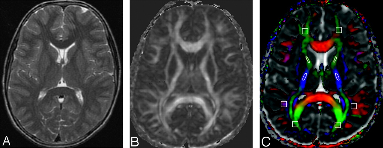
- Fig 1.
A 6-year-old healthy control. T2-weighted (A), FA (B), and color-coded FA images fused with apparent diffusion coefficient (ADC) maps (C) through the lateral ventricles show normal distribution of white matter. The rectangular and elliptic ROIs are placed (C) on the frontal, parietal, and occipital cerebral lobes and the posterior limb of the internal capsule for quantification of FA and MD values. The cutoff value for the color-coded FA for display is kept at 0.2 (C), above which the color-coded regions reflect the white matter only (red [right–left)], green [anteroposterior], and blue [superior–inferior)]).
- Fig 2.
A 12-year-old boy with clinical findings of SSPE shows normal appearance on the T2-weighted image (A). FA map (B) shows bilateral significantly low FA values in the white matter (right frontal white matter, 0.15; left frontal white matter, 0.18; right parietal white matter, 0.16; left parietal white matter, 0.15; right occipital white matter, 0.17; left occipital white matter, 0.12). Color-coded FA fused with the ADC map (C) shows the abnormality more clearly.
- Fig 3.
A 7-year-old boy with SSPE has hyperintensities on the T2-weighted (A) image (arrow) in the right frontal and parietooccipital region. The FA map (B) shows widespread bilateral abnormal white matter (right frontal white matter, 0.06; left frontal white matter, 0.14; right parietal white matter, 0.09; left parietal white matter, 0.13; right occipital white matter, 0.12; left occipital white matter, 0.17) and thinning of the genu and splenium of the corpus callosum. Color-coded FA fused with the ADC map (C) shows the abnormality more clearly.


Tables
- Table 1:
Summary of patient data for fractional anisotropy (FA) from the different regions of brain parenchyma collected from 10 age- and sex-matched controls and 21 patients of subacute sclerosing panencephalitis
Groups Corpus Callosum FWM PWM OWM TWM PLIC Genu Body Splenium a. Controls (n = 10) 0.55 ± 0.05 0.44 ± 0.04 0.61 ± 0.07 0.24 ± 0.04 0.25 ± 0.04 0.25 ± 0.02 0.25 ± 0.04 0.49 ± 0.06 b. PNCI (n ± 11) 0.42 ± 0.08 0.30 ± 0.08 0.43 ± 0.07 0.18 ± 0.02 0.18 ± 0.03 0.18 ± 0.04 0.19 ± 0.04 0.41 ± 0.06 c. PACI (n ± 10) 0.41 ± 0.09 0.32 ± 0.04 0.33 ± 0.11 0.15 ± 0.03 0.14 ± 0.03 0.15 ± 0.03 0.17 ± 0.02 0.39 ± 0.05 P (FA)* Pab = .00 Pab = .00 Pab = .00 Pab = .00 Pab = .00 Pab = .00 Pab = .00 pab = .01 Pac = .00 Pac = .00 Pac = .00 Pac = .00 Pac = .00 Pac = .00 Pac = .00 pac = .00 Pbc = .67 Pbc = .44 Pbc = .03 Pbc = .00 Pbc = .00 Pbc = .00 Pbc = .00 pbc = .48 Note:—PNCI indicates patients with normal conventional imaging; PACI, patients with abnormal conventional imaging; FWM, frontal white matter; PWM, parietal white matter; OWM, occipital white matter; TWM, temporal white matter; PLIC, posterior limb of internal capsule. Values are means ± SD.
* For FA values, Pab indicates a P value a vs b; Pbc, P value b vs c; Pac, P value a vs c.
- Table 2:
Summary of patient data for mean diffusivity (MD) from different regions of brain parenchyma collected from 10 age- and sex-matched controls and 21 patients of subacute sclerosing panencephalitis
Groups Corpus Callosum FWM PWM OWM TWM PLIC Genu Body Splenium a. Controls (n = 10) 0.80 ± 0.03 0.88 ± 0.05 0.79 ± 0.06 0.75 ± 0.05 0.70 ± 0.04 0.71 ± 0.04 0.74 ± 0.05 0.67 ± 0.03 b. PNCI (n = 11) 0.85 ± 0.05 0.96 ± 0.13 0.90 ± 0.11 0.80 ± 0.03 0.79 ± 0.06 0.78 ± 0.08 0.72 ± 0.05 0.71 ± 0.04 c. PACI (n = 10) 0.90 ± 0.24 0.94 ± 0.10 0.87 ± 0.20 0.85 ± 0.13 0.86 ± 0.17 0.87 ± 0.13 0.83 ± 0.13 0.70 ± 0.07 P (MD)* Pab = .02 Pab = .11 Pabt = .03 Pab = .00 Pab = .00 Pab = .00 Pab = .32 Pab = .07 Pac = .25 Pac = .14 Pac = .29 Pac = .01 Pac = .00 Pac = .00 Pac = .01 Pac = .40 Pbc = .45 Pbc = .75 Pbc = .73 Pbc = .17 Pbc = .15 Pbc = .01 Pbc = .00 Pbc = .61 Note:—PNCI indicates patients with normal conventional imaging; PACI, patients with abnormal conventional imaging; FWM, frontal white matter; PWM, parietal white matter; OWM, occipital white matter; TWM, temporal white matter; PLIC, posterior limb of internal capsule. Values are means ± SD.
* For MD values (expressed in ×10−3 mm2/s), Pab indicates a P value a vs b; Pbc, P value b vs c; Pac, P value a vs c.


