Advertisement

Main menu

  • Home
  • Content
    • Current Issue
    • Accepted Manuscripts
    • Article Preview
    • Past Issue Archive
    • Video Articles
    • AJNR Case Collection
    • Case of the Week Archive
    • Case of the Month Archive
    • Classic Case Archive
  • Special Collections
    • AJNR Awards
    • Low-Field MRI
    • Alzheimer Disease
    • ASNR Foundation Special Collection
    • Photon-Counting CT
    • View All
  • Multimedia
    • AJNR Podcasts
    • AJNR SCANtastic
    • Trainee Corner
    • MRI Safety Corner
    • Imaging Protocols
  • For Authors
    • Submit a Manuscript
    • Submit a Video Article
    • Submit an eLetter to the Editor/Response
    • Manuscript Submission Guidelines
    • Statistical Tips
    • Fast Publishing of Accepted Manuscripts
    • Graphical Abstract Preparation
    • Imaging Protocol Submission
    • Author Policies
  • About Us
    • About AJNR
    • Editorial Board
    • Editorial Board Alumni
  • More
    • Become a Reviewer/Academy of Reviewers
    • Subscribers
    • Permissions
    • Alerts
    • Feedback
    • Advertisers
    • ASNR Home

User menu

  • Alerts
  • Log in

Search

  • Advanced search
American Journal of Neuroradiology

American Journal of Neuroradiology

ASHNR American Society of Functional Neuroradiology ASHNR American Society of Pediatric Neuroradiology ASSR
  • Alerts
  • Log in

Advanced Search

  • Home
  • Content
    • Current Issue
    • Accepted Manuscripts
    • Article Preview
    • Past Issue Archive
    • Video Articles
    • AJNR Case Collection
    • Case of the Week Archive
    • Case of the Month Archive
    • Classic Case Archive
  • Special Collections
    • AJNR Awards
    • Low-Field MRI
    • Alzheimer Disease
    • ASNR Foundation Special Collection
    • Photon-Counting CT
    • View All
  • Multimedia
    • AJNR Podcasts
    • AJNR SCANtastic
    • Trainee Corner
    • MRI Safety Corner
    • Imaging Protocols
  • For Authors
    • Submit a Manuscript
    • Submit a Video Article
    • Submit an eLetter to the Editor/Response
    • Manuscript Submission Guidelines
    • Statistical Tips
    • Fast Publishing of Accepted Manuscripts
    • Graphical Abstract Preparation
    • Imaging Protocol Submission
    • Author Policies
  • About Us
    • About AJNR
    • Editorial Board
    • Editorial Board Alumni
  • More
    • Become a Reviewer/Academy of Reviewers
    • Subscribers
    • Permissions
    • Alerts
    • Feedback
    • Advertisers
    • ASNR Home
  • Follow AJNR on Twitter
  • Visit AJNR on Facebook
  • Follow AJNR on Instagram
  • Join AJNR on LinkedIn
  • RSS Feeds

AJNR Awards, New Junior Editors, and more. Read the latest AJNR updates

Research ArticleNeurointervention

Diffusion-Weighted MR Imaging Lesions after Filter-Protected Stenting of High-Grade Symptomatic Carotid Artery Stenoses

R. du Mesnil de Rochemont, S. Schneider, B. Yan, A. Lehr, M. Sitzer and J. Berkefeld
American Journal of Neuroradiology June 2006, 27 (6) 1321-1325;
R. du Mesnil de Rochemont
  • Find this author on Google Scholar
  • Find this author on PubMed
  • Search for this author on this site
S. Schneider
  • Find this author on Google Scholar
  • Find this author on PubMed
  • Search for this author on this site
B. Yan
  • Find this author on Google Scholar
  • Find this author on PubMed
  • Search for this author on this site
A. Lehr
  • Find this author on Google Scholar
  • Find this author on PubMed
  • Search for this author on this site
M. Sitzer
  • Find this author on Google Scholar
  • Find this author on PubMed
  • Search for this author on this site
J. Berkefeld
  • Find this author on Google Scholar
  • Find this author on PubMed
  • Search for this author on this site
  • Article
  • Figures & Data
  • Info & Metrics
  • Responses
  • References
  • PDF
Loading

Article Figures & Data

Figures

  • Tables
  • Fig. 1.
    • Download figure
    • Open in new tab
    • Download powerpoint
    Fig. 1.

    A, Number of new diffusion-weighted imaging (DWI) lesions after filter-protected carotid stent placement and median lesion load of DWI positive cases in different vascular territories.

    B, Individual numbers and vascular territory of DWI lesions. Note case 16, with severe aortic arch atherosclerosis exceeding the usual number of 1 to 4 lesions per positive case.
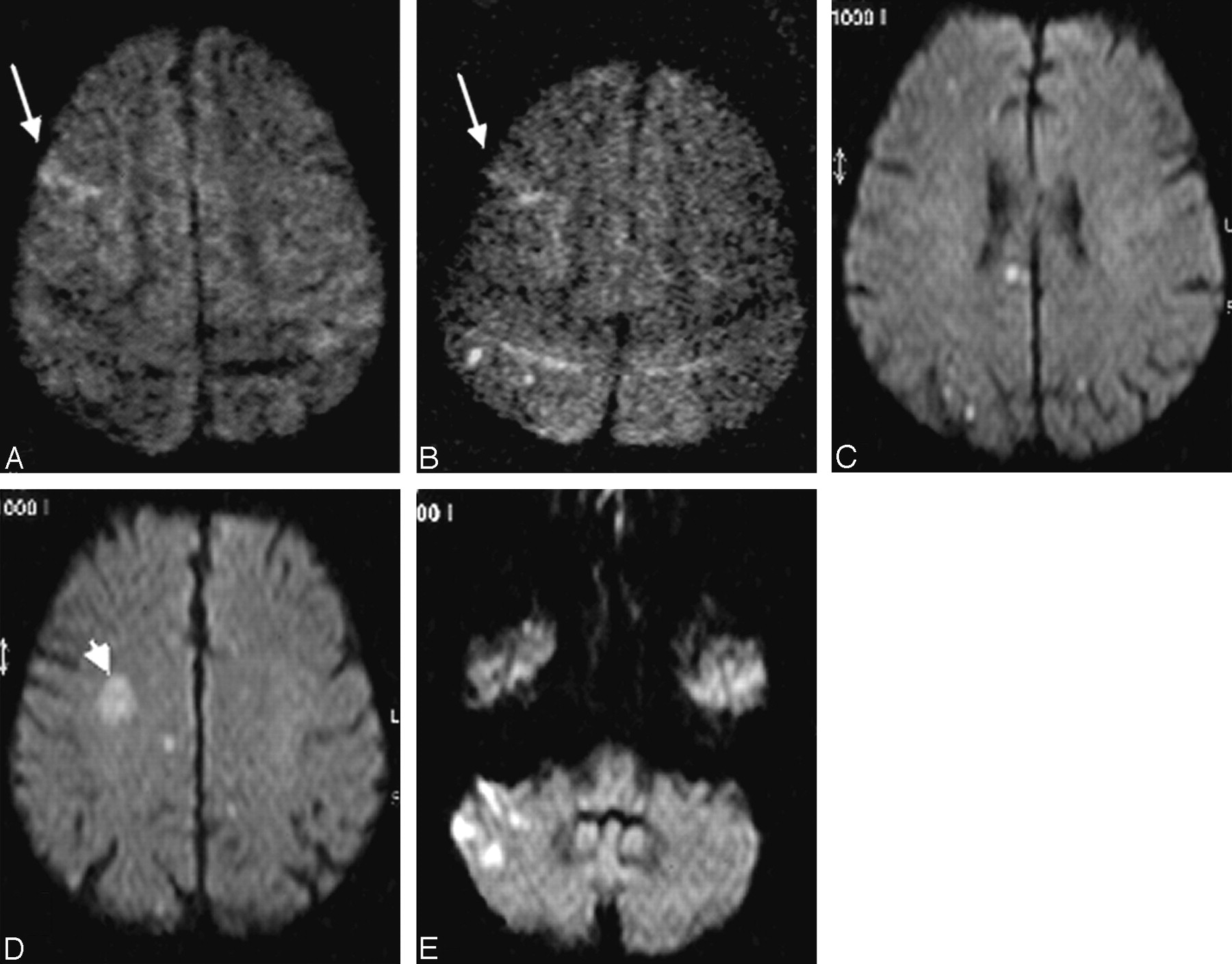
  • Fig. 2.
    • Download figure
    • Open in new tab
    • Download powerpoint
    Fig. 2.

    Range of expression of diffusion-weighted imaging (DWI) lesions after filter-protected internal carotid artery (ICA) stent placement.

    A and B, DWI the day before and the second day after stent placement of a right ICA stenosis. Upper limit of a small cortical infarct in the right middle cerebral artery (MCA) territory is visible on both images (arrow). Typical appearace of 2 new punctate lesions in the right parietal lobe (B).

    C–E, Poststent DWI after endovascular treatment of a right ICA stenosis in the patient who showed the maximal number and extension of new lesions in this study. New hyperintensities are detected in the ipsilateral ICA as well as in the contralateral ICA (C and D) and vertebrobasilar territory (E). Note a pre-existing subacute infarct in the right MCA territory (arrowhead, D).

  • Fig. 3.
    • Download figure
    • Open in new tab
    • Download powerpoint
    Fig. 3.

    Diagrams showing potential influence factors for the occurrence of diffusion-weighted imaging (DWI) lesions after carotid stent placement.

    A, Dependence between different age groups and DWI lesions was statistically significant (P = .01).

    B, Trend toward lower rates of lesions with increasing experience of operators (n.s.).

    C, Trend toward more DWI lesions with the use of segmented nitinol stents (Acculink) compared with Wallstents (n.s.).

Tables

  • Figures
  • Filter types used in this study

    Nitinol Filaments + Synthetic Membrane
    Material DesignConcentricEccentricNitinol Basket
    TypeAccunetAngioGuardEmboShieldEPI FilterWire EXFilterWire EZSpider
    ManufacturerGuidantCordisAbbottBoston ScientificBoston Scientificev3
    Porus size115 μm100 μm150 μm80 μm110 μm110 μm
    Crossing profile3.2F3.2F3.7F3.9F3.2F3.2F
    Number used in this study21517169
PreviousNext
Back to top

In this issue

American Journal of Neuroradiology: 27 (6)
American Journal of Neuroradiology
Vol. 27, Issue 6
June 2006
  • Table of Contents
  • Index by author
Advertisement
Print
Download PDF
Email Article

Thank you for your interest in spreading the word on American Journal of Neuroradiology.

NOTE: We only request your email address so that the person you are recommending the page to knows that you wanted them to see it, and that it is not junk mail. We do not capture any email address.

Enter multiple addresses on separate lines or separate them with commas.
Diffusion-Weighted MR Imaging Lesions after Filter-Protected Stenting of High-Grade Symptomatic Carotid Artery Stenoses
(Your Name) has sent you a message from American Journal of Neuroradiology
(Your Name) thought you would like to see the American Journal of Neuroradiology web site.
CAPTCHA
This question is for testing whether or not you are a human visitor and to prevent automated spam submissions.
Cite this article
R. du Mesnil de Rochemont, S. Schneider, B. Yan, A. Lehr, M. Sitzer, J. Berkefeld
Diffusion-Weighted MR Imaging Lesions after Filter-Protected Stenting of High-Grade Symptomatic Carotid Artery Stenoses
American Journal of Neuroradiology Jun 2006, 27 (6) 1321-1325;

Citation Manager Formats

  • BibTeX
  • Bookends
  • EasyBib
  • EndNote (tagged)
  • EndNote 8 (xml)
  • Medlars
  • Mendeley
  • Papers
  • RefWorks Tagged
  • Ref Manager
  • RIS
  • Zotero
0 Responses
Respond to this article
Share
Bookmark this article
Diffusion-Weighted MR Imaging Lesions after Filter-Protected Stenting of High-Grade Symptomatic Carotid Artery Stenoses
R. du Mesnil de Rochemont, S. Schneider, B. Yan, A. Lehr, M. Sitzer, J. Berkefeld
American Journal of Neuroradiology Jun 2006, 27 (6) 1321-1325;
del.icio.us logo Twitter logo Facebook logo Mendeley logo
  • Tweet Widget
  • Facebook Like
  • Google Plus One

Jump to section

  • Article
    • Abstract
    • Materials and Methods
    • Results
    • Discussion
    • Conclusions
    • References
  • Figures & Data
  • Info & Metrics
  • Responses
  • References
  • PDF

Related Articles

  • No related articles found.
  • PubMed
  • Google Scholar

Cited By...

  • Can Doppler Flow Parameters of Carotid Stenosis Predict the Occurrence of New Ischemic Brain Lesions Detected by Diffusion-Weighted MR Imaging after Filter-Protected Internal Carotid Artery Stenting?
  • New Ischemic Brain Lesions on Diffusion-Weighted MRI after Carotid Artery Stenting with Filter Protection: Frequency and Relationship with Plaque Morphology
  • The Influence of Carotid Artery Catheterization Technique on the Incidence of Thromboembolism during Carotid Artery Stenting
  • Late Cerebral Embolization After Emboli-Protected Carotid Artery Stenting Assessed by Sequential Diffusion-Weighted Magnetic Resonance Imaging
  • New Brain Lesions After Carotid Stenting Versus Carotid Endarterectomy: A Systematic Review of the Literature
  • Advances in Interventional Neuroradiology 2006
  • Crossref
  • Google Scholar

This article has not yet been cited by articles in journals that are participating in Crossref Cited-by Linking.

More in this TOC Section

  • Rescue Reentry in Carotid Near-Occlusion
  • Contour Neurovascular System: Five Year Follow Up
  • Effect of SARS-CoV2 on Endovascular Thrombectomy
Show more Neurointervention

Similar Articles

Advertisement

Indexed Content

  • Current Issue
  • Accepted Manuscripts
  • Article Preview
  • Past Issues
  • Editorials
  • Editor's Choice
  • Fellows' Journal Club
  • Letters to the Editor
  • Video Articles

Cases

  • Case Collection
  • Archive - Case of the Week
  • Archive - Case of the Month
  • Archive - Classic Case

More from AJNR

  • Trainee Corner
  • Imaging Protocols
  • MRI Safety Corner
  • Book Reviews

Multimedia

  • AJNR Podcasts
  • AJNR Scantastics

Resources

  • Turnaround Time
  • Submit a Manuscript
  • Submit a Video Article
  • Submit an eLetter to the Editor/Response
  • Manuscript Submission Guidelines
  • Statistical Tips
  • Fast Publishing of Accepted Manuscripts
  • Graphical Abstract Preparation
  • Imaging Protocol Submission
  • Evidence-Based Medicine Level Guide
  • Publishing Checklists
  • Author Policies
  • Become a Reviewer/Academy of Reviewers
  • News and Updates

About Us

  • About AJNR
  • Editorial Board
  • Editorial Board Alumni
  • Alerts
  • Permissions
  • Not an AJNR Subscriber? Join Now
  • Advertise with Us
  • Librarian Resources
  • Feedback
  • Terms and Conditions
  • AJNR Editorial Board Alumni

American Society of Neuroradiology

  • Not an ASNR Member? Join Now

© 2025 by the American Society of Neuroradiology All rights, including for text and data mining, AI training, and similar technologies, are reserved.
Print ISSN: 0195-6108 Online ISSN: 1936-959X

Powered by HighWire