
Article Figures & Data
Figures
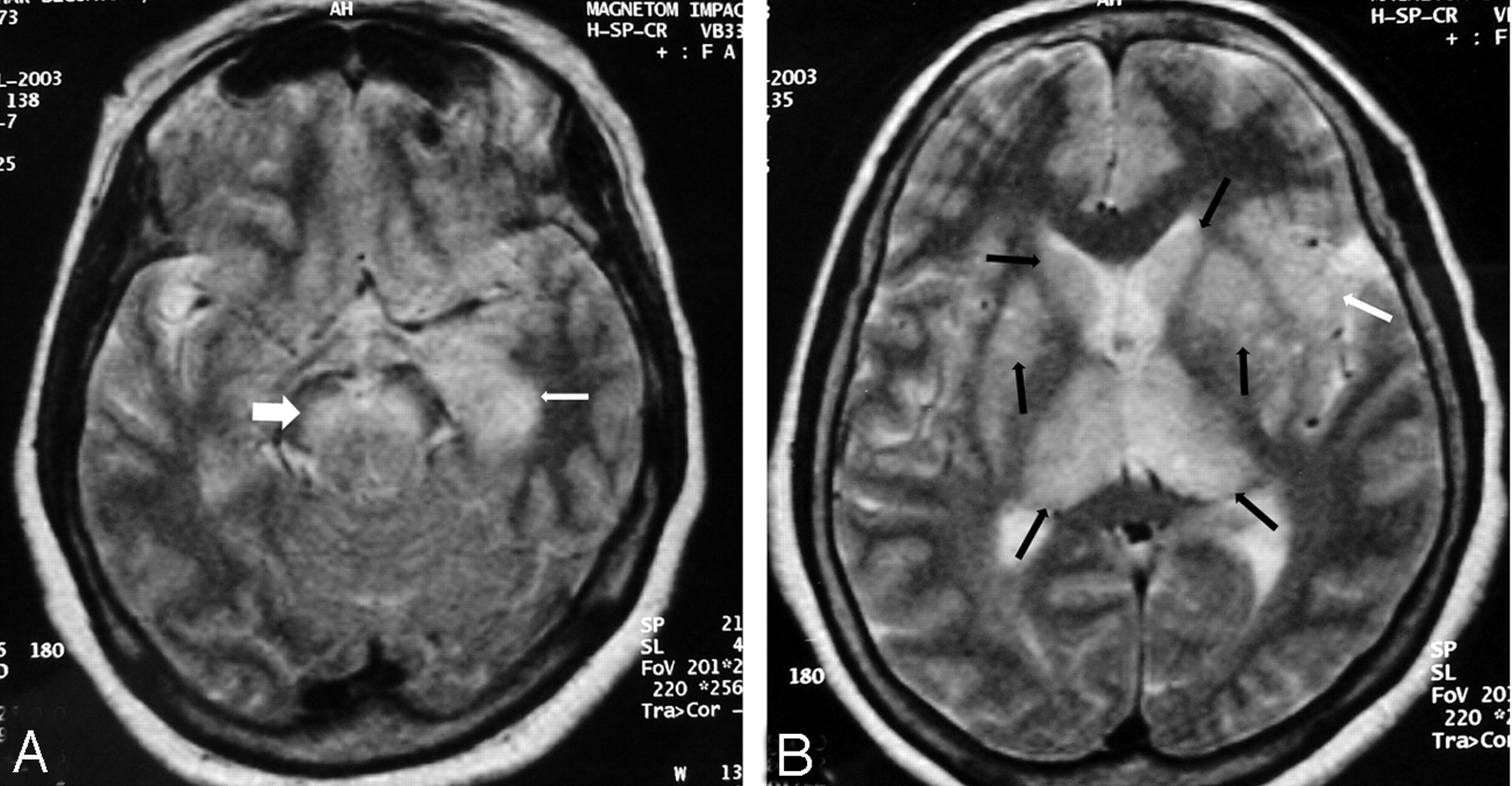
- Fig 1.
Patient 3. Fourteen-year-old boy.
A, T2-weighted axial image: bilateral thalamic lesions (black arrows). Note left hippocampal tail involvement (white arrow).
B, T2-weighted coronal image shows bilateral thalamic (black arrows), substantia nigra (white arrows), and left hippocampal body involvement (large white arrow).
C, Image more posterior than B shows hippocampal tail involvement on the left side (arrow).
D, Axial T2-weighted image shows bilateral substantia nigra lesions (arrows).
- Fig 2.
Patient 4. Eighteen-year-old man.
A, Axial T2-weighted image. Lesions are seen in both caudate heads and thalami (arrows).
B, Coronal T2-weighted image shows bilateral hippocampal body involvement (arrows).
C, Axial T2-weighted image shows the lesions involving the hippocampal tails (arrows). Note lesions in the caudate heads (white arrowheads).
D, Axial T2-weighted image done 3 months after C shows resolution of the hippocampal tail lesions (arrows). The caudate head lesions have resolved (white arrowheads).
E, CT scan done in the acute stage shows brain swelling and bilateral subtle thalamic lesions (arrows). The hippocampal and basal ganglia involvement was not apparent on this CT scan (not shown in the figure).
F, Follow-up CT scan, done 3 months after E, shows reduction in the edema and resolution of thalamic lesions.
- Fig 3.
Patient 7. Fifty-year-old woman.
A, Axial T2-weighted image shows left hippocampal head and body involvement (arrow). There is extension into the amygdala. Note bilateral substantia nigra lesions (large white arrow).
B, Axial T2-weighted image shows bilateral thalamic and basal ganglia lesions (black arrows). Note left-sided insular involvement (white arrow).
- Fig 4.
Patient 5. Twenty-nine-year-old man.
A, Coronal T2-weighted image shows bilateral hippocampal body involvement (black arrows). Note bilateral thalamic and substantia nigra involvement (white arrows).
B, Axial T2-weighted image shows bilateral hippocampal tail involvement (arrows).
C, Axial CT scan done at the same time as A and B shows hypoattenuated left mesial temporal lobe lesion. Note resemblance to Herpes simplex virus encephalitis. The right-sided involvement is not seen. The thalamic and substantia nigra lesions are not visible (not shown in the figure).
D and E, Follow-up axial and coronal T2-weighted MR done 2.5 months after A and B shows clearing up of the lesions in thalamus, substantia nigra, and hippocampus.


Tables
Patient No./Age (y)/Sex Day of MRI* Distribution of lesions on MRI Others Hippocampus Insula Neocortex Amygdala and Uncus Thalamus Substantia Nigra Basal Ganglia R L R L R L R L R L R L R L 1/28/F 5 T TB + + + + 2/58/F 3 TBH T + + + 17 Cerebral atrophy 3/14/M 4 TB + + + + + + 4/18/M 8 TBH TB + + + + + + 90 ↓ ↓ Cerebral atrophy 5/29/M 8 TBH TB + + + + 75 Normal 6/8/M 7 TBH TB + + + + + + 7/50/F 7 T TBH + + + + + + + + 8/29/M 9 TB + + + + 9/53/M 3 TBH + + + + + + 10/24/M 27 TBH TB + + + + + + 11/58/M 7 TB TB + + + + + + 16 ↓ ↓ ↓ ↓ Note:—T indicates tail of hippocampus; B, body of hippocampus; H, head of hippocampus; +, presence of lesions; ↓, decrease in the size of the lesions.
* After onset of symptoms.
Patient No./Age (y)/Sex Day of CT Distribution of lesions on CT Others Hippocampus Insula Neocortex Amygdala and Uncus Thalamus Substantia Nigra Basal Ganglia R L R L R L R L R L R L R L 1/28/F 4 2/58/F 2 3/14/M 3 + + 4/18/M 7 + + + + 3 mo 5/29/M 8 + Brain swelling 6/8/M Not done 7/50/F 4 Brain swelling 8/29/M 5 + + 9/53/M Not done 10/24/M 16 + 11/58/M 7 Cerebral atrophy Note:—+ indicates presence of lesions.
*After onset of symptoms.


