
Article Figures & Data
Figures
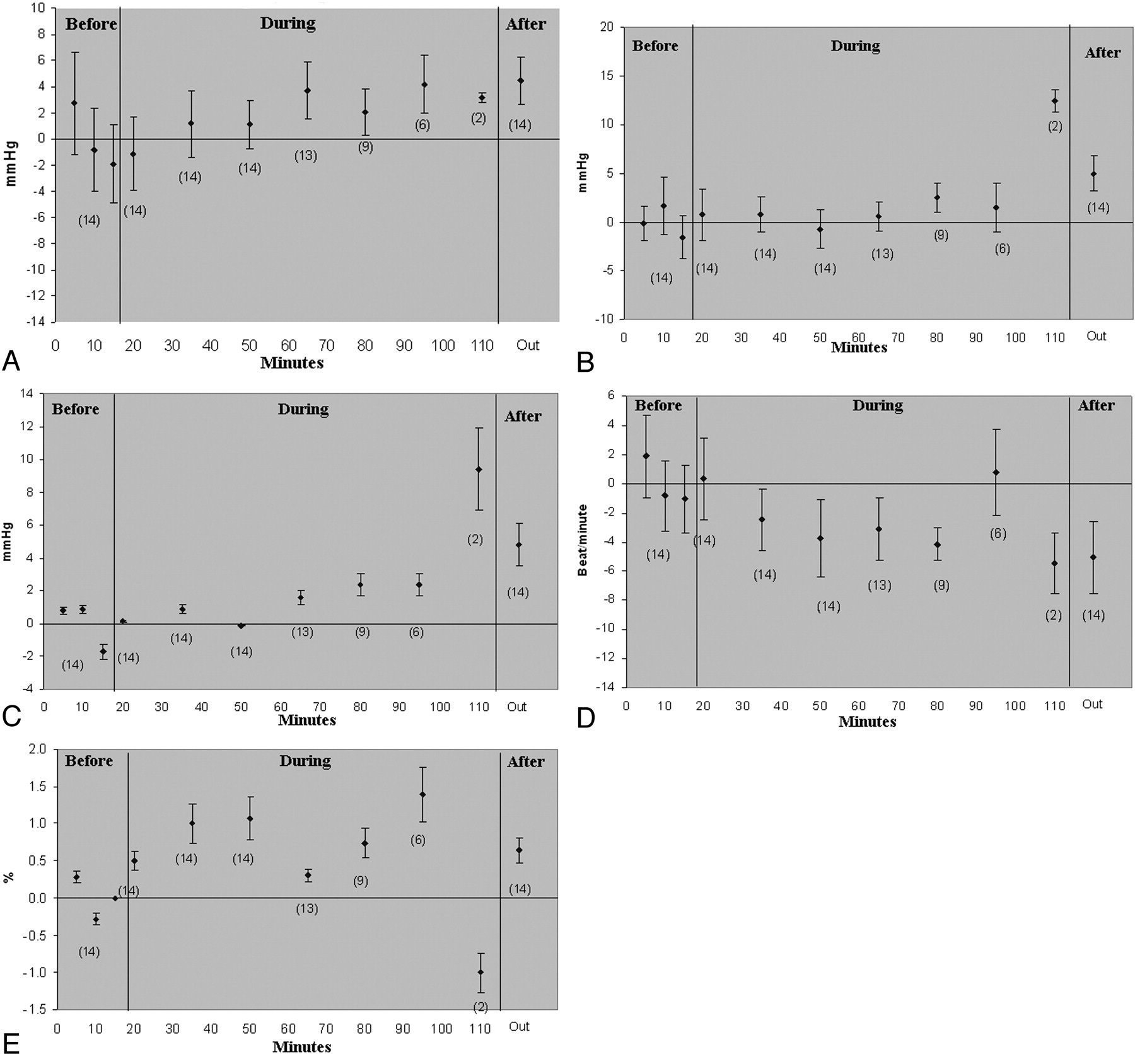
- Fig 1.
Temporal change of vital signs relative to a baseline values, before, during, and after imaging in 14 normal subjects. Mean values are indicated as solid diamonds with standard error bars. Y axis values indicate the change in the vital sign value relative to baseline mean obtained before scanning. Numbers in parentheses indicate total number of subjects at corresponding time spots. A, SBP; B, DBP; C, MAP; D, HR; E, O2 saturation.
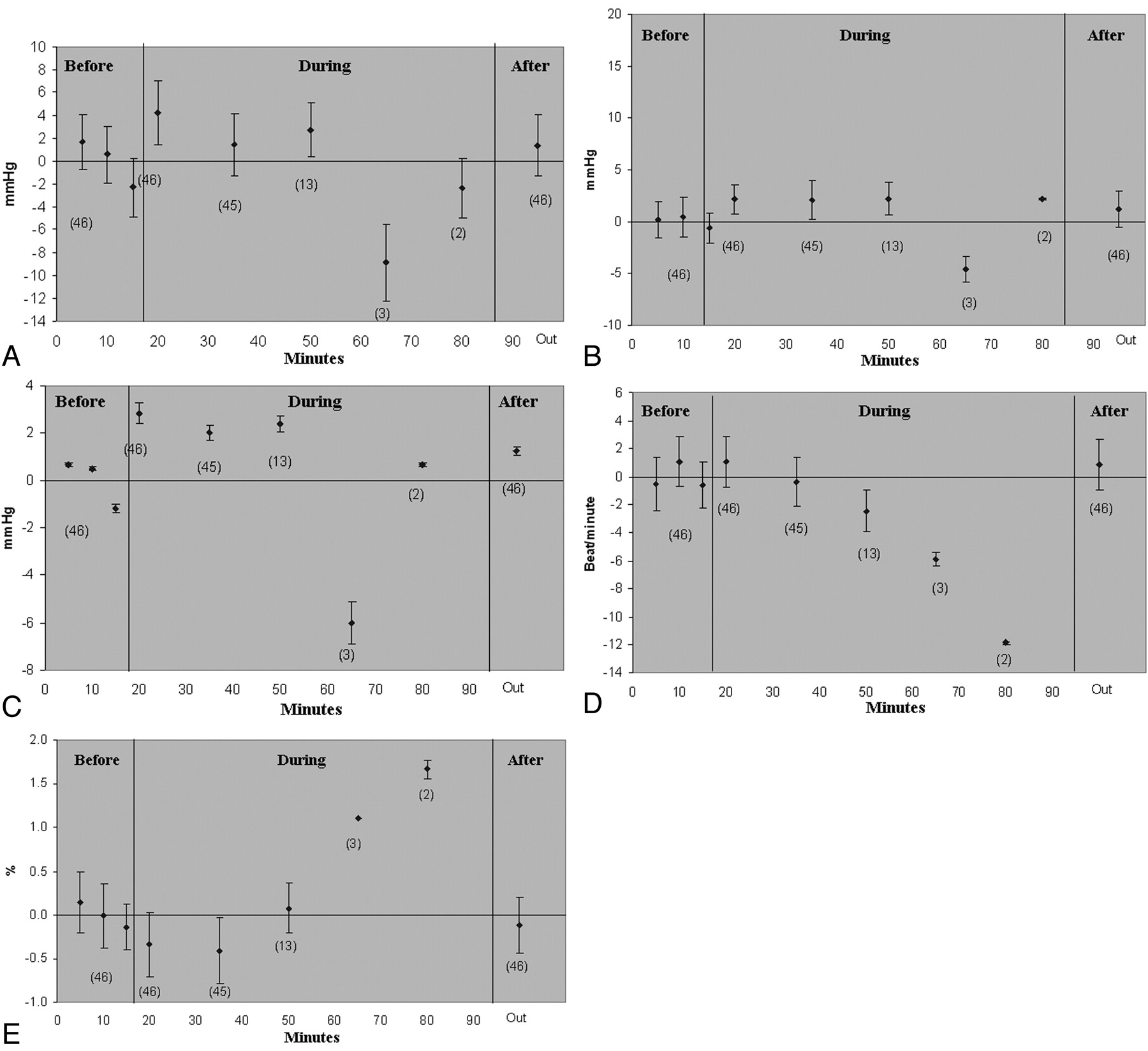
- Fig 2.
Temporal change of vital signs relative to a baseline values, before, during, and after imaging in 46 subjects with neurologic disease. Mean values are indicated as solid diamonds with standard error bars. Y axis values indicate the change in the vital sign value relative to baseline mean obtained before scanning. Numbers in parentheses indicate total number of subjects at corresponding time spots. A, SBP; B, DBP; C, MAP; D, HR; E, O2 saturation.


Tables
Vital signs measurements and comparison before and after imaging in normal and disease groups
Normal Group (18 Subjects) Disease Group (65 Subjects) Intergroup Before After Difference Before After Difference P value Respiratory rate (rpm) 14.3 14.8 0.5 ± 0.39 14.5 15.1 0.56 ± 0.18* .86 Temperature (°F) 97.1 97.2 −0.11 ± −0.24 97.3 97.2 −0.06 ± 0.06 .746 Heart rate (bpm) 65.2 60.3 −4.83 ± 1.16† 70.6 70.3 −0.32 ± 0.79 .006 SBP (mm Hg) 120.9 124.2 3.27 ± 2.78 130.3 132.2 189 ± 1.25 .621 DBP (mm Hg) 74.5 78 3.5 ± 1.46 79.5 91.1 1.62 ± 1.03 .376 Oxygenation (%) 97.2 97.4 0.21 ± 0.82 96.8 96.7 −0.09 ± 0.32 .685 MAP (mm Hg) 90 93.4 3.42 ± 1.63 96.4 98.1 1.71 ± 0.96 .372 Note:—SBP indicates systolic blood pressure; DBP, diastolic blood pressure; MAP, mean arterial blood pressure. Intergroup comparison was performed by Student t-text.
* Respiratory rate change in disease group before and after imaging is significant by paired t test (P < .001).
† Heart rate change in normal group before and after imaging is significant by paired t test (< .001).


