
Article Figures & Data
Figures
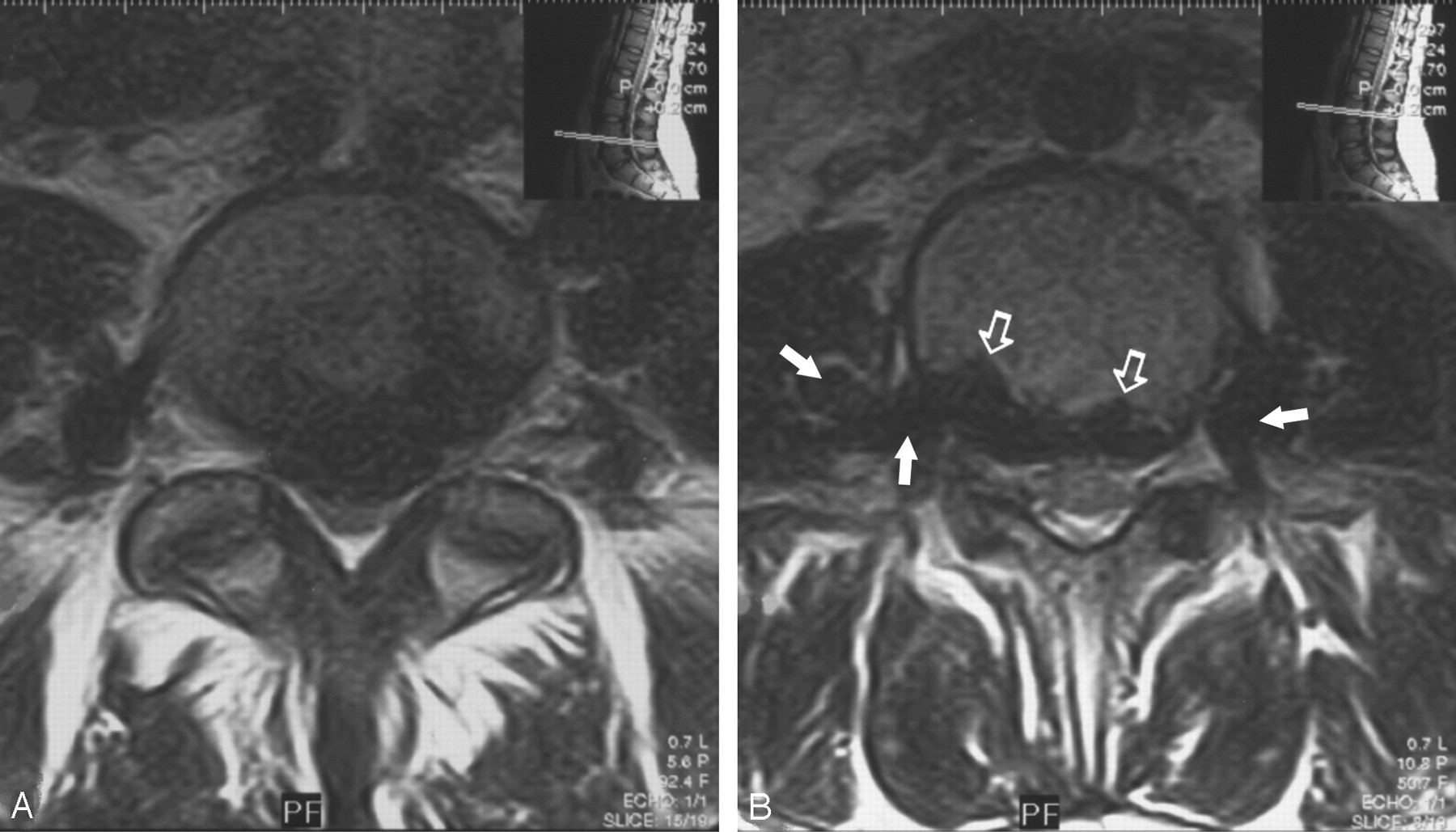
- Fig 1.
A, Axial T2-weighted spinal MR image at the level of L4–5 shows enlarged epidural veins characterized by signal void, causing a mass effect and displacing the thecal sac posteriorly. B, More cranial image depicts the scallopping of the posterior margin of L3 vertebra (open arrows) and the continuity of the epidural veins with paravertebral venous plexus via neural foraminae bilaterally (arrows).
- Fig 2.
A, Midsagittal T2-weighted scan shows the prominent epidural venous plexus below level L2 (arrowheads). B, Sagittal T2-weighted image shows the enlarged veins obliterating the neural foraminae of level L2–3 (arrows).
- Fig 3.
Venous phase of the 3D contrast-enhanced MR angiographic study, on a coronal MIP (maximum intensity projection) images.
A, Enlarged vertebral venous plexus (VVPx) consisting of epidural and paravertebral veins (arrows) extending up to the renal hilus level and consequently draining into the azygos-hemiazygous veins (open arrow).
B, Infrahepatic inferior vena cava cannot be distinguished as a separate structure. Common iliac veins appear at the conjunction level just behind the common iliac arteries (arrows) and drain into the VVPx eventually (not shown here). Enlarged left ovarian vein and a large-caliber tortuous right renal vein as a secondary drainage pathway of the lower body segments, as well as the VVPx, can also be recognized (asterisk). Arrowheads indicate the enlarged azygous vein.


In this issue


Jump to section
Related Articles
Cited By...
- No citing articles found.