Advertisement

Main menu

  • Home
  • Content
    • Current Issue
    • Accepted Manuscripts
    • Article Preview
    • Past Issue Archive
    • Video Articles
    • AJNR Case Collection
    • Case of the Week Archive
    • Case of the Month Archive
    • Classic Case Archive
  • Special Collections
    • AJNR Awards
    • Low-Field MRI
    • Alzheimer Disease
    • ASNR Foundation Special Collection
    • Photon-Counting CT
    • View All
  • Multimedia
    • AJNR Podcasts
    • AJNR SCANtastic
    • Trainee Corner
    • MRI Safety Corner
    • Imaging Protocols
  • For Authors
    • Submit a Manuscript
    • Submit a Video Article
    • Submit an eLetter to the Editor/Response
    • Manuscript Submission Guidelines
    • Statistical Tips
    • Fast Publishing of Accepted Manuscripts
    • Graphical Abstract Preparation
    • Imaging Protocol Submission
    • Author Policies
  • About Us
    • About AJNR
    • Editorial Board
    • Editorial Board Alumni
  • More
    • Become a Reviewer/Academy of Reviewers
    • Subscribers
    • Permissions
    • Alerts
    • Feedback
    • Advertisers
    • ASNR Home

User menu

  • Alerts
  • Log in

Search

  • Advanced search
American Journal of Neuroradiology

American Journal of Neuroradiology

ASHNR American Society of Functional Neuroradiology ASHNR American Society of Pediatric Neuroradiology ASSR
  • Alerts
  • Log in

Advanced Search

  • Home
  • Content
    • Current Issue
    • Accepted Manuscripts
    • Article Preview
    • Past Issue Archive
    • Video Articles
    • AJNR Case Collection
    • Case of the Week Archive
    • Case of the Month Archive
    • Classic Case Archive
  • Special Collections
    • AJNR Awards
    • Low-Field MRI
    • Alzheimer Disease
    • ASNR Foundation Special Collection
    • Photon-Counting CT
    • View All
  • Multimedia
    • AJNR Podcasts
    • AJNR SCANtastic
    • Trainee Corner
    • MRI Safety Corner
    • Imaging Protocols
  • For Authors
    • Submit a Manuscript
    • Submit a Video Article
    • Submit an eLetter to the Editor/Response
    • Manuscript Submission Guidelines
    • Statistical Tips
    • Fast Publishing of Accepted Manuscripts
    • Graphical Abstract Preparation
    • Imaging Protocol Submission
    • Author Policies
  • About Us
    • About AJNR
    • Editorial Board
    • Editorial Board Alumni
  • More
    • Become a Reviewer/Academy of Reviewers
    • Subscribers
    • Permissions
    • Alerts
    • Feedback
    • Advertisers
    • ASNR Home
  • Follow AJNR on Twitter
  • Visit AJNR on Facebook
  • Follow AJNR on Instagram
  • Join AJNR on LinkedIn
  • RSS Feeds

AJNR Awards, New Junior Editors, and more. Read the latest AJNR updates

LetterLetter

Reporting Terminology for Lumbar Disk Herniations: Axial Segmentation of the Preneural Foraminal Portion of the Lumbar Nerve Roots

Kimia Khalatbari, Mazyar Azar and Farid Kazemi Gazic
American Journal of Neuroradiology October 2005, 26 (9) 2430-2431;
Kimia Khalatbari
  • Find this author on Google Scholar
  • Find this author on PubMed
  • Search for this author on this site
Mazyar Azar
  • Find this author on Google Scholar
  • Find this author on PubMed
  • Search for this author on this site
Farid Kazemi Gazic
  • Find this author on Google Scholar
  • Find this author on PubMed
  • Search for this author on this site
  • Article
  • Figures & Data
  • Info & Metrics
  • Responses
  • References
  • PDF
Loading

Article Figures & Data

Figures

  • Fig 1.
    • Download figure
    • Open in new tab
    • Download powerpoint
    Fig 1.

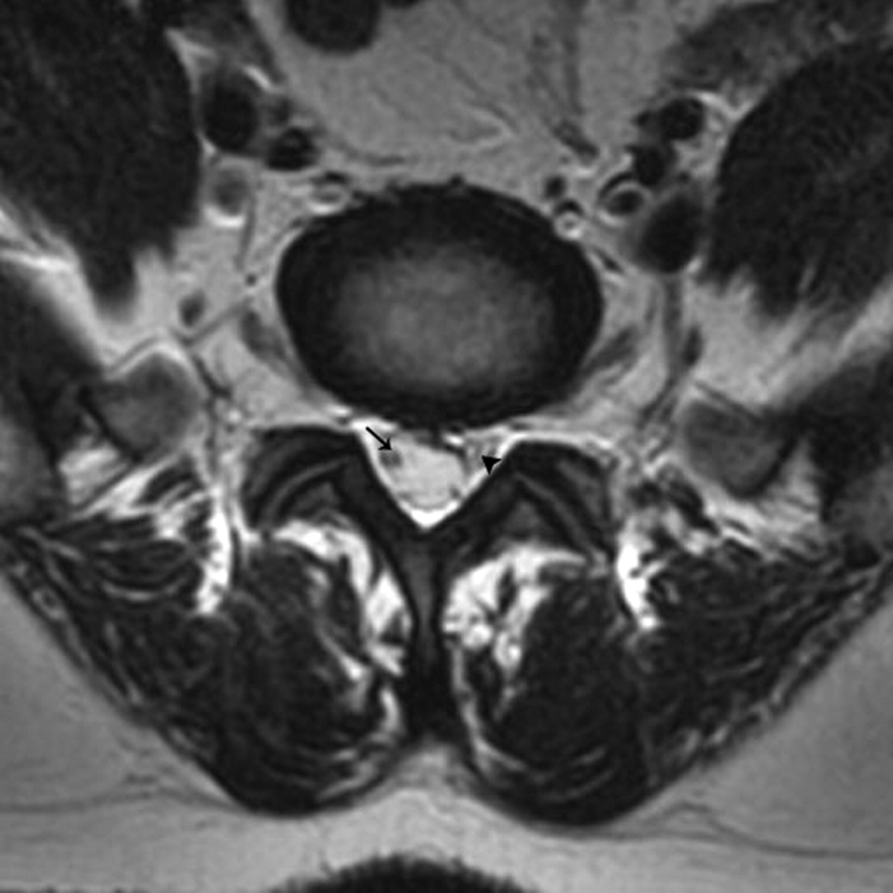
    Contiguous axial T2-weighted (3800/97.8) MR images through the L5–S1 level show the 3 segments of the preneural foraminal portion of the S1 nerve root. In this patient, individualization of all of the 3 segments is possible within a single zone.

    A, Cranial-most image demonstrates the lateral thecal sac segment of the right S1 nerve root (arrow) in the right central zone.

    B, Image shows the junctional zone segment of the right S1 nerve root (long arrow) in the right central zone. This segment, which is situated in the proximal portion of the dural root sleeve, is separated from the contents of the neighboring thecal sac by 2 adjacent layers of dura mater (arrowhead). The thickness of a single layer of dura mater is demarcated (short arrow) in the posterior portion of the thecal sac for comparison.

    C, More caudally, image demonstrates the dural root sleeve of the right S1 nerve root (arrow) in the right central zone. Individualization of the contained dural root sleeve segment of the S1 nerve root is not possible in this patient.

  • Fig 2.
    • Download figure
    • Open in new tab
    • Download powerpoint
    Fig 2.

    Contiguous axial T2-weighted (3700/106.7) MR images through the L5–S1 level show the varying appearances (also refer to Fig 1B) of the proposed “junctional segment” of a lumbar nerve root.

    A, Image demonstrates the junctional segment of the left S1 nerve root (long arrow) within the “pinched” portion (arrowheads) of the left ventrolateral angle of the thecal sac. The unrestricted contour of the right ventrolateral aspect of the thecal sac harbors the lateral thecal sac segment of the ipsilateral S1 nerve root (short arrow).

    B, Image at a slightly more caudal level shows the proximal-most portion of the left S1 dural nerve root sleeve (arrowheads), the medial wall of which is in close apposition to the ventrolateral wall of the neighboring thecal sac. This portion of the dural root sleeve is also regarded as housing the junctional segment of the S1 nerve root (arrow).

  • Fig 3.
    • Download figure
    • Open in new tab
    • Download powerpoint
    Fig 3.

    Axial T2-weighted (4000/98) MR image at the diskal-suprapedicular level of L5–S1 demonstrates the nonparallel segmentation of the S1 nerve roots. On the right, the lateral thecal sac segment (arrow), and on the left, the junctional segment (arrowhead), of the S1 nerve roots are visualized.

  • Fig 4.
    • Download figure
    • Open in new tab
    • Download powerpoint
    Fig 4.

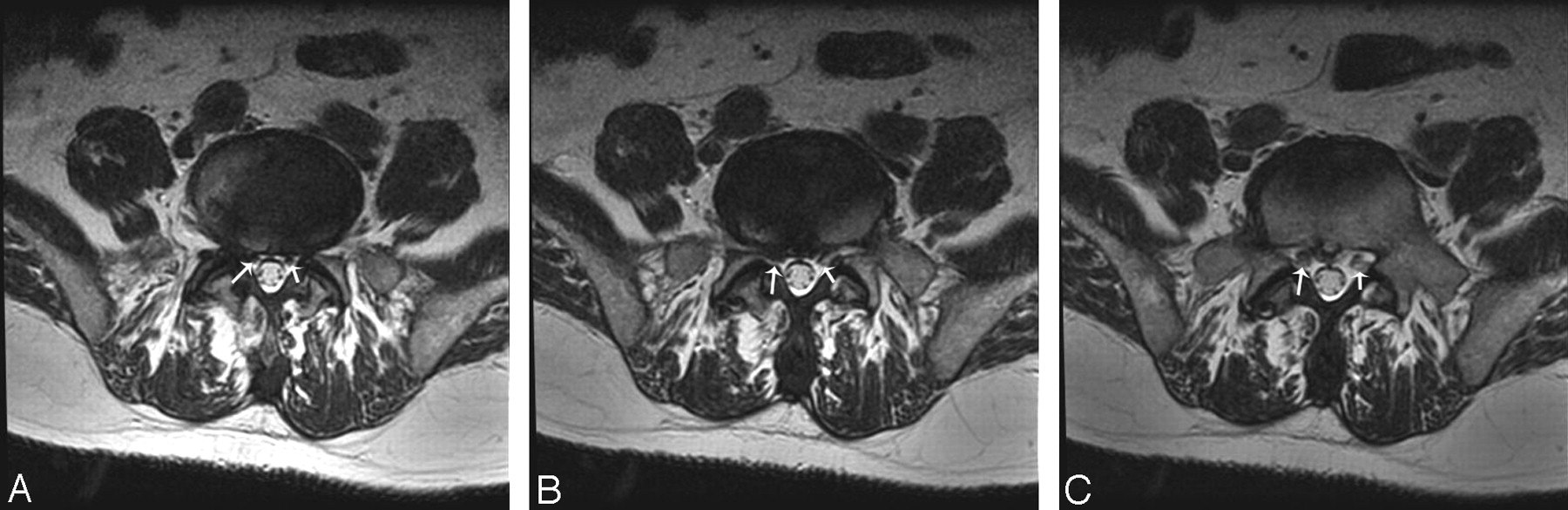
    Contiguous axial T2-weighted (3800/97.8) MR images through the L5–S1 level with the S1 dural nerve root sleeves (individualization of the dural root sleeve segments of the S1 nerve roots within their respective dural sleeves is not possible in this particular patient).

    A, Cranial-most image demonstrates the right (long arrow) and left (short arrow) S1 dural nerve root sleeves within the subarticular zones.

    B, Image at a slightly more caudal level shows a mild central-subarticular zone disk protrusion that causes a relatively severe compression of the right S1 dural nerve root sleeve (long arrow). The left-sided intact complex is demarcated (short arrow) for comparison.

    C, The S1 dural nerve root sleeves (long and short arrows) assume a symmetric appearance at a slightly more caudal level.

PreviousNext
Back to top

In this issue

American Journal of Neuroradiology: 26 (9)
American Journal of Neuroradiology
Vol. 26, Issue 9
1 Oct 2005
  • Table of Contents
  • Index by author
Advertisement
Print
Download PDF
Email Article

Thank you for your interest in spreading the word on American Journal of Neuroradiology.

NOTE: We only request your email address so that the person you are recommending the page to knows that you wanted them to see it, and that it is not junk mail. We do not capture any email address.

Enter multiple addresses on separate lines or separate them with commas.
Reporting Terminology for Lumbar Disk Herniations: Axial Segmentation of the Preneural Foraminal Portion of the Lumbar Nerve Roots
(Your Name) has sent you a message from American Journal of Neuroradiology
(Your Name) thought you would like to see the American Journal of Neuroradiology web site.
CAPTCHA
This question is for testing whether or not you are a human visitor and to prevent automated spam submissions.
Cite this article
Kimia Khalatbari, Mazyar Azar, Farid Kazemi Gazic
Reporting Terminology for Lumbar Disk Herniations: Axial Segmentation of the Preneural Foraminal Portion of the Lumbar Nerve Roots
American Journal of Neuroradiology Oct 2005, 26 (9) 2430-2431;

Citation Manager Formats

  • BibTeX
  • Bookends
  • EasyBib
  • EndNote (tagged)
  • EndNote 8 (xml)
  • Medlars
  • Mendeley
  • Papers
  • RefWorks Tagged
  • Ref Manager
  • RIS
  • Zotero
0 Responses
Respond to this article
Share
Bookmark this article
Reporting Terminology for Lumbar Disk Herniations: Axial Segmentation of the Preneural Foraminal Portion of the Lumbar Nerve Roots
Kimia Khalatbari, Mazyar Azar, Farid Kazemi Gazic
American Journal of Neuroradiology Oct 2005, 26 (9) 2430-2431;
del.icio.us logo Twitter logo Facebook logo Mendeley logo
  • Tweet Widget
  • Facebook Like
  • Google Plus One

Jump to section

  • Article
    • References
  • Figures & Data
  • Info & Metrics
  • Responses
  • References
  • PDF

Related Articles

  • No related articles found.
  • PubMed
  • Google Scholar

Cited By...

  • No citing articles found.
  • Crossref
  • Google Scholar

This article has not yet been cited by articles in journals that are participating in Crossref Cited-by Linking.

More in this TOC Section

  • Letter to the Editor regarding “Automated Volumetric Software in Dementia: Help or Hindrance to the Neuroradiologist?”
  • Reply:
  • Brain AVM’s Nidus: What if We Hadn’t Understood Anything?
Show more LETTERS

Similar Articles

Advertisement

Indexed Content

  • Current Issue
  • Accepted Manuscripts
  • Article Preview
  • Past Issues
  • Editorials
  • Editor's Choice
  • Fellows' Journal Club
  • Letters to the Editor
  • Video Articles

Cases

  • Case Collection
  • Archive - Case of the Week
  • Archive - Case of the Month
  • Archive - Classic Case

More from AJNR

  • Trainee Corner
  • Imaging Protocols
  • MRI Safety Corner
  • Book Reviews

Multimedia

  • AJNR Podcasts
  • AJNR Scantastics

Resources

  • Turnaround Time
  • Submit a Manuscript
  • Submit a Video Article
  • Submit an eLetter to the Editor/Response
  • Manuscript Submission Guidelines
  • Statistical Tips
  • Fast Publishing of Accepted Manuscripts
  • Graphical Abstract Preparation
  • Imaging Protocol Submission
  • Evidence-Based Medicine Level Guide
  • Publishing Checklists
  • Author Policies
  • Become a Reviewer/Academy of Reviewers
  • News and Updates

About Us

  • About AJNR
  • Editorial Board
  • Editorial Board Alumni
  • Alerts
  • Permissions
  • Not an AJNR Subscriber? Join Now
  • Advertise with Us
  • Librarian Resources
  • Feedback
  • Terms and Conditions
  • AJNR Editorial Board Alumni

American Society of Neuroradiology

  • Not an ASNR Member? Join Now

© 2025 by the American Society of Neuroradiology All rights, including for text and data mining, AI training, and similar technologies, are reserved.
Print ISSN: 0195-6108 Online ISSN: 1936-959X

Powered by HighWire