Advertisement

Main menu

  • Home
  • Content
    • Current Issue
    • Accepted Manuscripts
    • Article Preview
    • Past Issue Archive
    • Video Articles
    • AJNR Case Collection
    • Case of the Week Archive
    • Case of the Month Archive
    • Classic Case Archive
  • Special Collections
    • AJNR Awards
    • Low-Field MRI
    • Alzheimer Disease
    • ASNR Foundation Special Collection
    • Photon-Counting CT
    • View All
  • Multimedia
    • AJNR Podcasts
    • AJNR SCANtastic
    • Trainee Corner
    • MRI Safety Corner
    • Imaging Protocols
  • For Authors
    • Submit a Manuscript
    • Submit a Video Article
    • Submit an eLetter to the Editor/Response
    • Manuscript Submission Guidelines
    • Statistical Tips
    • Fast Publishing of Accepted Manuscripts
    • Graphical Abstract Preparation
    • Imaging Protocol Submission
    • Author Policies
  • About Us
    • About AJNR
    • Editorial Board
    • Editorial Board Alumni
  • More
    • Become a Reviewer/Academy of Reviewers
    • Subscribers
    • Permissions
    • Alerts
    • Feedback
    • Advertisers
    • ASNR Home

User menu

  • Alerts
  • Log in

Search

  • Advanced search
American Journal of Neuroradiology

American Journal of Neuroradiology

ASHNR American Society of Functional Neuroradiology ASHNR American Society of Pediatric Neuroradiology ASSR
  • Alerts
  • Log in

Advanced Search

  • Home
  • Content
    • Current Issue
    • Accepted Manuscripts
    • Article Preview
    • Past Issue Archive
    • Video Articles
    • AJNR Case Collection
    • Case of the Week Archive
    • Case of the Month Archive
    • Classic Case Archive
  • Special Collections
    • AJNR Awards
    • Low-Field MRI
    • Alzheimer Disease
    • ASNR Foundation Special Collection
    • Photon-Counting CT
    • View All
  • Multimedia
    • AJNR Podcasts
    • AJNR SCANtastic
    • Trainee Corner
    • MRI Safety Corner
    • Imaging Protocols
  • For Authors
    • Submit a Manuscript
    • Submit a Video Article
    • Submit an eLetter to the Editor/Response
    • Manuscript Submission Guidelines
    • Statistical Tips
    • Fast Publishing of Accepted Manuscripts
    • Graphical Abstract Preparation
    • Imaging Protocol Submission
    • Author Policies
  • About Us
    • About AJNR
    • Editorial Board
    • Editorial Board Alumni
  • More
    • Become a Reviewer/Academy of Reviewers
    • Subscribers
    • Permissions
    • Alerts
    • Feedback
    • Advertisers
    • ASNR Home
  • Follow AJNR on Twitter
  • Visit AJNR on Facebook
  • Follow AJNR on Instagram
  • Join AJNR on LinkedIn
  • RSS Feeds

AJNR Awards, New Junior Editors, and more. Read the latest AJNR updates

OtherSpine Imaging and Spine Image-Guided Interventions

Nonenhancing Spinal Subdural Metastatic Tumor

Zoran Rumboldt, Lara Lambert, Jasna Talan-Hranilovic, Domagoj Marjan and Tomislav Sajko
American Journal of Neuroradiology October 2005, 26 (9) 2406-2409;
Zoran Rumboldt
  • Find this author on Google Scholar
  • Find this author on PubMed
  • Search for this author on this site
Lara Lambert
  • Find this author on Google Scholar
  • Find this author on PubMed
  • Search for this author on this site
Jasna Talan-Hranilovic
  • Find this author on Google Scholar
  • Find this author on PubMed
  • Search for this author on this site
Domagoj Marjan
  • Find this author on Google Scholar
  • Find this author on PubMed
  • Search for this author on this site
Tomislav Sajko
  • Find this author on Google Scholar
  • Find this author on PubMed
  • Search for this author on this site
  • Article
  • Figures & Data
  • Info & Metrics
  • Responses
  • References
  • PDF
Loading

Article Figures & Data

Figures

  • Fig 1.
    • Download figure
    • Open in new tab
    • Download powerpoint
    Fig 1.

    MR images of a 59-year-old woman with a hemorrhagic spinal subdural metastasis.

    A, T2-weighted fast spin-echo image in the sagittal plane shows a mass (arrows) in the spinal canal that is of higher signal intensity in its central portion with peripheral hypointensity. The lesion is extramedullary and extends behind the C3 and C4 vertebral bodies, almost completely filling the spinal canal.

    B, T2*-weighted midsagittal gradient-echo image shows that the mass (arrows), which has a broad base anteriorly, consistent with extramedullary origin, is predominantly hypointense, indicative of a blood clot.

    C, T2*-weighted gradient-echo image in the axial plane at the C4-C5 level shows a mass (arrows) arising in the left anterior subdural space, extending into the left lateral recess, and causing severe spinal cord compression. The lesion is of mixed low and high signal intensity, indicative of partially clotted acute hemorrhage. The compressed spinal cord is hyperintense (arrowhead).

    D, T2*-weighted gradient-echo image at the C3-C4 level reveals at least 2 focal areas of very low signal intensity (arrows) within the spinal cord, corresponding to hemorrhagic cord lesions.

    E, Precontrast T1-weighted sagittal spin-echo image corresponding to A shows a lack of visualization of cerebrospinal fluid around the spinal cord at the C4 and C5 levels, indicating that the mass is isointense with the cord. The signal intensity pattern of this lesion is consistent with a combination of oxyhemoglobin (bright on T2-weighted images) with deoxyhemoglobin and early clot formation (dark areas on T2- and especially T2*-weighted images), corresponding to hyperacute-to-acute hemorrhage.

    F, Postcontrast T1-weighted sagittal spin-echo image corresponding to E demonstrates an absence of lesion enhancement. Note the presence of contrast enhancement in the anterior epidural space at the C1-C2 level and in the pharyngeal mucosa.

    G, Postcontrast axial T1-weighted spin-echo image corresponding to C shows a lack of lesion enhancement. The image is from the delayed set of postcontrast T1-weighted images acquired after postcontrast scanning of the brain. The absence of enhancement may be due to the mass effect of the hemorrhage and a relatively isolated location of the lesion. Contrast enhancement could perhaps have been observed with a higher (triple) dose of contrast agent.
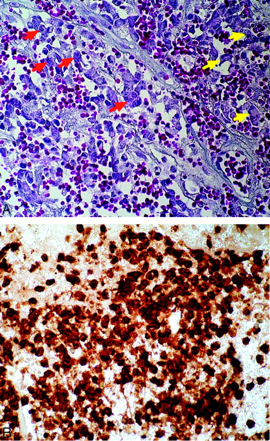
  • Fig 2.
    • Download figure
    • Open in new tab
    • Download powerpoint
    Fig 2.

    Histology of a hemorrhagic spinal subdural metastasis in a 59-year-old woman.

    A, Photomicrograph shows numerous small anaplastic epithelial cells with nuclear polymorphism (red arrows). These malignant cells are also present within the blood vessels (yellow arrows) (Mallory, original magnification ×400).

    B, Photomicrograph demonstrates strong carcinoembryonic antigen (CEA) reactivity in metastatic carcinoma cells (Immunohistochemistry for CEA, original magnification ×400).

PreviousNext
Back to top

In this issue

American Journal of Neuroradiology: 26 (9)
American Journal of Neuroradiology
Vol. 26, Issue 9
1 Oct 2005
  • Table of Contents
  • Index by author
Advertisement
Print
Download PDF
Email Article

Thank you for your interest in spreading the word on American Journal of Neuroradiology.

NOTE: We only request your email address so that the person you are recommending the page to knows that you wanted them to see it, and that it is not junk mail. We do not capture any email address.

Enter multiple addresses on separate lines or separate them with commas.
Nonenhancing Spinal Subdural Metastatic Tumor
(Your Name) has sent you a message from American Journal of Neuroradiology
(Your Name) thought you would like to see the American Journal of Neuroradiology web site.
CAPTCHA
This question is for testing whether or not you are a human visitor and to prevent automated spam submissions.
Cite this article
Zoran Rumboldt, Lara Lambert, Jasna Talan-Hranilovic, Domagoj Marjan, Tomislav Sajko
Nonenhancing Spinal Subdural Metastatic Tumor
American Journal of Neuroradiology Oct 2005, 26 (9) 2406-2409;

Citation Manager Formats

  • BibTeX
  • Bookends
  • EasyBib
  • EndNote (tagged)
  • EndNote 8 (xml)
  • Medlars
  • Mendeley
  • Papers
  • RefWorks Tagged
  • Ref Manager
  • RIS
  • Zotero
0 Responses
Respond to this article
Share
Bookmark this article
Nonenhancing Spinal Subdural Metastatic Tumor
Zoran Rumboldt, Lara Lambert, Jasna Talan-Hranilovic, Domagoj Marjan, Tomislav Sajko
American Journal of Neuroradiology Oct 2005, 26 (9) 2406-2409;
del.icio.us logo Twitter logo Facebook logo Mendeley logo
  • Tweet Widget
  • Facebook Like
  • Google Plus One

Jump to section

  • Article
    • Abstract
    • Case Report
    • Discussion
    • References
  • Figures & Data
  • Info & Metrics
  • Responses
  • References
  • PDF

Related Articles

  • No related articles found.
  • PubMed
  • Google Scholar

Cited By...

  • No citing articles found.
  • Crossref
  • Google Scholar

This article has not yet been cited by articles in journals that are participating in Crossref Cited-by Linking.

More in this TOC Section

  • Optimization of Photon Counting CT Myelography
  • Characteristics of SIH Type I Culprit Lesions
  • Management Outcomes For VO Spine Biopsy
Show more Spine Imaging and Spine Image-Guided Interventions

Similar Articles

Advertisement

Indexed Content

  • Current Issue
  • Accepted Manuscripts
  • Article Preview
  • Past Issues
  • Editorials
  • Editor's Choice
  • Fellows' Journal Club
  • Letters to the Editor
  • Video Articles

Cases

  • Case Collection
  • Archive - Case of the Week
  • Archive - Case of the Month
  • Archive - Classic Case

More from AJNR

  • Trainee Corner
  • Imaging Protocols
  • MRI Safety Corner
  • Book Reviews

Multimedia

  • AJNR Podcasts
  • AJNR Scantastics

Resources

  • Turnaround Time
  • Submit a Manuscript
  • Submit a Video Article
  • Submit an eLetter to the Editor/Response
  • Manuscript Submission Guidelines
  • Statistical Tips
  • Fast Publishing of Accepted Manuscripts
  • Graphical Abstract Preparation
  • Imaging Protocol Submission
  • Evidence-Based Medicine Level Guide
  • Publishing Checklists
  • Author Policies
  • Become a Reviewer/Academy of Reviewers
  • News and Updates

About Us

  • About AJNR
  • Editorial Board
  • Editorial Board Alumni
  • Alerts
  • Permissions
  • Not an AJNR Subscriber? Join Now
  • Advertise with Us
  • Librarian Resources
  • Feedback
  • Terms and Conditions
  • AJNR Editorial Board Alumni

American Society of Neuroradiology

  • Not an ASNR Member? Join Now

© 2025 by the American Society of Neuroradiology All rights, including for text and data mining, AI training, and similar technologies, are reserved.
Print ISSN: 0195-6108 Online ISSN: 1936-959X

Powered by HighWire