
Article Figures & Data
Figures
- Fig 1.
A single section from a typical examination after completion of IV-MTX demonstrating LE appearance on FLAIR image, segmented tissue map, quantitative T1 and quantitative T2 maps (left to right). LE is most evident in the frontal white matter.
- Fig 2.
Predicted probability of developing LE according to a general linear model previously reported for patients on the standard- or high-risk arm (light gray bars) and patients on the low-risk arm (black bars) of the treatment protocol. Quantitative MR measures were evaluated post 1, 4, and 7 courses of IV-MTX and at end of therapy (EOT).
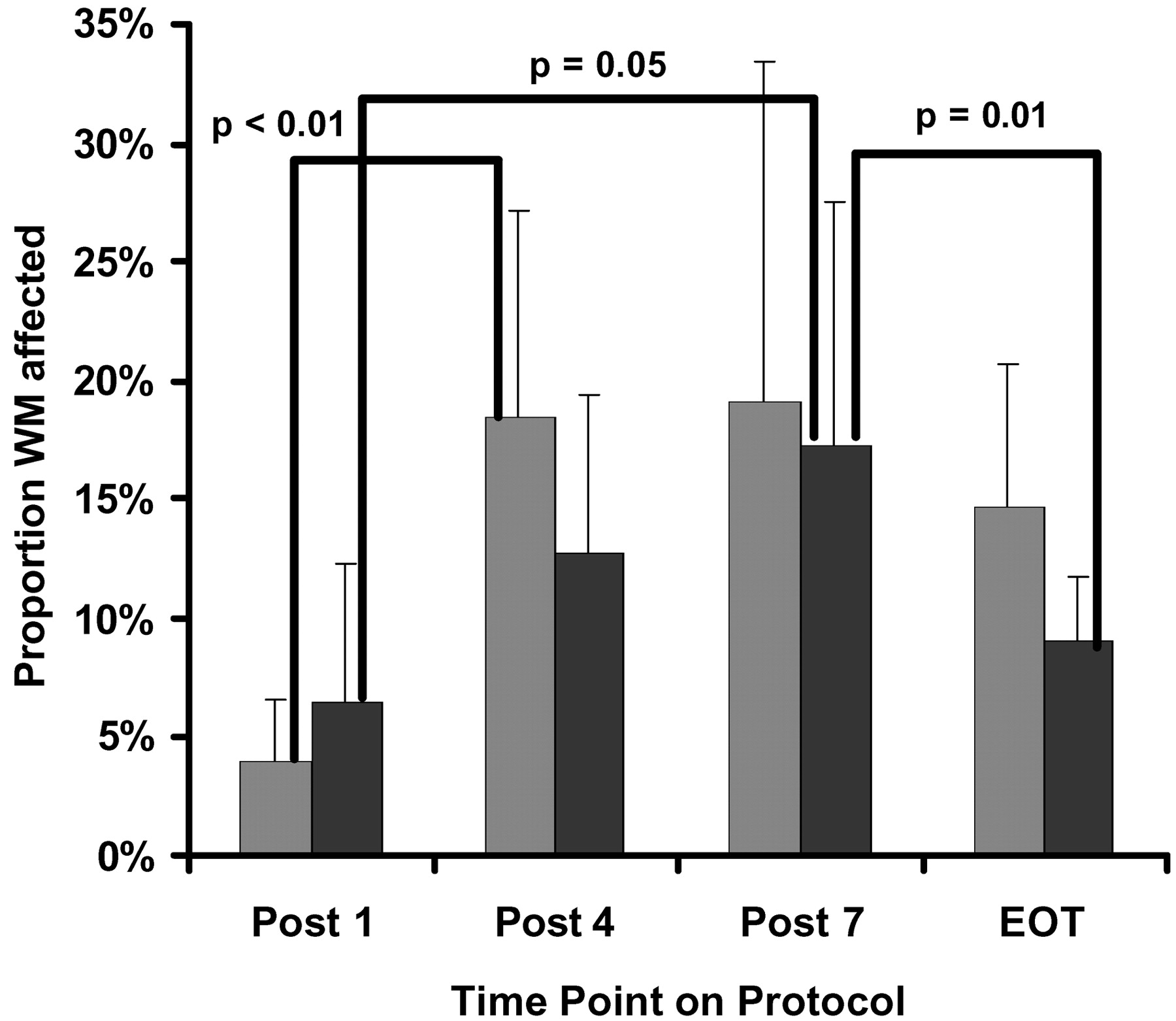
- Fig 3.
Proportion of white matter affected (extent of LE) for patients on the standard- or high-risk arm (light gray bars) and patients on the low-risk arm (black bars) of the treatment protocol. Statistical results from a nonparameteric one-sided Wilcoxon-Mann-Whitney test are shown for both within and between groups at each of the 4 time points. Quantitative MR measures were evaluated post 1, 4, and 7 courses of IVMTX and at end of therapy (EOT).
- Fig 4.
Increase in T1 relaxation rate of LE over NAWM (intensity of LE) for patients on the standard- or high-risk arm (light gray bars) and patients on the low-risk arm (black bars) of the treatment protocol. Statistical results from a nonparameteric one-sided Wilcoxon-Mann-Whitney test are shown for both within and between groups at each of the 4 time points. Quantitative MR measures were evaluated post 1, 4, and 7 courses of IV-MTX and at end of therapy (EOT).
- Fig 5.
Increase in T2 relaxation rate of LE over NAWM (intensity of LE) for patients on the standard- or high-risk arm (light gray bars) and patients on the low-risk arm (black bars) of the treatment protocol. Statistical results from a nonparameteric one-sided Wilcoxon-Mann-Whitney test are shown for both within and between groups at each of the 4 time points. Quantitative MR measures were evaluated post 1, 4, and 7 courses of IV-MTX and end of therapy (EOT).
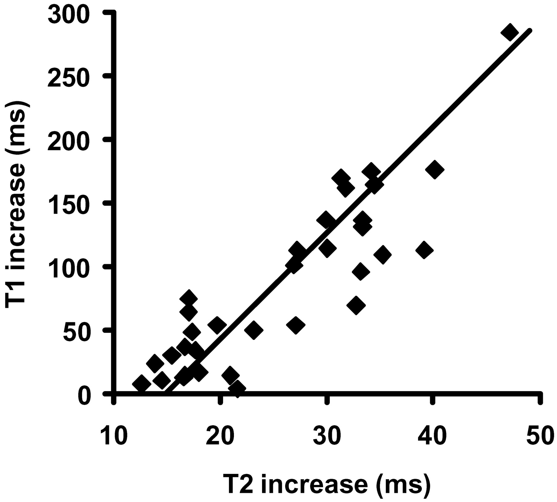
- Fig 6.
Scatter plot of increase in T1 and T2 relaxation rates for patients on both arms of the treatment protocol. The solid line is the result of a simple linear robust regression analysis. Increases in T1 and T2 relaxation relative to NAWM were highly correlated.


Tables
- TABLE 1:
Demographic and descriptive statistics for patients imaged at each time categorized by therapeutic risk arm
Low Risk (n) Standard/High Risk (n) Post 1 IV-MTX 21 23 Post 4 IV-MTX 20 21 Post 7 IV-MTX 21 21 End of therapy 20 17 Gender Male 10 11 Female 12 12 Age at diagnosis (y, ±SD) 5.0 ± 2.7 9.2 ± 4.8 Note.—IV-MTX indicates intravenous methotrexate.
- TABLE 2:
Total number of patients evaluable at each time and proportion with leukoencephalopathy*
Low Risk Standard/High Risk LE/Eval (n) T1 T2 LE/Eval (n) T1 T2 Post 1 IV-MTX 3/21 3 1 47/23 4 2 Post 4 IV-MTX 12/20 11 6 12/21 12 3 Post 7 IV-MTX 14/21 13 9 18/21 14 13 End of Therapy 8/20 7 6 8/17 6 5 Note.—LE indicates leukoencephalopathy; Eval, evaluated; IV-MTX, intravenous methotrexate.
* Note that quantitative T1 and T2 measurements were acquired at the end of the imaging studies and often suffered from motion of patients, making them inevaluable.


