
Article Figures & Data
Figures
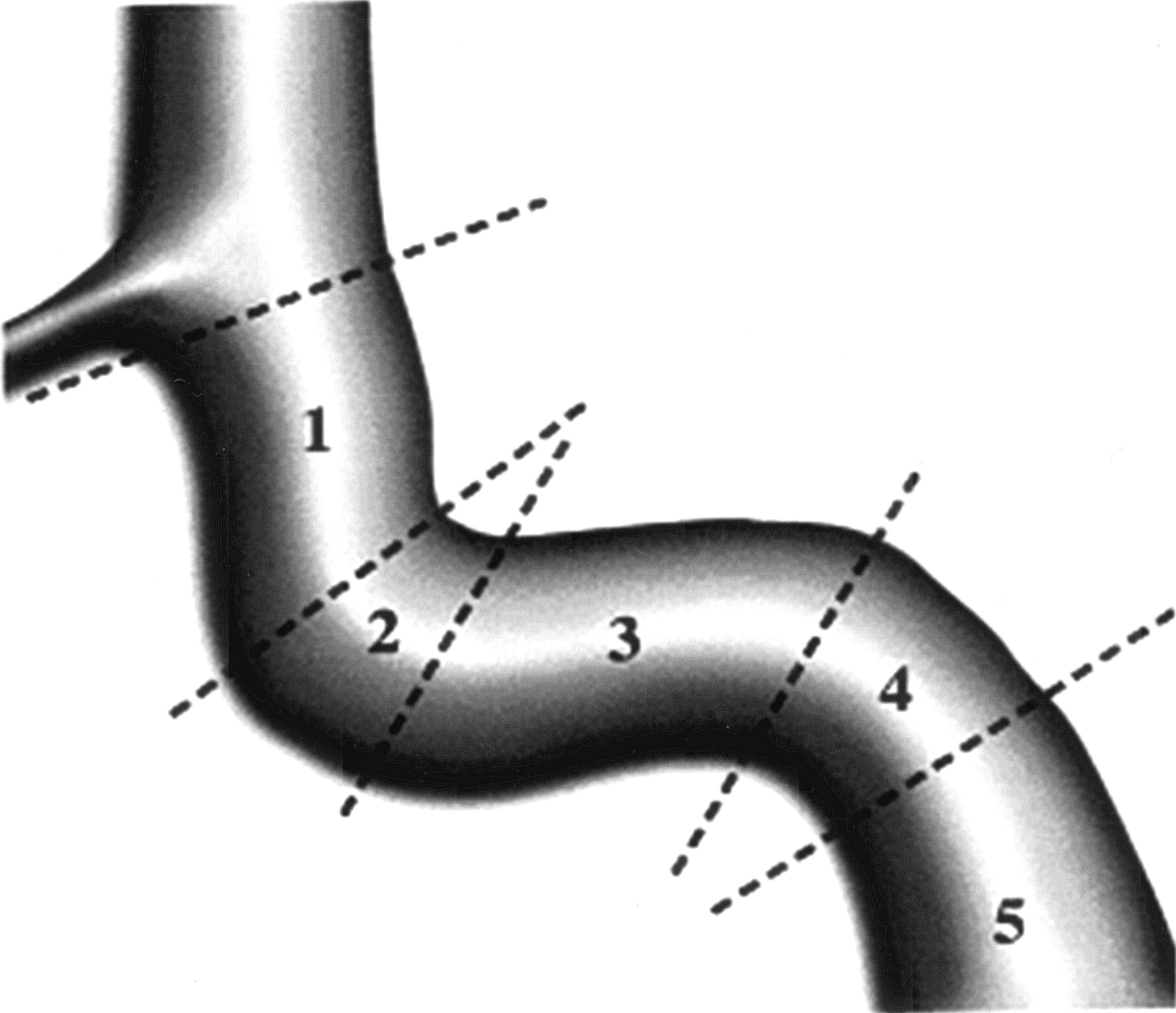
- Fig 1.
Segmental division of the cavernous carotid artery (after Debrun et al [10]).
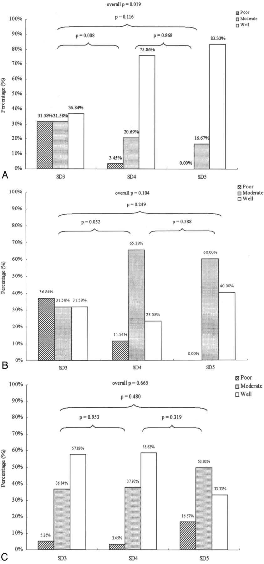
- Fig 2.
Detectability of CCFs by location according to segmental division (SD) of the ICA, by using each technique. Panels A, B, and C show results for CTA, MRA, and DSA, respectively. Bars indicate percentage of images having detectability ratings of poor (hatched), moderate (stippled), or good (open). P values indicate statistical significance for comparisons between locations by using the χ2 test, for each technique.
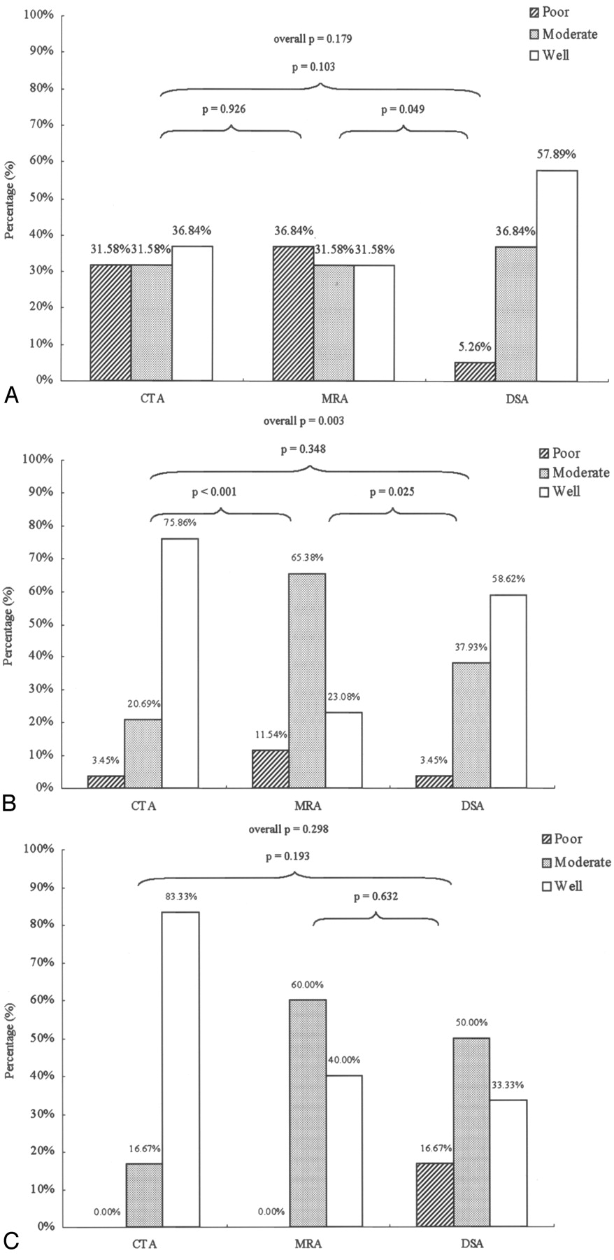
- Fig 3.
Detectability of CCFs by using CTA, MRA, and DSA, by location according to segmental division (SD) of the ICA. Panels A, B, and C show results for fistulas found at SD 3, SD 4, and SD 5, respectively. Bars indicate percentage of images having detectability ratings of poor (hatched), moderate (stippled) or good (open). P values indicate statistical significance for comparisons between modalities by using the χ2 test, for each location.
- Fig 4.
SD 3, DSA = CTA > MRA. Left CCF with left SOV drainage.
Images were made by using CTA (panels A–C), MRA (panels D–F), and vertebral DSA (posterior-anterior view in panel H, lateral view in panel I) before embolization. The fistula ostium (panels B and E), proximal portion (panels A and D), and distal portion (panels C and F) are shown. A CTA source image made following embolization (panel G) shows the detachable balloon located at the previous fistula site. CS, cavernous sinus; DB, detachable balloon; F, fistula tract; SD, segmental division of the ICA.
- Fig 5.
SD 3, DSA = CTA = MRA. Right CCF with transection of ICA.
Images were made by using CTA (panels A–C), MRA (panels D–F), and carotid DSA (lateral view, panel H) before embolization. The fistula ostium (panels B and E), proximal portion (panels A and D) and distal portion (panels C and F) are shown. Panel G shows an image made by using MIP reconstruction MRA. CS, cavernous sinus; F, fistula tract; MIP, maximal intensity projection; SD, segmental division of the ICA.


Tables
Comparison of imaging modalities for their ability to detect carotid cavernous fistula tracts
Modality (Fistula Tracts Identified) Ability to Detect Fistula Tracts, n (%)* P Value (Chi-square Test) Poor Moderate Well Overall CTA vs DSA CTA vs MRA MRA vs DSA CTA (n = 54) 7 (13.0) 13 (24.1) 34 (63.0) MRA (n = 50) 10 (20.0) 26 (52.0) 14 (28.0) .002 NS .001 .007 DSA (n = 54) 3 (5.6) 21 (38.9) 30 (55.6) Note.—CTA indicated computed tomography angiography; MRA, magnetic resonance angiography; DSA, digital subtraction angiography; NS, not significant.
* Poor indicates neither size nor location of fistula can be defined; moderate, either size or location of fistula can be defined; well, both size and location of fistula can be defined.


