Advertisement

Main menu

  • Home
  • Content
    • Current Issue
    • Accepted Manuscripts
    • Article Preview
    • Past Issue Archive
    • Video Articles
    • AJNR Case Collection
    • Case of the Week Archive
    • Case of the Month Archive
    • Classic Case Archive
  • Special Collections
    • AJNR Awards
    • Low-Field MRI
    • Alzheimer Disease
    • ASNR Foundation Special Collection
    • Photon-Counting CT
    • View All
  • Multimedia
    • AJNR Podcasts
    • AJNR SCANtastic
    • Trainee Corner
    • MRI Safety Corner
    • Imaging Protocols
  • For Authors
    • Submit a Manuscript
    • Submit a Video Article
    • Submit an eLetter to the Editor/Response
    • Manuscript Submission Guidelines
    • Statistical Tips
    • Fast Publishing of Accepted Manuscripts
    • Graphical Abstract Preparation
    • Imaging Protocol Submission
    • Author Policies
  • About Us
    • About AJNR
    • Editorial Board
    • Editorial Board Alumni
  • More
    • Become a Reviewer/Academy of Reviewers
    • Subscribers
    • Permissions
    • Alerts
    • Feedback
    • Advertisers
    • ASNR Home

User menu

  • Alerts
  • Log in

Search

  • Advanced search
American Journal of Neuroradiology

American Journal of Neuroradiology

ASHNR American Society of Functional Neuroradiology ASHNR American Society of Pediatric Neuroradiology ASSR
  • Alerts
  • Log in

Advanced Search

  • Home
  • Content
    • Current Issue
    • Accepted Manuscripts
    • Article Preview
    • Past Issue Archive
    • Video Articles
    • AJNR Case Collection
    • Case of the Week Archive
    • Case of the Month Archive
    • Classic Case Archive
  • Special Collections
    • AJNR Awards
    • Low-Field MRI
    • Alzheimer Disease
    • ASNR Foundation Special Collection
    • Photon-Counting CT
    • View All
  • Multimedia
    • AJNR Podcasts
    • AJNR SCANtastic
    • Trainee Corner
    • MRI Safety Corner
    • Imaging Protocols
  • For Authors
    • Submit a Manuscript
    • Submit a Video Article
    • Submit an eLetter to the Editor/Response
    • Manuscript Submission Guidelines
    • Statistical Tips
    • Fast Publishing of Accepted Manuscripts
    • Graphical Abstract Preparation
    • Imaging Protocol Submission
    • Author Policies
  • About Us
    • About AJNR
    • Editorial Board
    • Editorial Board Alumni
  • More
    • Become a Reviewer/Academy of Reviewers
    • Subscribers
    • Permissions
    • Alerts
    • Feedback
    • Advertisers
    • ASNR Home
  • Follow AJNR on Twitter
  • Visit AJNR on Facebook
  • Follow AJNR on Instagram
  • Join AJNR on LinkedIn
  • RSS Feeds

AJNR Awards, New Junior Editors, and more. Read the latest AJNR updates

Research ArticleBRAIN

Brain MR Imaging Abnormalities in Kidney Transplant Recipients

Nada Besenski, Zoran Rumboldt, Osemwegie Emovon, Joyce Nicholas, Sunil Kini, Jovan Milutinovic and Milos N. Budisavljevic
American Journal of Neuroradiology October 2005, 26 (9) 2282-2289;
Nada Besenski
  • Find this author on Google Scholar
  • Find this author on PubMed
  • Search for this author on this site
Zoran Rumboldt
  • Find this author on Google Scholar
  • Find this author on PubMed
  • Search for this author on this site
Osemwegie Emovon
  • Find this author on Google Scholar
  • Find this author on PubMed
  • Search for this author on this site
Joyce Nicholas
  • Find this author on Google Scholar
  • Find this author on PubMed
  • Search for this author on this site
Sunil Kini
  • Find this author on Google Scholar
  • Find this author on PubMed
  • Search for this author on this site
Jovan Milutinovic
  • Find this author on Google Scholar
  • Find this author on PubMed
  • Search for this author on this site
Milos N. Budisavljevic
  • Find this author on Google Scholar
  • Find this author on PubMed
  • Search for this author on this site
  • Article
  • Figures & Data
  • Info & Metrics
  • Responses
  • References
  • PDF
Loading

Article Figures & Data

Figures

  • Tables
  • Fig 1.
    • Download figure
    • Open in new tab
    • Download powerpoint
    Fig 1.

    A 52-year-old-man (group 3) with multiple acute embolic infarctions.

    A, Axial DWI shows small bilateral hyperintense lesions indicative of embolic infarcts.

    B, Axial ADC map reveals low signal intensity within the lesions, consistent with restricted diffusion of water molecules, confirming acute nature of embolic infarcts.

    C, FLAIR image at approximately the same level as A and B demonstrates faint hyperintensity of the lesions.

  • Fig 2.
    • Download figure
    • Open in new tab
    • Download powerpoint
    Fig 2.

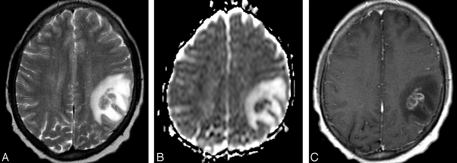
    A 48-year-old man (group 3) with cerebral fungal abscess.

    A, Axial T2-weighted image shows a relatively isointense intra-axial lesion with surrounding hyperintense vasogenic edema in the right frontoparietal region.

    B, Corresponding axial ADC map shows lower signal intensity of the lesion compared with the normal-appearing brain parenchyma, consistent with relatively decreased diffusion, as typically seen within brain abscesses. The lesion is surrounded by increased diffusion of vasogenic edema.

    C, Postcontrast axial T1-weighted image corresponding to A and B demonstrates irregular, predominantly peripheral enhancement of the mass.

  • Fig 3.
    • Download figure
    • Open in new tab
    • Download powerpoint
    Fig 3.

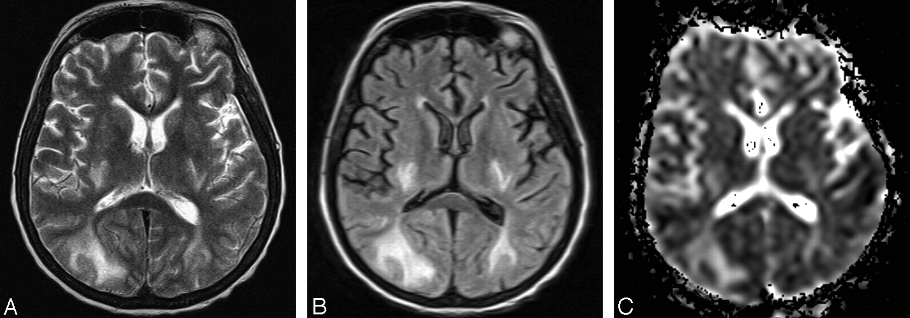
    A 45-year-old woman (group 2) with PRES.

    A, Axial T2-weighted image shows bilateral abnormal hyperintense signal intensity in the occipital subcortical white matter and in the posterior limb of the internal capsule.

    B, Corresponding axial FLAIR image shows the hyperintense abnormalities more clearly.

    C, On axial ADC map corresponding to A and B increased signal intensity of the abnormalities is seen, consistent with increased diffusion, which is indicative of vasogenic edema.

  • Fig 4.
    • Download figure
    • Open in new tab
    • Download powerpoint
    Fig 4.

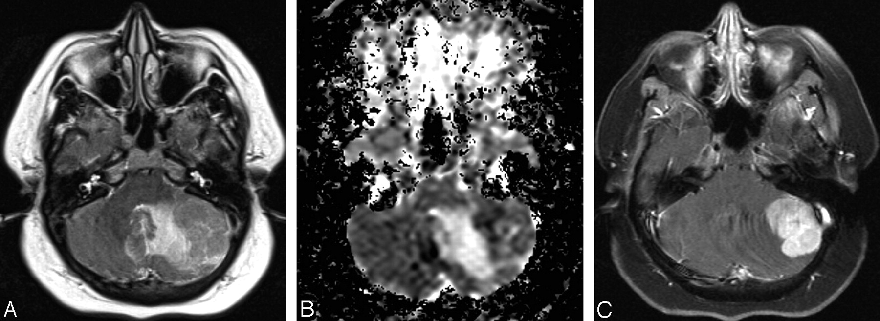
    A 39-year-old woman (group 3) with cerebellar lymphoma.

    A, Axial T2-weighted image shows an isointense left cerebellar mass surrounded by hyperintense area of vasogenic edema.

    B, Axial ADC map corresponding to A reveals relatively decreased diffusion within the mass with surrounding increased diffusion of the vasogenic edema. Relatively low diffusion rate is indicative of high cellularity of the mass.

    C, Postcontrast fat-suppressed axial T1-weighted corresponding to A and B images demonstrates attenuated homogenous enhancement of the mass.
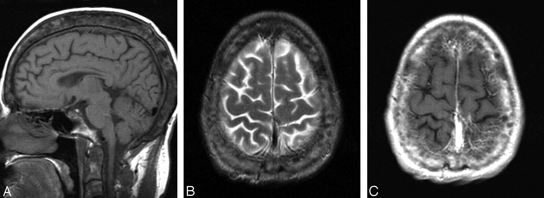
  • Fig 5.
    • Download figure
    • Open in new tab
    • Download powerpoint
    Fig 5.

    A 31-year-old woman (group 1) with heterogenous calvarial thickening.

    A, Midsagittal T1-weighted image demonstrates increased thickness of the calvaria with widening of the diploic space and abnormal heterogeneous signal intensity. This finding is presumably related to a long-standing chronic renal failure and associated chronic anemia and osteodistrophy before kidney transplantation.

    B, Axial T2-weighted image shows thickened and heterogenous diploic space of the calvaria.

    C, Axial postcontrast T1-weighted image corresponding to B reveals heterogenous enhancement of the calvaria.

Tables

  • Figures
    • View popup
    TABLE 1:

    Most common clinical presentations in kidney transplant recipients

    Group 0(n = 24)Group 1(n = 19)Group 2(n = 22)Group 3(n = 36)
    Altered mental status(13)11 (58)9 (41)9 (25)
    Headache4 (17)2 (11)4 (18)15 (42)
    Seizures4 (17)6 (32)1 (5)2 (6)
    Infection1 (4)2 (11)6 (27)4 (11)
    Vision disturbance001 (5)4 (11)
    Hemiparesis1 (4)003 (8)
    Weakness2 (8)2 (11)01 (3)
    Dizziness001 (5)3 (8)
    Ataxia1 (4)02 (9)1 (3)
    Aphasia01 (5)1 (5)2 (6)
    Syncope0002 (6)
    • Note.—Number of patients is followed by percentage in parentheses.

    • View popup
    TABLE 2:

    Location of lesions in 26 pre- and post-transplant MR imaging examinations

    NormalWhite MatterGray MatterBrain Stem
    FrontalParietalOccipitalTemporalDPVCerebellumBasal GangliaThalamusCortex
    Pretransplant8 (31)9 (35)1 (4)1 (4)1 (4)8 (31)01 (4)04 (15)1 (4)
    Post-transplant23 (23)13 (13)3 (3)4 (4)1 (1)38 (39)4 (4)15 (15)7 (7)9 (9)5 (5)
    • Note.—Number of patients with lesions is followed by percentage in parentheses. DPV indicates deep periventricular area.

    • View popup
    TABLE 3:

    Lesion type on MR image in patients before (group 0) and after kidney transplant (groups 1–3)

    Group(s)*NormalAcute ChangesChronic Changes
    AIIPRESLIMATDuraC
    08 (33)2 (8)01 (4)3 (13)10 (42)000
    14 (21)3 (16)2 (11)1 (5)4 (21)6 (32)01 (5)1 (5)
    27 (32)2 (9)01 (5)7 (32)10 (45)01 (5)2 (9)
    312 (33)4 (11)3 (8)2 (6)6 (17)13 (33)1 (3)2 (6)3 (8)
    1–323 (30)9 (12)5 (6)4 (5)17 (22)29 (38)1 (1)4 (5)6 (8)
    • Note.—Number of patients is followed by percentage in parentheses. AI indicates acute and subacute infarct; I, infection; PRES, posterior reversible encephalopathy syndrome; LI, chronic lacunar infarct; MA, microangiopathy; T, tumor; Dura, dural thickening; C, thickened calvaria.

    • * Group 0 (no. patients/no. MR imaging examinations), 24/26; group 1, 19/21; group 2, 22/26; group 3, 36/51.

PreviousNext
Back to top

In this issue

American Journal of Neuroradiology: 26 (9)
American Journal of Neuroradiology
Vol. 26, Issue 9
1 Oct 2005
  • Table of Contents
  • Index by author
Advertisement
Print
Download PDF
Email Article

Thank you for your interest in spreading the word on American Journal of Neuroradiology.

NOTE: We only request your email address so that the person you are recommending the page to knows that you wanted them to see it, and that it is not junk mail. We do not capture any email address.

Enter multiple addresses on separate lines or separate them with commas.
Brain MR Imaging Abnormalities in Kidney Transplant Recipients
(Your Name) has sent you a message from American Journal of Neuroradiology
(Your Name) thought you would like to see the American Journal of Neuroradiology web site.
CAPTCHA
This question is for testing whether or not you are a human visitor and to prevent automated spam submissions.
Cite this article
Nada Besenski, Zoran Rumboldt, Osemwegie Emovon, Joyce Nicholas, Sunil Kini, Jovan Milutinovic, Milos N. Budisavljevic
Brain MR Imaging Abnormalities in Kidney Transplant Recipients
American Journal of Neuroradiology Oct 2005, 26 (9) 2282-2289;

Citation Manager Formats

  • BibTeX
  • Bookends
  • EasyBib
  • EndNote (tagged)
  • EndNote 8 (xml)
  • Medlars
  • Mendeley
  • Papers
  • RefWorks Tagged
  • Ref Manager
  • RIS
  • Zotero
0 Responses
Respond to this article
Share
Bookmark this article
Brain MR Imaging Abnormalities in Kidney Transplant Recipients
Nada Besenski, Zoran Rumboldt, Osemwegie Emovon, Joyce Nicholas, Sunil Kini, Jovan Milutinovic, Milos N. Budisavljevic
American Journal of Neuroradiology Oct 2005, 26 (9) 2282-2289;
del.icio.us logo Twitter logo Facebook logo Mendeley logo
  • Tweet Widget
  • Facebook Like
  • Google Plus One

Jump to section

  • Article
    • Abstract
    • Methods
    • Results
    • Discussion
    • Conclusion
    • References
  • Figures & Data
  • Info & Metrics
  • Responses
  • References
  • PDF

Related Articles

  • No related articles found.
  • PubMed
  • Google Scholar

Cited By...

  • The Spectrum of Infectious Diseases in Kidney Transplantation: A Review of the Classification, Pathogens and Clinical Manifestations
  • Posterior reversible encephalopathy syndrome: the importance of early diagnosis
  • Crossref
  • Google Scholar

This article has not yet been cited by articles in journals that are participating in Crossref Cited-by Linking.

More in this TOC Section

  • Predictors of Reperfusion in Patients with Acute Ischemic Stroke
  • Enhanced Axonal Metabolism during Early Natalizumab Treatment in Relapsing-Remitting Multiple Sclerosis
  • Progression of Microstructural Damage in Spinocerebellar Ataxia Type 2: A Longitudinal DTI Study
Show more Brain

Similar Articles

Advertisement

Indexed Content

  • Current Issue
  • Accepted Manuscripts
  • Article Preview
  • Past Issues
  • Editorials
  • Editor's Choice
  • Fellows' Journal Club
  • Letters to the Editor
  • Video Articles

Cases

  • Case Collection
  • Archive - Case of the Week
  • Archive - Case of the Month
  • Archive - Classic Case

More from AJNR

  • Trainee Corner
  • Imaging Protocols
  • MRI Safety Corner
  • Book Reviews

Multimedia

  • AJNR Podcasts
  • AJNR Scantastics

Resources

  • Turnaround Time
  • Submit a Manuscript
  • Submit a Video Article
  • Submit an eLetter to the Editor/Response
  • Manuscript Submission Guidelines
  • Statistical Tips
  • Fast Publishing of Accepted Manuscripts
  • Graphical Abstract Preparation
  • Imaging Protocol Submission
  • Evidence-Based Medicine Level Guide
  • Publishing Checklists
  • Author Policies
  • Become a Reviewer/Academy of Reviewers
  • News and Updates

About Us

  • About AJNR
  • Editorial Board
  • Editorial Board Alumni
  • Alerts
  • Permissions
  • Not an AJNR Subscriber? Join Now
  • Advertise with Us
  • Librarian Resources
  • Feedback
  • Terms and Conditions
  • AJNR Editorial Board Alumni

American Society of Neuroradiology

  • Not an ASNR Member? Join Now

© 2025 by the American Society of Neuroradiology All rights, including for text and data mining, AI training, and similar technologies, are reserved.
Print ISSN: 0195-6108 Online ISSN: 1936-959X

Powered by HighWire