Advertisement

Main menu

  • Home
  • Content
    • Current Issue
    • Accepted Manuscripts
    • Article Preview
    • Past Issue Archive
    • Video Articles
    • AJNR Case Collection
    • Case of the Week Archive
    • Case of the Month Archive
    • Classic Case Archive
  • Special Collections
    • AJNR Awards
    • Low-Field MRI
    • Alzheimer Disease
    • ASNR Foundation Special Collection
    • Photon-Counting CT
    • View All
  • Multimedia
    • AJNR Podcasts
    • AJNR SCANtastic
    • Trainee Corner
    • MRI Safety Corner
    • Imaging Protocols
  • For Authors
    • Submit a Manuscript
    • Submit a Video Article
    • Submit an eLetter to the Editor/Response
    • Manuscript Submission Guidelines
    • Statistical Tips
    • Fast Publishing of Accepted Manuscripts
    • Graphical Abstract Preparation
    • Imaging Protocol Submission
    • Author Policies
  • About Us
    • About AJNR
    • Editorial Board
    • Editorial Board Alumni
  • More
    • Become a Reviewer/Academy of Reviewers
    • Subscribers
    • Permissions
    • Alerts
    • Feedback
    • Advertisers
    • ASNR Home

User menu

  • Alerts
  • Log in

Search

  • Advanced search
American Journal of Neuroradiology

American Journal of Neuroradiology

ASHNR American Society of Functional Neuroradiology ASHNR American Society of Pediatric Neuroradiology ASSR
  • Alerts
  • Log in

Advanced Search

  • Home
  • Content
    • Current Issue
    • Accepted Manuscripts
    • Article Preview
    • Past Issue Archive
    • Video Articles
    • AJNR Case Collection
    • Case of the Week Archive
    • Case of the Month Archive
    • Classic Case Archive
  • Special Collections
    • AJNR Awards
    • Low-Field MRI
    • Alzheimer Disease
    • ASNR Foundation Special Collection
    • Photon-Counting CT
    • View All
  • Multimedia
    • AJNR Podcasts
    • AJNR SCANtastic
    • Trainee Corner
    • MRI Safety Corner
    • Imaging Protocols
  • For Authors
    • Submit a Manuscript
    • Submit a Video Article
    • Submit an eLetter to the Editor/Response
    • Manuscript Submission Guidelines
    • Statistical Tips
    • Fast Publishing of Accepted Manuscripts
    • Graphical Abstract Preparation
    • Imaging Protocol Submission
    • Author Policies
  • About Us
    • About AJNR
    • Editorial Board
    • Editorial Board Alumni
  • More
    • Become a Reviewer/Academy of Reviewers
    • Subscribers
    • Permissions
    • Alerts
    • Feedback
    • Advertisers
    • ASNR Home
  • Follow AJNR on Twitter
  • Visit AJNR on Facebook
  • Follow AJNR on Instagram
  • Join AJNR on LinkedIn
  • RSS Feeds

AJNR Awards, New Junior Editors, and more. Read the latest AJNR updates

Research ArticleBRAIN

Diffusion Tensor Tractography of the Limbic System

Luis Concha, Donald W. Gross and Christian Beaulieu
American Journal of Neuroradiology October 2005, 26 (9) 2267-2274;
Luis Concha
  • Find this author on Google Scholar
  • Find this author on PubMed
  • Search for this author on this site
Donald W. Gross
  • Find this author on Google Scholar
  • Find this author on PubMed
  • Search for this author on this site
Christian Beaulieu
  • Find this author on Google Scholar
  • Find this author on PubMed
  • Search for this author on this site
  • Article
  • Figures & Data
  • Info & Metrics
  • Responses
  • References
  • PDF
Loading

Article Figures & Data

Figures

  • Tables
  • Fig 1.
    • Download figure
    • Open in new tab
    • Download powerpoint
    Fig 1.

    Diffusion tensor tractography of the fornix and cingulum in a healthy individual.

    3D fiber tracking of the fornix (green) and cingulum (orange) overlaid on an anatomical, T1-weighted 3D–magnetization-preparation rapid gradient echo (MPRAGE) volume. The anatomy of these 2 fiber bundles can be thought as 2 nested semicircles. The tracts shown were selected by brute force for display purposes only. The labeled portions of the tracts (indicated by yellow lines for the cingulum and black lines for the fornix) were analyzed subsequently by using judicious tract selection regions, as shown in Figs 2 and 3.
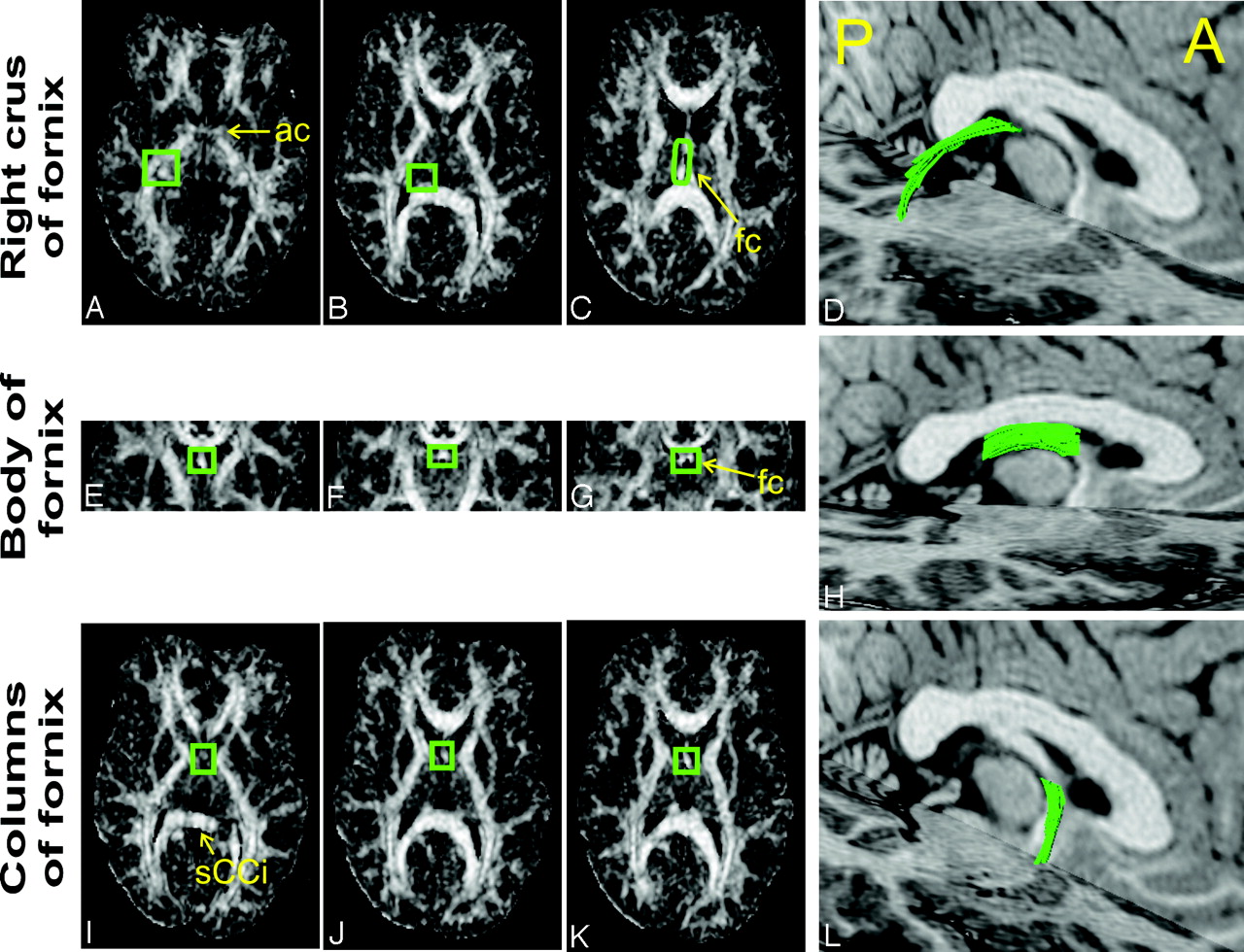
  • Fig 2.
    • Download figure
    • Open in new tab
    • Download powerpoint
    Fig 2.

    Region of interest placement for selection of individual portions of the fornix.

    The fornix was selected in 3 distinct portions, because this enhances the selection accuracy. For each portion, tract-selection regions were manually drawn on either FA or principal diffusivity color maps. To be selected, the tracts had to go through the middle tract-selection region (B, F, and J, green boxes) and through either one of the extreme regions (A or C; E or G; I or K, for the crus, body, and columns, respectively). The FA maps presented are derived from CSF-suppressed DTI. The same procedure was repeated for the standard DTI datasets by using identical tract selection regions. The resulting FLAIR DTI-derived tracts of a healthy subject are presented in 3D views overlaid on anatomic 3D-MPRAGE axial and sagittal sections (D, H, and L). Subsequent analyses of all segments were performed only between the 2 extreme tract selection regions. A, anterior; P, posterior; ac, anterior commissure; fc, fusion of the crura; sCCi, inferior border of the splenium of the corpus callosum. Panel E is 0.5 cm posterior to the vertical AC line. Panel K is parallel to the inferior border of the body of the fornix.

  • Fig 3.
    • Download figure
    • Open in new tab
    • Download powerpoint
    Fig 3.

    Region of interest placement for selection of individual portions of the cingulum.

    Three portions of the cingulum—namely, the descending, superior, and anterior portions—were selected after fiber tracking of the entire volume in both standard and FLAIR DTI datasets. For each portion, tract-selection regions were manually drawn on either FA or principal diffusivity color maps. To be selected, the tracts had to go through the middle tract-selection region (B, F, and J, orange boxes) and through either one of the extreme regions (A or C; E or G; I or K, for the descending, superior, and anterior portions, respectively). The FA maps presented are derived from CSF-suppressed DTI. The same procedure was repeated for the standard DTI datasets by using identical tract selection regions. The resulting FLAIR DTI-derived tracts of a healthy subject are presented in 3D views overlaid on anatomic 3D-MPRAGE axial and sagittal sections (D, H, and L). Subsequent analyses of all segments were performed only between the 2 extreme tract selection regions. A, anterior; P, posterior; ac, anterior commissure; bf, body of fornix; gCCp, posterior border of the genu of the corpus callosum; sCCa, anterior border of the callosal splenium; gCCi, inferior border of the callosal genu.

  • Fig 4.
    • Download figure
    • Open in new tab
    • Download powerpoint
    Fig 4.

    Qualitative assessment of the role of CSF suppression in tractography.

    Because of CSF signal intensity contamination, the crus of the fornix is difficult to depict with fiber tracking derived from standard DTI datasets. Tissue voxels belonging to the top portion of the crus of the fornix (yellow arrow) suffer partial volume averaging and their diffusion anisotropy drops below the fiber tracking FA threshold (0.3) to values that range from 0.15 to 0.25. When CSF suppression is performed, the same region contains higher anisotropy values (∼0.30–0.40) and its voxels are included by the tracking algorithm, which results in a complete depiction of the structure. The axial nondiffusion-weighted images (b = 0 s/mm2) show the bright CSF signal intensity in standard DTI, which is suppressed with FLAIR DTI. The portions of the fornix are overlaid on T1-weighted, anatomical 3D-MPRAGE axial and sagittal sections.

Tables

  • Figures
  • Diffusion parameters and volume of limbic connections

    CingulumFornix
    DescendingSuperiorAnteriorCrusBodyColumns
    FA
        Standard0.48 ± 0.020.66 ± 0.020.44 ± 0.020.49 ± 0.020.54 ± 0.050.52 ± 0.02
        FLAIR0.50 ± 0.020.65 ± 0.030.48 ± 0.030.53 ± 0.020.61 ± 0.030.54 ± 0.03
        Difference0.02 ± 0.01−0.01 ± 0.010.05 ± 0.030.03 ± 0.020.07 ± 0.050.01 ± 0.04
        P.011.04.027.011.015.29
    Mean ADC (×10−3mm2/s)
        Standard0.75 ± 0.030.71 ± 0.020.72 ± 0.030.96 ± 0.11.45 ± 0.10.99 ± 0.07
        FLAIR0.75 ± 0.020.72 ± 0.010.73 ± 0.030.89 ± 0.041.0 ± 0.040.95 ± 0.06
        Difference0 ± 0.020.01 ± 0.010.01 ± 0.01−0.06 ± 0.08−0.44 ± 0.13−0.04 ± 0.08
        P1.0.14.16.05.008.16
    Volume (mm3)
        Standard504 ± 1361196 ± 504168 ± 30244 ± 136572 ± 96174 ± 44
        FLAIR600 ± 1961508 ± 492260 ± 72540 ± 130724 ± 126292 ± 110
        Difference110 ± 120312 ± 391120 ± 43196 ± 87150 ± 138118 ± 81
        P.123.036.018.008.015.011
    • Note.—FA indicates fractional anisotropy; ADC, apparent diffusion coefficient; FLAIR, fluid-attenuated inversion recovery.

      FA, mean ADC, and volume (mean ± SD) are presented for 3 portions of the fornix and cingulum depicted with tractography derived from diffusion tensor imaging datasets with and without CSF suppression (FLAIR and Standard, respectively). Except for the superior portion of the cingulum, all structures showed no interhemispheric asymmetry and are therefore presented as collapsed data; that is (left + right)/2. FLAIR diffusion tensor imaging resulted in significant increases of fractional anisotropy in the descending and anterior portions of the cingulum, crus, and body of the fornix, as well as decreases of mean ADC in the crus and body of the fornix. A significant increase in tract volume with CSF suppression was also observed in 5 of 6 tract portions.

PreviousNext
Back to top

In this issue

American Journal of Neuroradiology: 26 (9)
American Journal of Neuroradiology
Vol. 26, Issue 9
1 Oct 2005
  • Table of Contents
  • Index by author
Advertisement
Print
Download PDF
Email Article

Thank you for your interest in spreading the word on American Journal of Neuroradiology.

NOTE: We only request your email address so that the person you are recommending the page to knows that you wanted them to see it, and that it is not junk mail. We do not capture any email address.

Enter multiple addresses on separate lines or separate them with commas.
Diffusion Tensor Tractography of the Limbic System
(Your Name) has sent you a message from American Journal of Neuroradiology
(Your Name) thought you would like to see the American Journal of Neuroradiology web site.
CAPTCHA
This question is for testing whether or not you are a human visitor and to prevent automated spam submissions.
Cite this article
Luis Concha, Donald W. Gross, Christian Beaulieu
Diffusion Tensor Tractography of the Limbic System
American Journal of Neuroradiology Oct 2005, 26 (9) 2267-2274;

Citation Manager Formats

  • BibTeX
  • Bookends
  • EasyBib
  • EndNote (tagged)
  • EndNote 8 (xml)
  • Medlars
  • Mendeley
  • Papers
  • RefWorks Tagged
  • Ref Manager
  • RIS
  • Zotero
0 Responses
Respond to this article
Share
Bookmark this article
Diffusion Tensor Tractography of the Limbic System
Luis Concha, Donald W. Gross, Christian Beaulieu
American Journal of Neuroradiology Oct 2005, 26 (9) 2267-2274;
del.icio.us logo Twitter logo Facebook logo Mendeley logo
  • Tweet Widget
  • Facebook Like
  • Google Plus One

Jump to section

  • Article
    • Abstract
    • Methods
    • Results
    • Discussion
    • Conclusions
    • Acknowledgments
    • Footnotes
    • References
  • Figures & Data
  • Info & Metrics
  • Responses
  • References
  • PDF

Related Articles

  • No related articles found.
  • PubMed
  • Google Scholar

Cited By...

  • Structural connectivity matures along a sensorimotor-association connectional axis in youth
  • Sex, racial, and APOE-{varepsilon}4 allele differences in longitudinal white matter microstructure in multiple cohorts of aging and Alzheimers disease
  • Relationship between temporal projections and cognition in patients with putaminal hemorrhage
  • Selective medial septum lesions in healthy rats induce longitudinal changes in microstructure of limbic regions, behavioral alterations, and increased susceptibility to status epilepticus
  • Conduction velocity along a key white matter tract is associated with autobiographical memory recall ability
  • A ketogenic supplement improves white matter energy supply and processing speed in mild cognitive impairment
  • Diffusion MRI free water is a sensitive marker of age-related changes in the cingulum
  • The role of the pre-commissural fornix in episodic autobiographical memory and simulation
  • Hippocampal subfields and limbic white matter jointly predict learning rate in older adults
  • Bundle-specific fornix reconstruction for dual-tracer PET-tractometry
  • The role of the fornix in human navigational learning
  • Posterior cingulate epilepsy: clinical and neurophysiological analysis
  • Effects of early-life adversity on white matter diffusivity changes in patients at risk for major depression
  • Frontotemporal Connections in Episodic Memory and Aging: A Diffusion MRI Tractography Study
  • Longitudinal changes of structural connectivity in traumatic axonal injury
  • In Vivo Diffusion Tensor Imaging and Histopathology of the Fimbria-Fornix in Temporal Lobe Epilepsy
  • White-matter diffusion abnormalities in temporal-lobe epilepsy with and without mesial temporal sclerosis
  • Crossref
  • Google Scholar

This article has not yet been cited by articles in journals that are participating in Crossref Cited-by Linking.

More in this TOC Section

  • Evaluating the Effects of White Matter Multiple Sclerosis Lesions on the Volume Estimation of 6 Brain Tissue Segmentation Methods
  • Quiet PROPELLER MRI Techniques Match the Quality of Conventional PROPELLER Brain Imaging Techniques
  • Predictors of Reperfusion in Patients with Acute Ischemic Stroke
Show more Brain

Similar Articles

Advertisement

Indexed Content

  • Current Issue
  • Accepted Manuscripts
  • Article Preview
  • Past Issues
  • Editorials
  • Editor's Choice
  • Fellows' Journal Club
  • Letters to the Editor
  • Video Articles

Cases

  • Case Collection
  • Archive - Case of the Week
  • Archive - Case of the Month
  • Archive - Classic Case

More from AJNR

  • Trainee Corner
  • Imaging Protocols
  • MRI Safety Corner
  • Book Reviews

Multimedia

  • AJNR Podcasts
  • AJNR Scantastics

Resources

  • Turnaround Time
  • Submit a Manuscript
  • Submit a Video Article
  • Submit an eLetter to the Editor/Response
  • Manuscript Submission Guidelines
  • Statistical Tips
  • Fast Publishing of Accepted Manuscripts
  • Graphical Abstract Preparation
  • Imaging Protocol Submission
  • Evidence-Based Medicine Level Guide
  • Publishing Checklists
  • Author Policies
  • Become a Reviewer/Academy of Reviewers
  • News and Updates

About Us

  • About AJNR
  • Editorial Board
  • Editorial Board Alumni
  • Alerts
  • Permissions
  • Not an AJNR Subscriber? Join Now
  • Advertise with Us
  • Librarian Resources
  • Feedback
  • Terms and Conditions
  • AJNR Editorial Board Alumni

American Society of Neuroradiology

  • Not an ASNR Member? Join Now

© 2025 by the American Society of Neuroradiology All rights, including for text and data mining, AI training, and similar technologies, are reserved.
Print ISSN: 0195-6108 Online ISSN: 1936-959X

Powered by HighWire