
Article Figures & Data
Figures
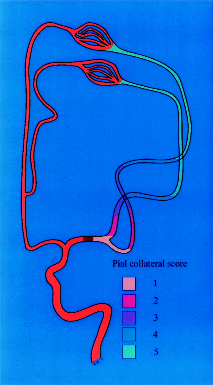
- Fig 1.
Scoring of the anatomic extent of pial collateral blood flow from the ACA territory to the MCA territory during occlusion of the M1 segment. Scoring corresponds to the angiographically visible retrograde reconstitution of the MCA segments on the delayed venous phase. Each color is depicts the furthest extent of retrograde opacification depicted on anteroposterior cerebral angiograms for each pial collateral score.
- Fig 2.
Anteroposterior (A and C) and lateral (B and C) images from a left internal carotid artery angiogram obtained during the early (A and B) and delayed angiographic phases (C and D) in a patient with acute ischemic stroke due to occlusion at the M1 segment (arrow). Note the retrograde opacification of the MCA branches via pial collateral vessels extending from the ACA (arrowheads). Because there is reverse opacification of the MCA extending to the distal M1 segment, a pial collateral formation score of 1 was assigned.


Tables
- TABLE 1:
Infarct volumes (cm3) distributed according to pial collateral score and recanalization result
Collateral Score Completea Recanalization Mean (σ, n) Partial and Nob Recanalization Mean (σ, n) Total Mean (σ, n) 1 2.53 (2.92, 12) 68.55 (53.59, 15) 39.23 (51.67, 27) 2 32.47 (12.90, 3) 41.93 (25.09, 6) 38.78 (21.38, 9) 3 81.66 (31.13, 5) 122.04 (54.83, 5) 101.85 (47.11, 10) 4 and 5 154.28 (136.04, 4) 392.9 (125.209, 3) 256.54 (175.35, 7) 1 and 2 8.52 (13.57, 15) 60.98 (48.17, 21) 39.12 (45.69, 36) 3–5 113.93 (94.27, 9) 223.61 (160.78, 8) 165.55 (137.61, 17) Total 48.05 (76.95, 24) 105.84 (116.58, 29) 79.67 (103.83, 53) a P = .003, Wilcoxon rank sum test for statistically significant increase in infarct volume with increase in pial collateral score.
b P =.0067, Wilcoxon rank sum test for statistically significant increase in infarct volume with increase in pial collateral score.
- TABLE 2:
Clinical outcome (presentation NIHSS, change in NIHSS, discharge NIHSS, and modified Rankin) distribution according to pial collateral score and recanalization
Pial Collateral Score Recanalization No. Presentation NIHSS Score Median (Range) Change in NIHSS Score Median (Range) Discharge NIHSS Score Median (Range) Discharge Modified Rankin Scale Score Median (Range) 1 Complete 12 13.5 (4–23) 9.5 (0–15) 1.5 (0–19) 1.5 (0.5) Partial/none 15 11 (5–30) 2 (−12–11) 10 (0–30) 4 (0–5) 2 Complete 3 16 (14–16) 12 (10–16) 2 (0–6) 2 (0–3) Partial/none 6 8.5 (5–21) 0 (−6–6) 10 (3–20) 4 (1–5) 3 Complete 5 16 (12–21) 9 (−10–15) 6 (3–22) 3 (1–5) Partial/none 5 17 (15–22) 5 (1–9) 14 (8–18) 4 (4–4) 4 and 5 Complete 4 13.5 (7–26) 1 (−16–5) 10.5 (6–42) 4.5 (4–6) Partial/none 3 19 (17–29) 0 (−25–4) 25 (19–42) 5 (4–6) Total Complete 24 15 (4–26) 9 (−16–16) 5 (0–42) 3 (0–6) Partial/none 29 15 (0–30) 2 (−25–11) 11 (0–42) 4 (0–6) - TABLE 3:
Infarct volume (cm3) distribution according to pial collateral score, occlusion site, and recanalization
Collateral Score Complete Recanalization Partial/No Recanalization Combined Proximal Distal Proximal Distal Proximal Distal Mean (σ, n) Mean (σ, n) Mean (σ, n) Mean (σ, n) Mean (σ, n) Mean (σ, n) 1 and 2 *9.4 †6.2 ‡68.3 a49.2 41.3 34.83 (14.7, 11) (11.3, 4) (50.7, 13) (44.4, 8) (48.3, 24) (41.7, 12) 3–5 *153.0 †82.7 ‡247.7 a55.1 213.3 78.1 (66.7, 4) (40.00, 5) (157.3, 7) (na, 1) (149.9, 11) (38.4, 6) Total 47.7 48.7 131.1 49.8 95.3 49.2 (91.1, 15) (50.1, 9) (131.0, 20) (41.5, 9) (121.4, 35) (44.7, 18) a Unable to prove statistical significance.
* P = .0040, Wilcoxon rank sum test.
† P = .0139, Wilcoxon rank sum test.
‡ P = .0023, Wilcoxon rank sum test.
- TABLE 4:
Linear regression analysis model performed with significant predictive factors for infarct volume (age per year, high collateral score, distal occlusion site, and complete recanalization)
Predictive Factor Estimatea Standard Error t ratio P > |t| Age per year 1.907 0.66 2.88 0.0059 High collateral score 130.73 21.21 6.16 <0.0001 Distal occlusion site −46.53 20.80 −2.24 0.030 Complete recanalization −72.49 20.80 −3.84 0.0007 Intercept 45.71 50.72 0.90 0.37 Note.—F ratio, 15.43; P > F < 0.0001; root square = 0.563; root mean square error = 71.48.
a Estimates indicate change in infarct volume based on indicated predictive factors. For example, high collateral score (3–5) is estimated to increase infarct value by 130.73 cm3.
- TABLE 5:
Linear regression analysis model performed only with significant predictive factors for discharge NIHSS score (age per year, high collateral score, and complete recanalization)
Predictive Factor Estimate Standard Error t ratio P > |t| Age per year 0.247 0.068 3.63 .0007 High collateral score 7.73 2.18 3.54 .0009 Complete recanalization 7.53 2.05 3.68 .0006 Intercept 3.00 4.95 0.61 .547 Note.—NIHSS was considered as a continuous variable for the purpose of this analysis.
- TABLE 6:
Multivariate logistic regression analysis model for poor outcome (modified Rankin scale score > 2) performed with predictive factors (age per year, high collateral score, and complete recanalization)
Predictive Factor Estimate Standard Error χ2 P > χ2 Age per year −0.0620 0.0306 4.10 .0429 High collateral score 3.27 1.299 6.33 .0119 Complete recanalization 2.65 0.890 8.88 .0029 Intercept −1.019 1.87 0.30 .586 Note.—Whole model test: R2 = 0.3476; P < .0001.
In this issue


Jump to section
Related Articles
Cited By...
- A Method for Imaging the Ischemic Penumbra with MRI Using Intravoxel Incoherent Motion
- Quantification of Collateral Supply with Local-AIF Dynamic Susceptibility Contrast MRI Predicts Infarct Growth
- Intracerebral arterial blood pressure in the vasculature distal to large vessel occlusions in patients with ischemic stroke: correlation with clinical and imaging parameters
- Perfusion Collateral Index versus Hypoperfusion Intensity Ratio in Assessment of Collaterals in Patients with Acute Ischemic Stroke
- Endovascular recanalization for symptomatic non-acute middle cerebral artery occlusion: proposal of a new angiographic classification
- Systematic review protocol to assess artificial intelligence diagnostic accuracy performance in detecting acute ischaemic stroke and large-vessel occlusions on CT and MR medical imaging
- Distal Vessel Imaging via Intra-arterial Flat Panel Detector CTA during Mechanical Thrombectomy
- Significance of angiographic clot meniscus sign in mechanical thrombectomy of basilar artery stroke
- Identifying Severe Stroke Patients Likely to Benefit From Thrombectomy Despite Delays of up to a Day
- Predictors of malignant brain edema after mechanical thrombectomy for acute ischemic stroke
- Insula stroke: the weird and the worrisome
- Inter- and intraobserver reliability for angiographic leptomeningeal collateral flow assessment by the American Society of Interventional and Therapeutic Neuroradiology/Society of Interventional Radiology (ASITN/SIR) scale
- Guidelines for evaluation and management of cerebral collateral circulation in ischaemic stroke 2017
- Is bridging therapy still required in stroke due to carotid artery terminus occlusions?
- Value of Quantitative Collateral Scoring on CT Angiography in Patients with Acute Ischemic Stroke
- Pretreatment predictors of malignant evolution in patients with ischemic stroke undergoing mechanical thrombectomy
- CT angiography-based collateral flow and time to reperfusion are strong predictors of outcome in endovascular treatment of patients with stroke
- General Anesthesia Versus Conscious Sedation for Endovascular Treatment of Acute Ischemic Stroke: The AnStroke Trial (Anesthesia During Stroke)
- Complete reperfusion mitigates influence of treatment time on outcomes after acute stroke
- Impact of Pial Collaterals on Infarct Growth Rate in Experimental Acute Ischemic Stroke
- CT perfusion and angiographic assessment of pial collateral reperfusion in acute ischemic stroke: the CAPRI study
- Comparison of four different collateral scores in acute ischemic stroke by CT angiography
- Good Intracranial Collaterals Trump Poor ASPECTS (Alberta Stroke Program Early CT Score) for Intravenous Thrombolysis in Anterior Circulation Acute Ischemic Stroke
- How temporal evolution of intracranial collaterals in acute stroke affects clinical outcomes
- Comparing Vessel Imaging: Noncontrast Computed Tomography/Computed Tomographic Angiography Should Be the New Minimum Standard in Acute Disabling Stroke
- Poor Collateral Circulation Assessed by Multiphase Computed Tomographic Angiography Predicts Malignant Middle Cerebral Artery Evolution After Reperfusion Therapies
- Predictors of Reperfusion in Patients with Acute Ischemic Stroke
- Relative Influence of Capillary Index Score, Revascularization, and Time on Stroke Outcomes From the Interventional Management of Stroke III Trial
- Differential Effect of Baseline Computed Tomographic Angiography Collaterals on Clinical Outcome in Patients Enrolled in the Interventional Management of Stroke III Trial
- Early Mobilization After Stroke: Early Adoption but Limited Evidence
- Diabetic Microangiopathy: Impact of Impaired Cerebral Vasoreactivity and Delayed Angiogenesis After Permanent Middle Cerebral Artery Occlusion on Stroke Damage and Cerebral Repair in Mice
- Impact of Time-to-Reperfusion on Outcome in Patients with Poor Collaterals
- Prediction of Infarction and Reperfusion in Stroke by Flow- and Volume-Weighted Collateral Signal in MR Angiography
- Collateral Score Complements Clot Location in Predicting the Outcome of Intravenous Thrombolysis
- Time and Diffusion Lesion Size in Major Anterior Circulation Ischemic Strokes
- Relative Filling Time Delay Based on CT Perfusion Source Imaging: A Simple Method to Predict Outcome in Acute Ischemic Stroke
- CTA Collateral Status and Response to Recanalization in Patients with Acute Ischemic Stroke
- Thrombus length estimation in acute ischemic stroke: a potential role for delayed contrast enhanced CT
- Arterial Spin-Labeled Perfusion Imaging in Acute Ischemic Stroke
- Relative Cerebral Blood Volume as a Marker of Durable Tissue-at-Risk Viability in Hyperacute Ischemic Stroke
- Expanding the role of NCCT in acute stroke imaging: thrombus length measurement and its potential impact on current practice
- Admission Insular Infarction >25% Is the Strongest Predictor of Large Mismatch Loss in Proximal Middle Cerebral Artery Stroke
- Prognostic Evaluation Based on Cortical Vein Score Difference in Stroke
- Recommendations on Angiographic Revascularization Grading Standards for Acute Ischemic Stroke: A Consensus Statement
- Pathologic validation of clot length determined using thin section non-contrast CT
- Imaging-based selection for intra-arterial stroke therapies
- The capillary index score: rethinking the acute ischemic stroke treatment algorithm. Results from the Borgess Medical Center Acute Ischemic Stroke Registry
- Incidence and outcome of procedural distal emboli using the Penumbra thrombectomy for acute stroke
- Factors Influencing Clinically Meaningful Recanalization after IV-rtPA in Acute Ischemic Stroke
- Factors associated with rapid neurological improvement 24 h following intra-arterial thrombolytic treatment for acute ischemic stroke
- Revascularization grading in endovascular acute ischemic stroke therapy
- Reperfusion Rates Following Intra-Arterial Thrombolysis for Acute Ischemic Stroke: The Influence of the Method for Alteplase Delivery
- Comparison of Arterial Spin Labeling and Bolus Perfusion-Weighted Imaging for Detecting Mismatch in Acute Stroke
- Clot Characterization by Noncontrast CT to Predict IV tPA Failure
- Systematic Review of Methods for Assessing Leptomeningeal Collateral Flow
- Extending the Time Window for Endovascular Procedures According to Collateral Pial Circulation
- Effect of Time to Reperfusion on Clinical Outcome of Anterior Circulation Strokes Treated With Thrombectomy: Pooled Analysis of the MERCI and Multi MERCI Trials
- Regional Leptomeningeal Score on CT Angiography Predicts Clinical and Imaging Outcomes in Patients with Acute Anterior Circulation Occlusions
- Arterial Spin-Labeling MRI Can Identify the Presence and Intensity of Collateral Perfusion in Patients With Moyamoya Disease
- The impact of diabetes on the extent of pial collaterals in acute ischemic stroke patients
- Collateral Flow Averts Hemorrhagic Transformation After Endovascular Therapy for Acute Ischemic Stroke
- Collateral Flow Predicts Response to Endovascular Therapy for Acute Ischemic Stroke
- MRI-Based Selection for Intra-Arterial Stroke Therapy: Value of Pretreatment Diffusion-Weighted Imaging Lesion Volume in Selecting Patients With Acute Stroke Who Will Benefit From Early Recanalization
- Distal hyperintense vessels on FLAIR: An MRI marker for collateral circulation in acute stroke?
- Intra-Arterial Stroke Therapy: An Assessment of Demand and Available Work Force
- Predictors of Hemorrhage Following Intra-Arterial Thrombolysis for Acute Ischemic Stroke: The Role of Pial Collateral Formation
- Determinants of the distribution and severity of hypoperfusion in patients with ischemic stroke
- Impact of collateral flow on tissue fate in acute ischaemic stroke
- Size Matters: Hemorrhage Volume as an Objective Measure to Define Significant Intracranial Hemorrhage Associated With Thrombolysis
- Accuracy of Pre- and Postcontrast 3D Time-of-Flight MR Angiography in Patients with Acute Ischemic Stroke: Correlation with Catheter Angiography