Advertisement

Main menu

  • Home
  • Content
    • Current Issue
    • Accepted Manuscripts
    • Article Preview
    • Past Issue Archive
    • Video Articles
    • AJNR Case Collection
    • Case of the Week Archive
    • Case of the Month Archive
    • Classic Case Archive
  • Special Collections
    • AJNR Awards
    • Low-Field MRI
    • Alzheimer Disease
    • ASNR Foundation Special Collection
    • Photon-Counting CT
    • View All
  • Multimedia
    • AJNR Podcasts
    • AJNR SCANtastic
    • Trainee Corner
    • MRI Safety Corner
    • Imaging Protocols
  • For Authors
    • Submit a Manuscript
    • Submit a Video Article
    • Submit an eLetter to the Editor/Response
    • Manuscript Submission Guidelines
    • Statistical Tips
    • Fast Publishing of Accepted Manuscripts
    • Graphical Abstract Preparation
    • Imaging Protocol Submission
    • Author Policies
  • About Us
    • About AJNR
    • Editorial Board
    • Editorial Board Alumni
  • More
    • Become a Reviewer/Academy of Reviewers
    • Subscribers
    • Permissions
    • Alerts
    • Feedback
    • Advertisers
    • ASNR Home

User menu

  • Alerts
  • Log in

Search

  • Advanced search
American Journal of Neuroradiology

American Journal of Neuroradiology

ASHNR American Society of Functional Neuroradiology ASHNR American Society of Pediatric Neuroradiology ASSR
  • Alerts
  • Log in

Advanced Search

  • Home
  • Content
    • Current Issue
    • Accepted Manuscripts
    • Article Preview
    • Past Issue Archive
    • Video Articles
    • AJNR Case Collection
    • Case of the Week Archive
    • Case of the Month Archive
    • Classic Case Archive
  • Special Collections
    • AJNR Awards
    • Low-Field MRI
    • Alzheimer Disease
    • ASNR Foundation Special Collection
    • Photon-Counting CT
    • View All
  • Multimedia
    • AJNR Podcasts
    • AJNR SCANtastic
    • Trainee Corner
    • MRI Safety Corner
    • Imaging Protocols
  • For Authors
    • Submit a Manuscript
    • Submit a Video Article
    • Submit an eLetter to the Editor/Response
    • Manuscript Submission Guidelines
    • Statistical Tips
    • Fast Publishing of Accepted Manuscripts
    • Graphical Abstract Preparation
    • Imaging Protocol Submission
    • Author Policies
  • About Us
    • About AJNR
    • Editorial Board
    • Editorial Board Alumni
  • More
    • Become a Reviewer/Academy of Reviewers
    • Subscribers
    • Permissions
    • Alerts
    • Feedback
    • Advertisers
    • ASNR Home
  • Follow AJNR on Twitter
  • Visit AJNR on Facebook
  • Follow AJNR on Instagram
  • Join AJNR on LinkedIn
  • RSS Feeds

AJNR Awards, New Junior Editors, and more. Read the latest AJNR updates

LetterLetter

Transient Infundibular Mass Associated with Temporary Functional Deficiency

Duncan Browne, Mary Armitage, Jeremy Small and Peter Lees
American Journal of Neuroradiology June 2005, 26 (6) 1605-1606;
Duncan Browne
  • Find this author on Google Scholar
  • Find this author on PubMed
  • Search for this author on this site
Mary Armitage
  • Find this author on Google Scholar
  • Find this author on PubMed
  • Search for this author on this site
Jeremy Small
  • Find this author on Google Scholar
  • Find this author on PubMed
  • Search for this author on this site
Peter Lees
  • Find this author on Google Scholar
  • Find this author on PubMed
  • Search for this author on this site
  • Article
  • Figures & Data
  • Info & Metrics
  • Responses
  • References
  • PDF
Loading

Pituitary tumors present because of either mass effect or functional abnormalities or, increasingly, as incidental findings on MR imaging. Although most infundibular masses are benign, the differential diagnosis includes germ cell tumors, CNS lymphoma, and cerebral inflammatory lesions, including neurosarcoidosis and histiocytosis (1–3). Invasive biopsy, with its associated risks, is required for definitive diagnosis of infundibular lesions. A patient with an infundibular mass associated with pituitary hormonal deficiency—both of which spontaneously regressed, enabling conservative management—is described in this letter.

A 17-year old male patient was referred to the endocrine clinic with a 7-week history of thirst, polydipsia, and polyuria. He was otherwise well, with no significant medical history, and diabetes had been excluded. The initial differential diagnosis lay between psychogenic polydipsia and diabetes insipidus. Initial investigations demonstrated normal serum osmolality (293 mosmol/kg) yet dilute urine (89 mosmol/kg), while other pituitary function tests were unremarkable. Consequently, a water deprivation test was performed, which confirmed a diagnosis of cranial diabetes insipidus. Oral desmopressin was commenced, with immediate symptomatic improvement.

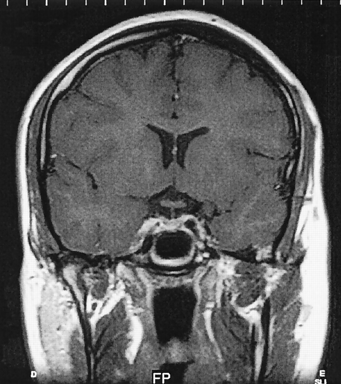
MR imaging of his pituitary gland demonstrated loss of high signal intensity within the posterior pituitary gland and an associated 6-mm focal enhancing infundibular mass following contrast (Fig 1). The radiologic appearance of the anterior pituitary was unremarkable, and there was no involvement of the optic chiasm. Further examination of the MR images revealed no meningeal enhancement, whereas a chest radiograph, autoimmune profile, anti-neutrophil cytoplasmic antibody, c-reactive protein, erythrocyte sedimentation rate, beta human chorionic gonadotrophin, and alpha fetoprotein did not suggest sarcoidosis or inflammatory or malignant disease. Invasive biopsy was deferred because visual fields were normal, and the patient was studying for examinations.

In the 3 months following the initial endocrine assessment, repeat hormone testing revealed progressively rising prolactin (656 mU/L, 1366 mU/L) and diminishing testosterone (5.5 nmol/L, 0.6 nmol/L), consistent with an evolving pituitary stalk lesion (Table). He was commenced on testosterone replacement therapy. A second MR imaging performed 3 months following the initial imaging demonstrated the infundibular mass to be fractionally smaller. Subsequent MR images confirmed shrinkage of the infundibular lesion until it completely resolved a year after initial imaging (Fig 2); however, the loss of high signal intensity in the posterior pituitary seen on initial imaging remained unaltered. Further endocrine tests reflected the radiologic regression of the pituitary lesion with prolactin and gonadotrophin levels normalizing (Table). Testosterone replacement therapy was successfully withdrawn a year after initiation. Gonadotrophin and prolactin levels have remained within the normal range for 4 years, and the radiologic improvement has been sustained over this period. In keeping with the persistent loss of the normal high signal intensity seen in the posterior pituitary, however, repeated attempts to wean him from desmopressin acetate replacement have failed, which has resulted in an immediate return of polyuria.

Regression of pituitary region tumors following administration of steroids is well recognized in germ cell tumors, lymphomas, and neurosarcoidosis (1–3). Similar regression of a pituitary mass due to lymphocytic adenohypophysitis has been reported, but only during the postpartum period (4). We are unaware of other cases with spontaneous radiologic and hormonal recovery of an infundibular mass outside of the postpartum period. Furthermore, we can demonstrate the acute deterioration of pituitary function followed by functional recovery mirrored by the disappearance of the pituitary mass on MR imaging. Although Rathke cleft cysts have been reported to regress spontaneously, MR imaging appearances in these patients have been of a cystic mass with ring enhancement.

The explanation for regression of the infundibular mass is unclear. Lymphocytic infundibuloneurohypophytis can cause transient abnormalities of pituitary function, yet the radiologic appearances are of thickened pituitary stalk as opposed to the discrete mass seen in this case. Our findings challenge the necessity for tissue diagnosis in small infundibular masses, and in similar circumstances a “wait-and-see” policy of close clinical and radiologic surveillance may negate the potential hazards of pituitary biopsy. We are curious whether others have seen this type of apparent spontaneous mass regression.

Fig 1.
  • Download figure
  • Open in new tab
  • Download powerpoint
Fig 1.

Coronal T1-weighted image following intravenous gadolinium, showing discrete enhancing infundibular mass at presentation.

Fig 2.
  • Download figure
  • Open in new tab
  • Download powerpoint
Fig 2.

Coronal T1-weighted image following intravenous gadolinium 2 years after presentation, showing normal appearances of the pituitary gland, infundibulum, and suprasella cistern.

View this table:
  • View inline
  • View popup

Summary of hormonal and radiologic results

References

  1. ↵
    Mascalchi M, Roncaroli F, Salvi F, Frank G. Transient regression of an intracranial germ cell tumour after intravenous steroid administration: a case report. J Neurol Neurosurg Psychiatry 1998;64:670–672
    Abstract/FREE Full Text
  2. Alderson L, Fetell MR, Sisti M, et al. Sentinel lesions of primary CNS lymphoma. J Neurol Neurosurg Psychiatry 1996;60:102–105
    Abstract/FREE Full Text
  3. ↵
    Lexa FJ, Grossman RI. MR of sarcoidosis in the head and spine: spectrum of manifestations and radiographic response to steroid therapy. AJNR Am J Neuroradiol 1994;15:973–982
    Abstract/FREE Full Text
  4. ↵
    Yamakita N, Iwamura M, Murai T, et al. Spontaneous recovery from pathologically confirmed lymphocytic adenohypophysitis with a dramatic reduction of hypophyseal size. Intern Med 1999;38:865–870
    PubMed
  • Copyright © American Society of Neuroradiology
PreviousNext
Back to top

In this issue

American Journal of Neuroradiology: 26 (6)
American Journal of Neuroradiology
Vol. 26, Issue 6
1 Jun 2005
  • Table of Contents
  • Index by author
Advertisement
Print
Download PDF
Email Article

Thank you for your interest in spreading the word on American Journal of Neuroradiology.

NOTE: We only request your email address so that the person you are recommending the page to knows that you wanted them to see it, and that it is not junk mail. We do not capture any email address.

Enter multiple addresses on separate lines or separate them with commas.
Transient Infundibular Mass Associated with Temporary Functional Deficiency
(Your Name) has sent you a message from American Journal of Neuroradiology
(Your Name) thought you would like to see the American Journal of Neuroradiology web site.
CAPTCHA
This question is for testing whether or not you are a human visitor and to prevent automated spam submissions.
Cite this article
Duncan Browne, Mary Armitage, Jeremy Small, Peter Lees
Transient Infundibular Mass Associated with Temporary Functional Deficiency
American Journal of Neuroradiology Jun 2005, 26 (6) 1605-1606;

Citation Manager Formats

  • BibTeX
  • Bookends
  • EasyBib
  • EndNote (tagged)
  • EndNote 8 (xml)
  • Medlars
  • Mendeley
  • Papers
  • RefWorks Tagged
  • Ref Manager
  • RIS
  • Zotero
0 Responses
Respond to this article
Share
Bookmark this article
Transient Infundibular Mass Associated with Temporary Functional Deficiency
Duncan Browne, Mary Armitage, Jeremy Small, Peter Lees
American Journal of Neuroradiology Jun 2005, 26 (6) 1605-1606;
del.icio.us logo Twitter logo Facebook logo Mendeley logo
  • Tweet Widget
  • Facebook Like
  • Google Plus One

Jump to section

  • Article
    • References
  • Figures & Data
  • Info & Metrics
  • Responses
  • References
  • PDF

Related Articles

  • No related articles found.
  • PubMed
  • Google Scholar

Cited By...

  • No citing articles found.
  • Crossref
  • Google Scholar

This article has not yet been cited by articles in journals that are participating in Crossref Cited-by Linking.

More in this TOC Section

  • Letter to the Editor regarding “Automated Volumetric Software in Dementia: Help or Hindrance to the Neuroradiologist?”
  • Reply:
  • Brain AVM’s Nidus: What if We Hadn’t Understood Anything?
Show more Letter

Similar Articles

Advertisement

Indexed Content

  • Current Issue
  • Accepted Manuscripts
  • Article Preview
  • Past Issues
  • Editorials
  • Editor's Choice
  • Fellows' Journal Club
  • Letters to the Editor
  • Video Articles

Cases

  • Case Collection
  • Archive - Case of the Week
  • Archive - Case of the Month
  • Archive - Classic Case

More from AJNR

  • Trainee Corner
  • Imaging Protocols
  • MRI Safety Corner
  • Book Reviews

Multimedia

  • AJNR Podcasts
  • AJNR Scantastics

Resources

  • Turnaround Time
  • Submit a Manuscript
  • Submit a Video Article
  • Submit an eLetter to the Editor/Response
  • Manuscript Submission Guidelines
  • Statistical Tips
  • Fast Publishing of Accepted Manuscripts
  • Graphical Abstract Preparation
  • Imaging Protocol Submission
  • Evidence-Based Medicine Level Guide
  • Publishing Checklists
  • Author Policies
  • Become a Reviewer/Academy of Reviewers
  • News and Updates

About Us

  • About AJNR
  • Editorial Board
  • Editorial Board Alumni
  • Alerts
  • Permissions
  • Not an AJNR Subscriber? Join Now
  • Advertise with Us
  • Librarian Resources
  • Feedback
  • Terms and Conditions
  • AJNR Editorial Board Alumni

American Society of Neuroradiology

  • Not an ASNR Member? Join Now

© 2025 by the American Society of Neuroradiology All rights, including for text and data mining, AI training, and similar technologies, are reserved.
Print ISSN: 0195-6108 Online ISSN: 1936-959X

Powered by HighWire