Advertisement

Main menu

  • Home
  • Content
    • Current Issue
    • Accepted Manuscripts
    • Article Preview
    • Past Issue Archive
    • Video Articles
    • AJNR Case Collection
    • Case of the Week Archive
    • Case of the Month Archive
    • Classic Case Archive
  • Special Collections
    • AJNR Awards
    • Low-Field MRI
    • Alzheimer Disease
    • ASNR Foundation Special Collection
    • Photon-Counting CT
    • View All
  • Multimedia
    • AJNR Podcasts
    • AJNR SCANtastic
    • Trainee Corner
    • MRI Safety Corner
    • Imaging Protocols
  • For Authors
    • Submit a Manuscript
    • Submit a Video Article
    • Submit an eLetter to the Editor/Response
    • Manuscript Submission Guidelines
    • Statistical Tips
    • Fast Publishing of Accepted Manuscripts
    • Graphical Abstract Preparation
    • Imaging Protocol Submission
    • Author Policies
  • About Us
    • About AJNR
    • Editorial Board
    • Editorial Board Alumni
  • More
    • Become a Reviewer/Academy of Reviewers
    • Subscribers
    • Permissions
    • Alerts
    • Feedback
    • Advertisers
    • ASNR Home

User menu

  • Alerts
  • Log in

Search

  • Advanced search
American Journal of Neuroradiology

American Journal of Neuroradiology

ASHNR American Society of Functional Neuroradiology ASHNR American Society of Pediatric Neuroradiology ASSR
  • Alerts
  • Log in

Advanced Search

  • Home
  • Content
    • Current Issue
    • Accepted Manuscripts
    • Article Preview
    • Past Issue Archive
    • Video Articles
    • AJNR Case Collection
    • Case of the Week Archive
    • Case of the Month Archive
    • Classic Case Archive
  • Special Collections
    • AJNR Awards
    • Low-Field MRI
    • Alzheimer Disease
    • ASNR Foundation Special Collection
    • Photon-Counting CT
    • View All
  • Multimedia
    • AJNR Podcasts
    • AJNR SCANtastic
    • Trainee Corner
    • MRI Safety Corner
    • Imaging Protocols
  • For Authors
    • Submit a Manuscript
    • Submit a Video Article
    • Submit an eLetter to the Editor/Response
    • Manuscript Submission Guidelines
    • Statistical Tips
    • Fast Publishing of Accepted Manuscripts
    • Graphical Abstract Preparation
    • Imaging Protocol Submission
    • Author Policies
  • About Us
    • About AJNR
    • Editorial Board
    • Editorial Board Alumni
  • More
    • Become a Reviewer/Academy of Reviewers
    • Subscribers
    • Permissions
    • Alerts
    • Feedback
    • Advertisers
    • ASNR Home
  • Follow AJNR on Twitter
  • Visit AJNR on Facebook
  • Follow AJNR on Instagram
  • Join AJNR on LinkedIn
  • RSS Feeds

AJNR Awards, New Junior Editors, and more. Read the latest AJNR updates

Getting new auth cookie, if you see this message a lot, tell someone!
Research ArticleBrain

Retrograde Cortical and Deep Venous Drainage in Patients with Intracranial Dural Arteriovenous Fistulas: Comparison of MR Imaging and Angiographic Findings

Mika Kitajima, Toshinori Hirai, Yukunori Korogi, Masayuki Yamura, Koichi Kawanaka, Ichiro Ikushima, Yoshiko Hayashida, Yasuyuki Yamashita and Junichi Kuratsu
American Journal of Neuroradiology June 2005, 26 (6) 1532-1538;
Mika Kitajima
  • Find this author on Google Scholar
  • Find this author on PubMed
  • Search for this author on this site
Toshinori Hirai
  • Find this author on Google Scholar
  • Find this author on PubMed
  • Search for this author on this site
Yukunori Korogi
  • Find this author on Google Scholar
  • Find this author on PubMed
  • Search for this author on this site
Masayuki Yamura
  • Find this author on Google Scholar
  • Find this author on PubMed
  • Search for this author on this site
Koichi Kawanaka
  • Find this author on Google Scholar
  • Find this author on PubMed
  • Search for this author on this site
Ichiro Ikushima
  • Find this author on Google Scholar
  • Find this author on PubMed
  • Search for this author on this site
Yoshiko Hayashida
  • Find this author on Google Scholar
  • Find this author on PubMed
  • Search for this author on this site
Yasuyuki Yamashita
  • Find this author on Google Scholar
  • Find this author on PubMed
  • Search for this author on this site
Junichi Kuratsu
  • Find this author on Google Scholar
  • Find this author on PubMed
  • Search for this author on this site
  • Article
  • Figures & Data
  • Info & Metrics
  • Responses
  • References
  • PDF
Loading

Article Figures & Data

Figures

  • Tables
  • Fig 1.
    • Download figure
    • Open in new tab
    • Download powerpoint
    Fig 1.

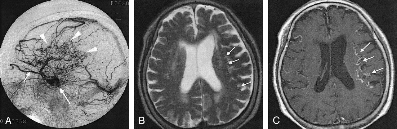
    Case 19. A 78-year-old woman with dAVF in the left CS.

    A, Lateral intra-arterial DSA of the left ECA shows the arteriovenous shunt in the left CS (long arrow) and retrograde venous drainage to the superior ophthalmic vein (short arrow) and cerebral cortical veins (arrowheads). Severity of the drainage was grade 2 (26%–50% of cortical veins on angiography).

    B, Axial T2-weighted SE MR image shows flow voids (arrows) in the subarachnoid space, suggesting dilated cortical veins in bilateral frontal and parietal lobes.

    C, Abnormal dilated cortical veins (arrows) are more prominent on this axial enhanced T1-weighted SE image than in B.

  • Fig 2.
    • Download figure
    • Open in new tab
    • Download powerpoint
    Fig 2.

    Case 13. A 70-year-old woman with dAVF in the left CS. Enhanced 3D MP-RAGE image clearly shows marked dilatation and thrombus in the anterior part of the left superior ophthalmic vein (arrows).

  • Fig 3.
    • Download figure
    • Open in new tab
    • Download powerpoint
    Fig 3.

    Case 17. A 50-year-old woman with dAVF in the left transverse sinus.

    A, Lateral intra-arterial DSA of the left ECA shows obstruction of the left transverse sinus (arrows). Abnormal vessels (arrowheads) are observed around the obstructed left transverse sinus. Severity of the drainage was grade 2 (26%–50% of cortical veins on angiography).

    B, Enhanced 3D MP-RAGE image shows abnormal dilated veins at the base of the cerebrum (arrow) and in the posterior fossa (arrowhead). Bilateral posterior cerebral arteries were identified on other sections (not shown).

  • Fig 4.
    • Download figure
    • Open in new tab
    • Download powerpoint
    Fig 4.

    Case 4. A 62-year-old woman with dAVF in the left CS.

    A, Lateral intra-arterial DSA of the left ICA shows retrograde venous drainage to the superior ophthalmic (arrow) and pontomedullary (arrowheads) veins.

    B, Enhanced 3D MP RAGE image shows cephalocaudal, contiguous enhancing structures (arrow) at the ventral side of midbrain corresponding to retrograde venous drainage to the pontomedullary vein.

  • Fig 5.
    • Download figure
    • Open in new tab
    • Download powerpoint
    Fig 5.

    Case 3. A 58-year-old woman with dAVF in the bilateral CSs. Enhanced 3D MP-RAGE image (and 3D FISP image, not shown) depicts a filling defect in the right inferior petrosal sinus (arrow). This finding suggests thrombosis in the right inferior petrosal sinus.

Tables

  • Figures
    • View popup
    TABLE 1:

    Angiographic findings

    Patient/Age (years)/SexFistula SiteFeeding ArteryObstructed Petrosal SinusRetrograde Drainage Veins
    1/69/FR/L CSR/L ECA, ICAR/L superior, inferiorCortical, deep
    2/66/FR/L CSR/L ECA, ICAR/L superior, inferiorCortical, deep
    3/58/FR/L CSR/L ECA, ICAR/L superior, inferiorCortical, deep, superior ophthalmic, PF
    4/62/FL CSR/L ECA, ICAL superior, R/L inferiorDeep, superior ophthalmic, PF, SV
    5/77/ML CSR/L ICAR/L superior, inferiorCortical, deep
    6/51/FR CSR/L ECA, ICAR/L superior, inferiorSuperior ophthalmic
    7/62/FR CSR/L ECA, ICAR/L superior, inferiorDeep, superior ophthalmic
    8/62/FL CSR/L ECA, ICAR/L superior, inferiorSuperior ophthalmic
    9/76/FR/L CSR ICA, R/L ECAR/L superior, inferiorCortical, deep, superior ophthalmic, PF
    10.52/MR CSR/L ECANot applicableCortical
    11/50/FL CSL ECANot applicableSuperior ophthalmic
    12/52/FL CSL ECAR/L superior, inferiorDeep
    13/70/FL CSL ECA, ICAR/L superior, inferiorSuperior ophthalmic
    14/77/FL CSL ECA, ICAL superior, R/L inferiorCortical, deep
    15/70/MTentL ECA, ICA; R/L VAaCortical, deep
    16/64/ML CSL ECA, ICAR/L superior, inferiorPF
    17/50/FL transverse sinusL ECA, ICA; LaCortical, deep, PF, SV, superior ophthalmic
    18/70/FL CSR ECA, ICANot applicableSuperior ophthalmic, PF
    19/78/FL CSR/L ECA, R, ICAL inferiorCortical, superior ophthalmic, SV
    20/70/ML sigmoid sinusL ECAaCortical, PF
    21/71/FR CSR ECA, ICAL inferiorSuperior ophthalmic
    • Note.—PF, leptomeningeal vein of the posterior fossa; SV, spinal vein; VA, verbetral arteries.

    • a Obstruction was in the L sigmoid sinus and superior sagittal sinus (patient 15), R transverse sinus (patient 17), or R transverse sinus (patient 20).

    • View popup
    TABLE 2:

    Mean sensitivity, specificity, and accuracy judged by two observers

    VeinaSensitivity(%)Specificity(%)Accuracy(%)
    Cortical
        T1 weighted5010073.8
        T2 weighted77.310088.1
        CE T1 weighted100100100
        3D FISP77.386.785.7
        3D CE MP-RAGE1009095.3
    Ophthalmic
        T1 weighted91.710095.2
        T2 weighted66.794.578.6
        CE T1 weighted40.010068.5
        3D FISP45.910069.1
        3D CE MP-RAGE91.783.488.1
    Deep
        T1 weighted13.410054.8
        T2 weighted47.310069.1
        CE T1 weighted34.795.068.4
        3D FISP46.895.569.1
        3D CE MP-RAGE51.485.069.1
    Posterior
        T1 weighted25.910073.8
        T2 weighted25.910073.8
        CE T1 weighted50.096.581.6
        3D FISP18.896.569.1
        3D CE MP-RAGE59.082.376.2
    Spinal
        T1 weighted010085.7
        T2 weighted16.710085.7
        CE T1 weighted75.010094.8
        3D FISP010085.7
        3D CE MP-RAGE100100100
    • a CE = contrast enhanced.

PreviousNext
Back to top

In this issue

American Journal of Neuroradiology: 26 (6)
American Journal of Neuroradiology
Vol. 26, Issue 6
1 Jun 2005
  • Table of Contents
  • Index by author
Advertisement
Print
Download PDF
Email Article

Thank you for your interest in spreading the word on American Journal of Neuroradiology.

NOTE: We only request your email address so that the person you are recommending the page to knows that you wanted them to see it, and that it is not junk mail. We do not capture any email address.

Enter multiple addresses on separate lines or separate them with commas.
Retrograde Cortical and Deep Venous Drainage in Patients with Intracranial Dural Arteriovenous Fistulas: Comparison of MR Imaging and Angiographic Findings
(Your Name) has sent you a message from American Journal of Neuroradiology
(Your Name) thought you would like to see the American Journal of Neuroradiology web site.
CAPTCHA
This question is for testing whether or not you are a human visitor and to prevent automated spam submissions.
Cite this article
Mika Kitajima, Toshinori Hirai, Yukunori Korogi, Masayuki Yamura, Koichi Kawanaka, Ichiro Ikushima, Yoshiko Hayashida, Yasuyuki Yamashita, Junichi Kuratsu
Retrograde Cortical and Deep Venous Drainage in Patients with Intracranial Dural Arteriovenous Fistulas: Comparison of MR Imaging and Angiographic Findings
American Journal of Neuroradiology Jun 2005, 26 (6) 1532-1538;

Citation Manager Formats

  • BibTeX
  • Bookends
  • EasyBib
  • EndNote (tagged)
  • EndNote 8 (xml)
  • Medlars
  • Mendeley
  • Papers
  • RefWorks Tagged
  • Ref Manager
  • RIS
  • Zotero
0 Responses
Respond to this article
Share
Bookmark this article
Retrograde Cortical and Deep Venous Drainage in Patients with Intracranial Dural Arteriovenous Fistulas: Comparison of MR Imaging and Angiographic Findings
Mika Kitajima, Toshinori Hirai, Yukunori Korogi, Masayuki Yamura, Koichi Kawanaka, Ichiro Ikushima, Yoshiko Hayashida, Yasuyuki Yamashita, Junichi Kuratsu
American Journal of Neuroradiology Jun 2005, 26 (6) 1532-1538;
del.icio.us logo Twitter logo Facebook logo Mendeley logo
  • Tweet Widget
  • Facebook Like
  • Google Plus One

Jump to section

  • Article
    • Abstract
    • Methods
    • Results
    • Discussion
    • Conclusion
    • References
  • Figures & Data
  • Info & Metrics
  • Responses
  • References
  • PDF

Related Articles

  • No related articles found.
  • PubMed
  • Google Scholar

Cited By...

  • Susceptibility-Weighted MR Phase Imaging Can Demonstrate Retrograde Leptomeningeal Venous Drainage in Patients with Dural Arteriovenous Fistula
  • Crossref
  • Google Scholar

This article has not yet been cited by articles in journals that are participating in Crossref Cited-by Linking.

More in this TOC Section

  • White Matter Alterations in the Brains of Patients with Active, Remitted, and Cured Cushing Syndrome: A DTI Study
  • Qualitative and Quantitative Analysis of MR Imaging Findings in Patients with Middle Cerebral Artery Stroke Implanted with Mesenchymal Stem Cells
  • Fast Contrast-Enhanced 4D MRA and 4D Flow MRI Using Constrained Reconstruction (HYPRFlow): Potential Applications for Brain Arteriovenous Malformations
Show more Brain

Similar Articles

Advertisement

Indexed Content

  • Current Issue
  • Accepted Manuscripts
  • Article Preview
  • Past Issues
  • Editorials
  • Editor's Choice
  • Fellows' Journal Club
  • Letters to the Editor
  • Video Articles

Cases

  • Case Collection
  • Archive - Case of the Week
  • Archive - Case of the Month
  • Archive - Classic Case

More from AJNR

  • Trainee Corner
  • Imaging Protocols
  • MRI Safety Corner
  • Book Reviews

Multimedia

  • AJNR Podcasts
  • AJNR Scantastics

Resources

  • Turnaround Time
  • Submit a Manuscript
  • Submit a Video Article
  • Submit an eLetter to the Editor/Response
  • Manuscript Submission Guidelines
  • Statistical Tips
  • Fast Publishing of Accepted Manuscripts
  • Graphical Abstract Preparation
  • Imaging Protocol Submission
  • Evidence-Based Medicine Level Guide
  • Publishing Checklists
  • Author Policies
  • Become a Reviewer/Academy of Reviewers
  • News and Updates

About Us

  • About AJNR
  • Editorial Board
  • Editorial Board Alumni
  • Alerts
  • Permissions
  • Not an AJNR Subscriber? Join Now
  • Advertise with Us
  • Librarian Resources
  • Feedback
  • Terms and Conditions
  • AJNR Editorial Board Alumni

American Society of Neuroradiology

  • Not an ASNR Member? Join Now

© 2025 by the American Society of Neuroradiology All rights, including for text and data mining, AI training, and similar technologies, are reserved.
Print ISSN: 0195-6108 Online ISSN: 1936-959X

Powered by HighWire