
Article Figures & Data
Figures
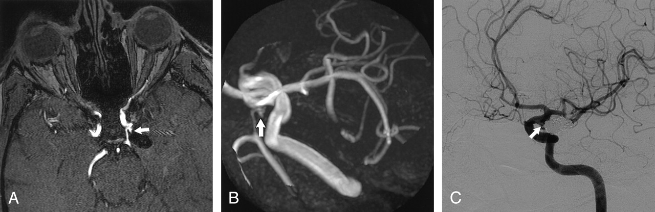
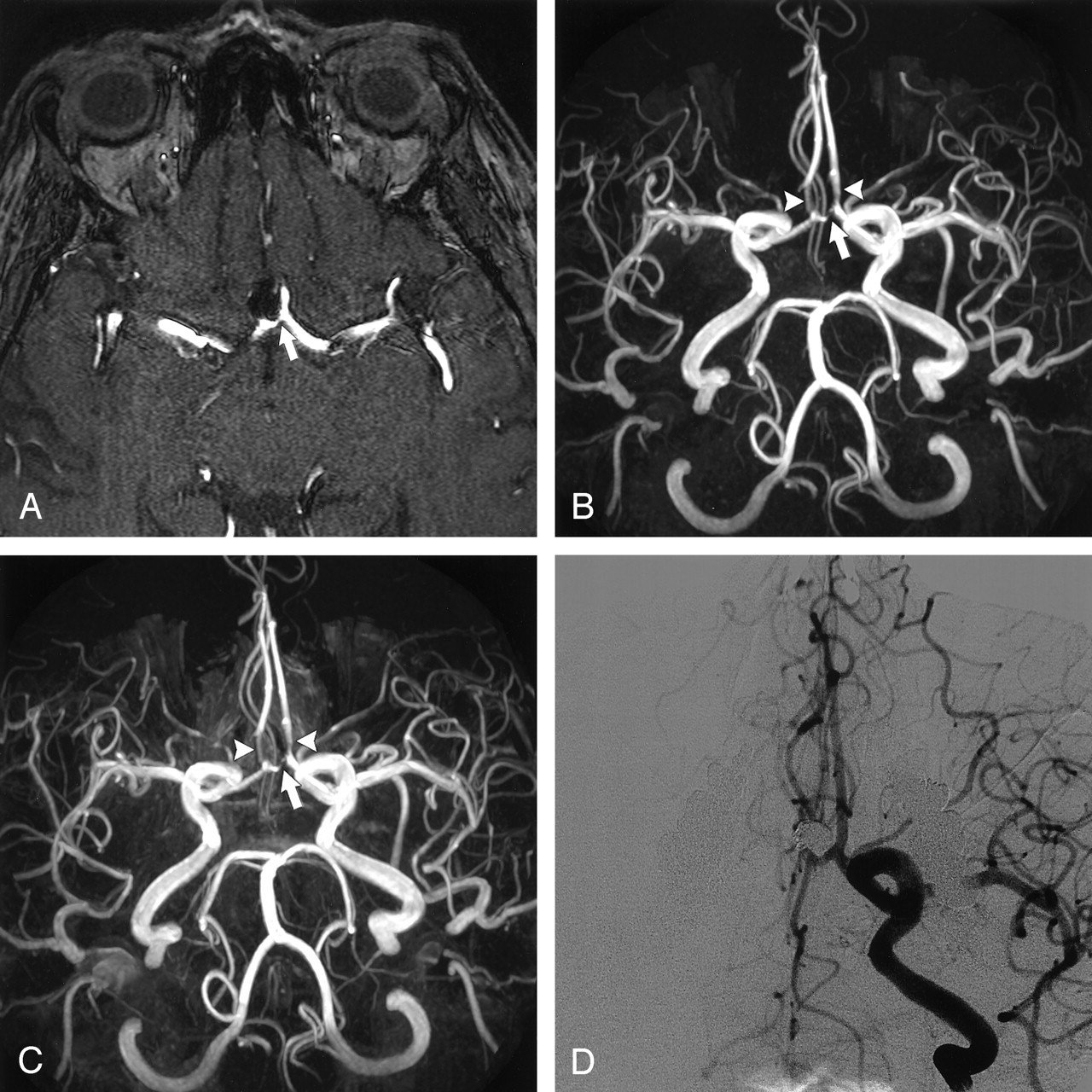
- Fig 1.
Interobserver disagreement on MRA and disagreement between MRA and DSA on the occlusion of a basilar tip aneurysm after treatment with coils.
A, DSA image obtained immediately after treatment with coils shows complete aneurysm occlusion.
B, Nonenhanced MOTSA 3D TOF MRA image obtained 5 months after treatment shows filling of the aneurysm neck (arrow), which was interpreted by one observer as a 2-mm remnant and by the other observer as a 2-mm recurrence. During the consensus reading, it was scored as a remnant. Note minor signal intensity loss in the proximal P1 segment of the left posterior cerebral artery.
C, DSA image obtained 5 months after treatment with coils. This image was interpreted as occlusion.
- Fig 2.
Posterior communicating artery aneurysm with small neck remnant 7 months after treatment with coils.
A, Axial nonenhanced MOTSA 3D TOF MRA source image demonstrates a 2-mm neck remnant (arrow).
B, Nonenhanced MOTSA 3D TOF MR target maximum intensity projection image also shows a small neck remnant (arrow).
C, DSA image confirms the presence of a small neck remnant (arrow).
- Fig 3.
Middle cerebral artery aneurysm with high-signal-intensity rim artifact and recurrence 6 months after treatment with detachable coils.
A, Axial fast spin-echo T2-weighted MR image (3394/80) shows a 2-mm rim of increased signal intensity around the coils (arrow).
B, Axial nonenhanced MOTSA 3D TOF MRA source image demonstrates recurrence of the aneurysm (arrow).
C, Nonenhanced MOTSA 3D TOF MRA image shows recurrence (arrow).
D, Contrast-enhanced MOTSA 3D TOF MRA image shows the same recurrence (arrow).
E, DSA image obtained 6 months after treatment confirms the presence of recurrence (arrow).
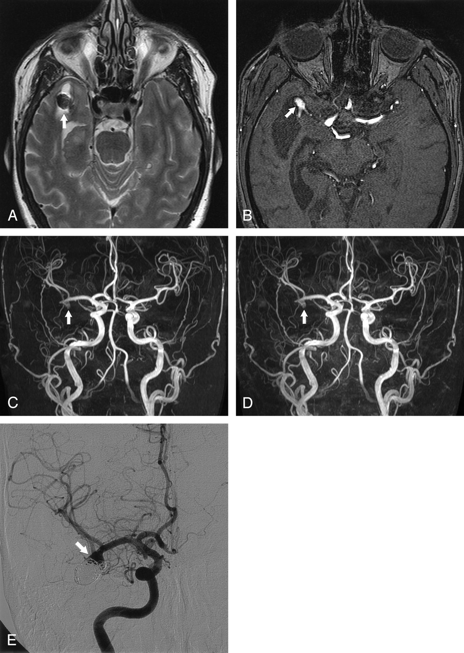
- Fig 4.
Anterior communicating artery aneurysm with recurrence 8 months after treatment with coils.
A, DSA image shows large anterior communicating artery aneurysm.
B, DSA image shows complete occlusion after coiling.
C, Axial nonenhanced MOTSA 3D TOF MRA source image obtained 8 months after treatment demonstrates recurrence (arrow).
D, Nonenhanced MOTSA 3D TOF MRA image demonstrates recurrence (arrow).
E, DSA image obtained 8 months after treatment confirms the presence of recurrence due to coil compaction (arrow).
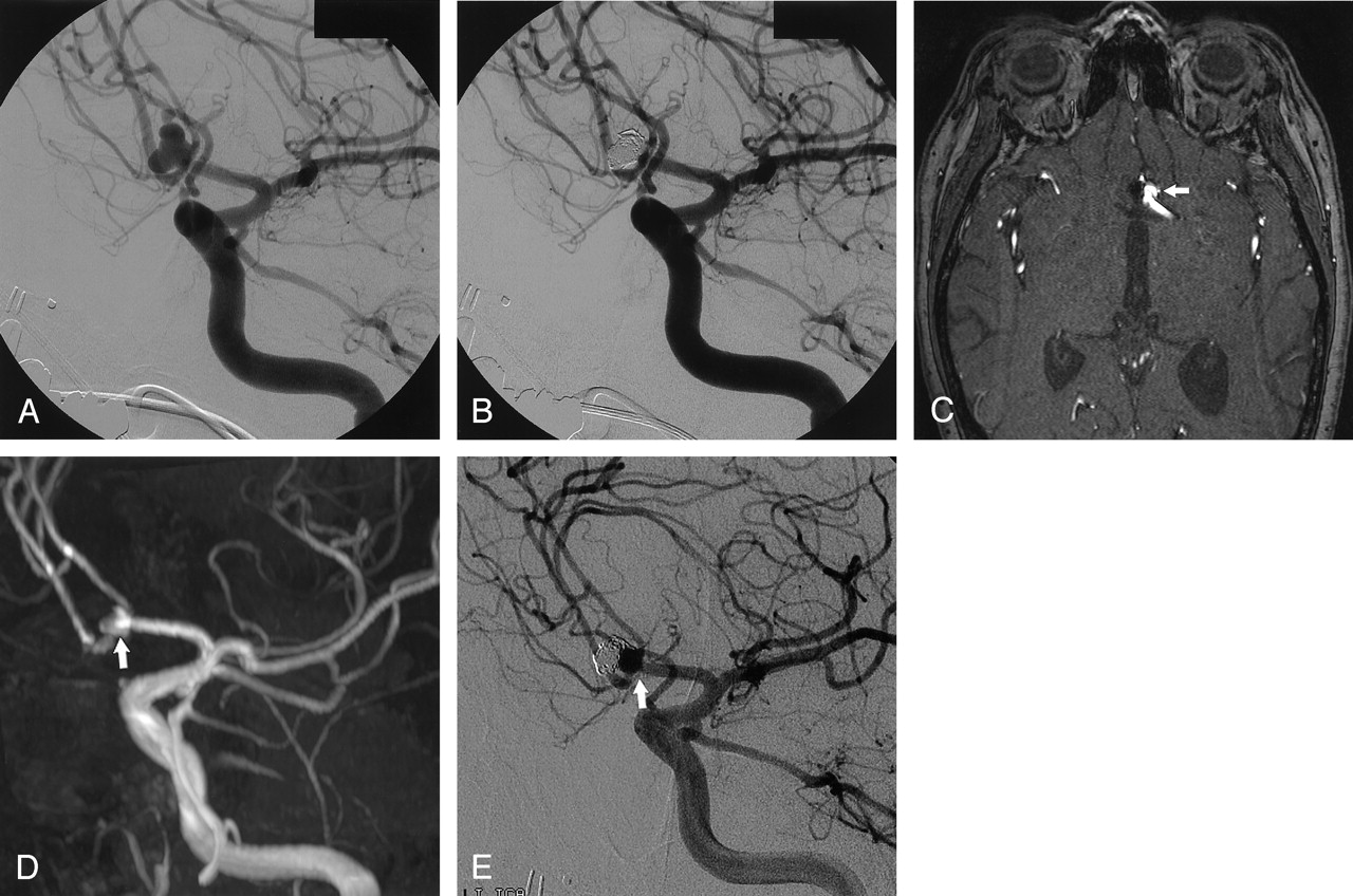
- Fig 5.
Disagreement between MRA and DSA on the occlusion status of a posterior inferior cerebellar artery aneurysm 14 months after treatment with coils.
A, DSA image obtained immediately after coiling shows a small area of residual filling (arrow).
B, Nonenhanced MOTSA 3D TOF MRA image obtained 14 months after treatment shows flow in the aneurysm neck (arrow), which was interpreted as a 2-mm remnant by both observers.
C, DSA image shows filling of the aneurysm neck (arrow), which was interpreted as a 2-mm recurrence (including a 1-mm remnant) due to coil compaction. Both observers thought that additional treatment for this small recurrence was not indicated.
- Fig 6.
Anterior communicating artery aneurysm with coil-related signal intensity loss in parent and branch vessels.
A, Axial nonenhanced MOTSA 3D TOF MRA source image obtained 8 months after treatment shows narrowing of the anterior communicating artery (arrow). No neck remnant or aneurysm recurrence was found.
B, Nonenhanced MOTSA 3D TOF MRA image shows narrowing of the anterior communicating artery (arrow) and the proximal part of both A2 segments of the anterior cerebral arteries (arrowheads).
C, Enhanced MOTSA 3D TOF MRA image shows similar narrowings as described in B.
D, DSA image shows complete occlusion of the aneurysm without narrowing of parent and branch vessels.


Tables
Results of MRA at 3.0 T versus DSA in 21 intracranial aneurysms after treatment with detachable coils
MRA DSA Recurrence Remnant Occlusion Total Recurrence 3 0 0 3 Remnant 1* 5 3 9 Occlusion 0 0 9 9 Total 4 5 12 21 Note—κ=0.70 (95% CI: 0.44–0.95); full agreement=81% (17/21)
* Coil-treated aneurysm was scored as a 2-mm recurrence on DSA image, and as a 2-mm remnant on MRA images. Additional treatment was not required.
In this issue


Jump to section
Related Articles
Cited By...
- MRA versus DSA for the follow-up imaging of intracranial aneurysms treated using endovascular techniques: a meta-analysis
- In vitro accuracy and inter-observer reliability of CT angiography in detecting intracranial aneurysm enlargement
- Usefulness of Silent MR Angiography for Intracranial Aneurysms Treated with a Flow-Diverter Device
- The Neuroform Atlas stent to assist coil embolization of intracranial aneurysms: a multicentre experience
- The New Low-Profile WEB 17 System for Treatment of Intracranial Aneurysms: First Clinical Experiences
- Is Visual Evaluation of Aneurysm Coiling a Reliable Study End Point?: Systematic Review and Meta-Analysis
- Efficacy of Skull Plain Films in Follow-up Evaluation of Cerebral Aneurysms Treated with Detachable Coils: Quantitative Assessment of Coil Mass
- Contrast-Enhanced Time-Resolved MRA for Follow-Up of Intracranial Aneurysms Treated with the Pipeline Embolization Device
- MRA Versus DSA for Follow-Up of Coiled Intracranial Aneurysms: A Meta-Analysis
- Standard of practice: embolization of ruptured and unruptured intracranial aneurysms
- Long-term effects of antiplatelet drugs on aneurysm occlusion after endovascular treatment
- Outcomes of Endovascular Treatments of Aneurysms: Observer Variability and Implications for Interpreting Case Series and Planning Randomized Trials
- Residual Flow After Cerebral Aneurysm Coil Occlusion: Diagnostic Accuracy of MR Angiography
- Late Reopening of Adequately Coiled Intracranial Aneurysms: Frequency and Risk Factors in 400 Patients With 440 Aneurysms
- Reporting standards for endovascular repair of saccular intracranial cerebral aneurysms
- Two-Year Follow-Up of Contrast Stasis within the Sac in Unruptured Aneurysm Coil Embolization: Progressive Thrombosis or Enlargement?
- Long-Term Prospective Follow-Up of Intracranial Aneurysms Treated with Endovascular Coiling Using Contrast-Enhanced MR Angiography
- A Prospective Trial of 3T and 1.5T Time-of-Flight and Contrast-Enhanced MR Angiography in the Follow-Up of Coiled Intracranial Aneurysms
- Reporting Standards for Endovascular Repair of Saccular Intracranial Cerebral Aneurysms
- Evaluation of the Occlusion Status of Coiled Intracranial Aneurysms with MR Angiography at 3T: Is Contrast Enhancement Necessary?
- MR Angiographic Follow-Up of Intracranial Aneurysms Treated with Detachable Coils: Evaluation of a Blood-Pool Contrast Medium
- Reporting Standards for Endovascular Repair of Saccular Intracranial Cerebral Aneurysms
- MR Angiography Follow-Up 5 Years after Coiling: Frequency of New Aneurysms and Enlargement of Untreated Aneurysms