Advertisement

Main menu

  • Home
  • Content
    • Current Issue
    • Accepted Manuscripts
    • Article Preview
    • Past Issue Archive
    • Video Articles
    • AJNR Case Collection
    • Case of the Week Archive
    • Case of the Month Archive
    • Classic Case Archive
  • Special Collections
    • AJNR Awards
    • Low-Field MRI
    • Alzheimer Disease
    • ASNR Foundation Special Collection
    • Photon-Counting CT
    • View All
  • Multimedia
    • AJNR Podcasts
    • AJNR SCANtastic
    • Trainee Corner
    • MRI Safety Corner
    • Imaging Protocols
  • For Authors
    • Submit a Manuscript
    • Submit a Video Article
    • Submit an eLetter to the Editor/Response
    • Manuscript Submission Guidelines
    • Statistical Tips
    • Fast Publishing of Accepted Manuscripts
    • Graphical Abstract Preparation
    • Imaging Protocol Submission
    • Author Policies
  • About Us
    • About AJNR
    • Editorial Board
    • Editorial Board Alumni
  • More
    • Become a Reviewer/Academy of Reviewers
    • Subscribers
    • Permissions
    • Alerts
    • Feedback
    • Advertisers
    • ASNR Home

User menu

  • Alerts
  • Log in

Search

  • Advanced search
American Journal of Neuroradiology

American Journal of Neuroradiology

ASHNR American Society of Functional Neuroradiology ASHNR American Society of Pediatric Neuroradiology ASSR
  • Alerts
  • Log in

Advanced Search

  • Home
  • Content
    • Current Issue
    • Accepted Manuscripts
    • Article Preview
    • Past Issue Archive
    • Video Articles
    • AJNR Case Collection
    • Case of the Week Archive
    • Case of the Month Archive
    • Classic Case Archive
  • Special Collections
    • AJNR Awards
    • Low-Field MRI
    • Alzheimer Disease
    • ASNR Foundation Special Collection
    • Photon-Counting CT
    • View All
  • Multimedia
    • AJNR Podcasts
    • AJNR SCANtastic
    • Trainee Corner
    • MRI Safety Corner
    • Imaging Protocols
  • For Authors
    • Submit a Manuscript
    • Submit a Video Article
    • Submit an eLetter to the Editor/Response
    • Manuscript Submission Guidelines
    • Statistical Tips
    • Fast Publishing of Accepted Manuscripts
    • Graphical Abstract Preparation
    • Imaging Protocol Submission
    • Author Policies
  • About Us
    • About AJNR
    • Editorial Board
    • Editorial Board Alumni
  • More
    • Become a Reviewer/Academy of Reviewers
    • Subscribers
    • Permissions
    • Alerts
    • Feedback
    • Advertisers
    • ASNR Home
  • Follow AJNR on Twitter
  • Visit AJNR on Facebook
  • Follow AJNR on Instagram
  • Join AJNR on LinkedIn
  • RSS Feeds

AJNR Awards, New Junior Editors, and more. Read the latest AJNR updates

Research ArticlePediatric Neuroimaging

Proton MR Spectroscopic Imaging Depicts Diffuse Axonal Injury in Children with Traumatic Brain Injury

Barbara A. Holshouser, Karen A. Tong and Stephen Ashwal
American Journal of Neuroradiology May 2005, 26 (5) 1276-1285;
Barbara A. Holshouser
  • Find this author on Google Scholar
  • Find this author on PubMed
  • Search for this author on this site
Karen A. Tong
  • Find this author on Google Scholar
  • Find this author on PubMed
  • Search for this author on this site
Stephen Ashwal
  • Find this author on Google Scholar
  • Find this author on PubMed
  • Search for this author on this site
  • Article
  • Figures & Data
  • Info & Metrics
  • Responses
  • References
  • PDF
Loading

Article Figures & Data

Figures

  • Tables
  • Fig 1.
    • Download figure
    • Open in new tab
    • Download powerpoint
    Fig 1.

    Mean NAA/Cr and Cho/Cr ratios from all voxels plotted as a function of the neurologic outcome assigned by a pediatric neurologist based on the PCPC score 6–12 months after injury. CTL indicates control; MOD, moderate disability; NL, normal; SEV, severe disability; and VS, vegetative state.

  • Fig 2.
    • Download figure
    • Open in new tab
    • Download powerpoint
    Fig 2.

    Total mean metabolite ratios plotted by neurologic outcomes or control (CNTL). Ratios were calculated from all voxels including those containing hemorrhagic and nonhemorrhagic DAI lesions for patients with TBI. Asterisk indicates P = .01; double asterisk, P = .000.

  • Fig 3.
    • Download figure
    • Open in new tab
    • Download powerpoint
    Fig 3.

    Regional mean metabolite ratios from MRSI data collected in a transverse plane through the level of the CC plotted by neurologic outcomes compared with control (CNTL); * indicates P = .00–.05; +, P < .02.
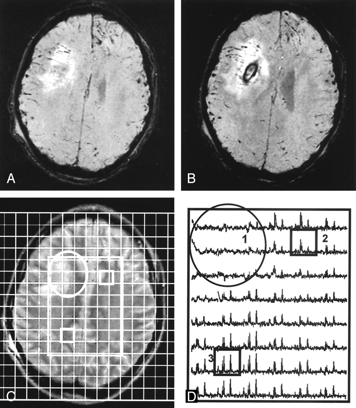
  • Fig 4.
    • Download figure
    • Open in new tab
    • Download powerpoint
    Fig 4.

    15-year-old female adolescent ejected from a car. Patient had a good outcome (GOS score = 1, normal) at 12 months after injury. Total mean metabolite ratios are 1.63 (normal) for NAA/Cr, and 1.67 for NAA/Cho, and 1.06 (increased) for Cho/Cr.

    A and B, T2-weighted MR image (A) corresponding SWI (B) show hemorrhagic lesions in the body of the CC and bifrontal extra-axial collections. A with grid overlay shows the 160-mm FOV and 54 (6 × 9)-voxel volume of interest (rectangle). Boxes indicate corresponding voxels in C.

    C, Spectral map from 54 voxels in the volume of interest shows a (1) spectrum from normal-appearing brain in the anterior CC with decreased NAA (2.0 ppm), (2) a spectrum with reduced metabolite signal intensity due to a small hemorrhagic lesion in the mid CC, and (3) a spectrum from parietal white matter with normal metabolite ratios.

  • Fig 5.
    • Download figure
    • Open in new tab
    • Download powerpoint
    Fig 5.

    12-year-old boy hit by a car. Patient had a poor outcome (GOS score = 4, severe disabilities) at 12-month follow-up. Total mean metabolite ratios from MRSI were 1.14 (decreased) for NAA/Cr, 1.13 for NAA/Cho, and 1.11 (increased) Cho/Cr.

    A and B, Contiguous SWI images show a moderately large hemorrhagic lesion in the deep right frontal lobe.

    C, Corresponding T2-weighted MR image shows the lesion (circle) with an overlay of the volume of interest (rectangle). Boxes indicate corresponding voxels in D.

    D, Spectral map shows loss of metabolite signal intensity in area of the lesion (circle) and (2) a spectrum from normal-appearing left FWM with markedly decreased NAA/Cr and increased Cho/Cr, as compared with (3) a spectrum from normal-appearing parietal white matter with decreased NAA/Cr and normal Cho/Cr.

  • Fig 6.
    • Download figure
    • Open in new tab
    • Download powerpoint
    Fig 6.

    Mean metabolite ratios from visibly injured (hemorrhagic, Hem) brain and normal-appearing (Nrm) brain were significantly different from those in control subjects (CNTL). Asterisk indicates P < .02. NAA/Cr from normal-appearing brain was the only ratio that differentiates between good- and poor- outcome groups; + indicates P = .01.

Tables

  • Figures
    • View popup
    TABLE 1:

    Clinical variables in dichotomized outcome groups

    VariableGood-Outcome Group* (n = 30)Poor-Outcome Group† (n = 10)P Value
    Age (y)11.7 ± 5.99.7 ± 3.3.20
    GCS score7 ± 44 ± 1.03‡
    Duration (d)
        In coma5 ± 611 ± 9.02‡
        Ventilated6 ± 612 ± 6.01‡
        Hospitalized27 ± 2550 ± 30.03‡
        Unconsciousness before MRS4 ± 47 ± 3.01‡
        To MRS7 ± 48 ± 4.20
    Hemorrhagic Lesions
        Total number116 ± 146302 ± 199.002‡
        Total volume (mm3)11271 ± 1573036007 ± 31936.002‡
    • * Normal or mild disability.

    • † Moderate or severe disability or vegetative state, as based on the Pediatric Cerebral Performance Category Scaled score.

    • ‡ P ≤ .05, 2-tailed Mann-Whitney test.

    • View popup
    TABLE 2:

    Mean metabolite ratios

    MRSI RatioControl (n = 9)TBI (n = 40)Normal-Appearing Brain (n = 40)Visible Hemorrhage (n = 33)P Value*
    NAA/Cr1.86 ± 0.111.50 ± 0.29†1.55 ± 0.52‡1.33 ± 0.56†.46
    NAA/Cho2.10 ± 0.191.44 ± 0.34†1.52 ± 0.58†1.16 ± 0.50†.001†
    Cho/Cr0.93 ± 0.071.12 ± 0.13†1.10 ± 0.39‡1.24 ± 0.51†.00†
    • Note.—Ratios are average of mean ratios from each patient. Seven patients did not have voxels containing hemorrhage in the brain section sampled with MRSI.

    • * Normal versus hemorrhage.

    • † P ≤ .001 compared with controls, one-way ANOVA and post-hoc Bonferroni test.

    • ‡ P ≤ 0.05 level compared to controls, one-way ANOVA and post-hoc Bonferroni test.

    • View popup
    TABLE 3:

    Results of logistic regression analyses to predict dichotomized GOS outcomes at 6–12 mo

    VariablesAccuracy (%)Predictionsχ2P Value
    False-Positive (%)False-Negative Rate (%)
    GCS score7501005.2.40
    Days unconscious before MRS and days ventilated783804.5.81
    Days in coma, unconscious before MRS, and ventilated807608.4.39
    MRS ratios*
        Normal-appearing brain8535010.7.22
        Hemorrhagic brain6713807.3.50
        Total brain853505.4.72
    Total NAA/Cr, days unconscious before MRS and ventilated8834011.2.19
    • * Total mean NAA/Cr, NAA/Cho, and Cho/Cr.

PreviousNext
Back to top

In this issue

American Journal of Neuroradiology: 26 (5)
American Journal of Neuroradiology
Vol. 26, Issue 5
1 May 2005
  • Table of Contents
  • Index by author
Advertisement
Print
Download PDF
Email Article

Thank you for your interest in spreading the word on American Journal of Neuroradiology.

NOTE: We only request your email address so that the person you are recommending the page to knows that you wanted them to see it, and that it is not junk mail. We do not capture any email address.

Enter multiple addresses on separate lines or separate them with commas.
Proton MR Spectroscopic Imaging Depicts Diffuse Axonal Injury in Children with Traumatic Brain Injury
(Your Name) has sent you a message from American Journal of Neuroradiology
(Your Name) thought you would like to see the American Journal of Neuroradiology web site.
CAPTCHA
This question is for testing whether or not you are a human visitor and to prevent automated spam submissions.
Cite this article
Barbara A. Holshouser, Karen A. Tong, Stephen Ashwal
Proton MR Spectroscopic Imaging Depicts Diffuse Axonal Injury in Children with Traumatic Brain Injury
American Journal of Neuroradiology May 2005, 26 (5) 1276-1285;

Citation Manager Formats

  • BibTeX
  • Bookends
  • EasyBib
  • EndNote (tagged)
  • EndNote 8 (xml)
  • Medlars
  • Mendeley
  • Papers
  • RefWorks Tagged
  • Ref Manager
  • RIS
  • Zotero
0 Responses
Respond to this article
Share
Bookmark this article
Proton MR Spectroscopic Imaging Depicts Diffuse Axonal Injury in Children with Traumatic Brain Injury
Barbara A. Holshouser, Karen A. Tong, Stephen Ashwal
American Journal of Neuroradiology May 2005, 26 (5) 1276-1285;
del.icio.us logo Twitter logo Facebook logo Mendeley logo
  • Tweet Widget
  • Facebook Like
  • Google Plus One

Jump to section

  • Article
    • Abstract
    • Methods
    • Results
    • Discussion
    • Conclusion
    • Acknowledgments
    • References
  • Figures & Data
  • Info & Metrics
  • Responses
  • References
  • PDF

Related Articles

  • No related articles found.
  • PubMed
  • Google Scholar

Cited By...

  • Imaging Evidence and Recommendations for Traumatic Brain Injury: Advanced Neuro- and Neurovascular Imaging Techniques
  • Crossref
  • Google Scholar

This article has not yet been cited by articles in journals that are participating in Crossref Cited-by Linking.

More in this TOC Section

  • Frontal Paraventricular Cysts
  • Sodium MRI in Pediatric Brain Tumors
  • FRACTURE MR in Congenital Vertebral Anomalies
Show more Pediatric Neuroimaging

Similar Articles

Advertisement

Indexed Content

  • Current Issue
  • Accepted Manuscripts
  • Article Preview
  • Past Issues
  • Editorials
  • Editor's Choice
  • Fellows' Journal Club
  • Letters to the Editor
  • Video Articles

Cases

  • Case Collection
  • Archive - Case of the Week
  • Archive - Case of the Month
  • Archive - Classic Case

More from AJNR

  • Trainee Corner
  • Imaging Protocols
  • MRI Safety Corner
  • Book Reviews

Multimedia

  • AJNR Podcasts
  • AJNR Scantastics

Resources

  • Turnaround Time
  • Submit a Manuscript
  • Submit a Video Article
  • Submit an eLetter to the Editor/Response
  • Manuscript Submission Guidelines
  • Statistical Tips
  • Fast Publishing of Accepted Manuscripts
  • Graphical Abstract Preparation
  • Imaging Protocol Submission
  • Evidence-Based Medicine Level Guide
  • Publishing Checklists
  • Author Policies
  • Become a Reviewer/Academy of Reviewers
  • News and Updates

About Us

  • About AJNR
  • Editorial Board
  • Editorial Board Alumni
  • Alerts
  • Permissions
  • Not an AJNR Subscriber? Join Now
  • Advertise with Us
  • Librarian Resources
  • Feedback
  • Terms and Conditions
  • AJNR Editorial Board Alumni

American Society of Neuroradiology

  • Not an ASNR Member? Join Now

© 2025 by the American Society of Neuroradiology All rights, including for text and data mining, AI training, and similar technologies, are reserved.
Print ISSN: 0195-6108 Online ISSN: 1936-959X

Powered by HighWire