
Article Figures & Data
Figures
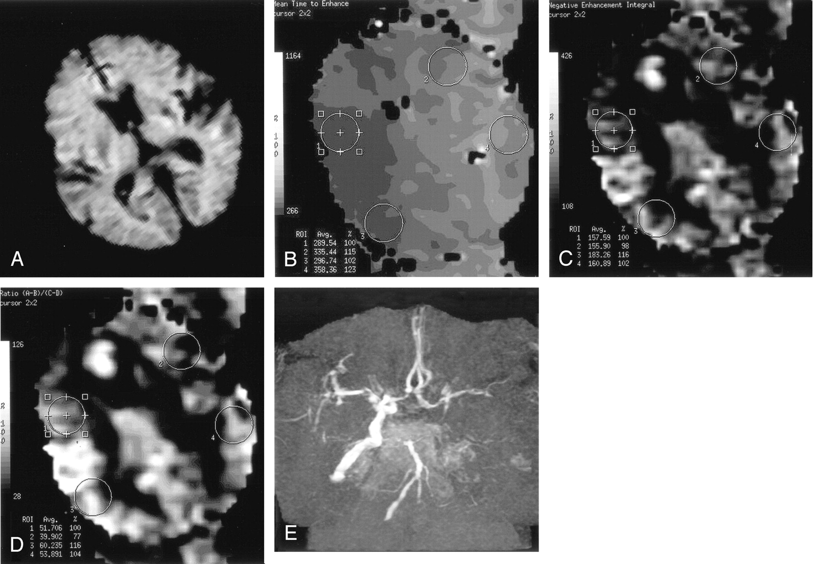
- Fig 1.
Images obtained on admission in case 1.
A, DW image reveals slightly high signal intensity in the cortex of the left parietal lobe.
B, The rMTT map of PW image shows delay of 15–25% in the territory of the left MCA (ROI 2 and 4).
C, The rCBV map of PW image shows slight decrease of 2–12% in the territory of the left MCA (ROI 2 and 4).
D, The rCBF map of PW image reveals reduction of 11–23% in the territory of the left MCA (ROI 2 and 4).
E, MR angiogram shows poor visualization of the left ICA.
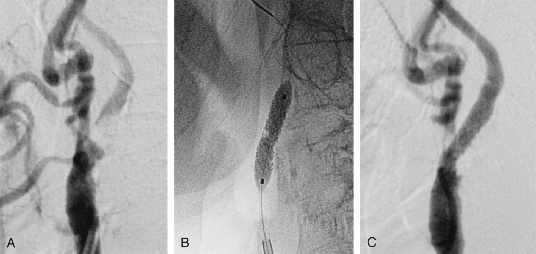
- Fig 2.
Periprocedural images obtained in case 1.
A, Lateral left carotid angiogram obtained before the procedure shows severe stenosis at the origin of the ICA.
B, Lateral left carotid angiogram obtained during the procedure demonstrates stent implantation and postdilation.
C, Lateral left carotid angiogram obtained after the procedure shows an excellent angiographic result.
- Fig 3.
Images obtained on admission in case 4.
A, DW image reveals no area of high signal intensity in the left cerebral hemisphere.
B, The rMTT map of PW image shows delay of 15% in the territory of the left MCA (ROI 4).
C, The rCBV map of PW image shows slight decrease of 11.2% in the territory of the left MCA (ROI 4).
D, The rCBF map of PW image reveals reduction of 43% in the territory of the left MCA (ROI 4).
E, MR angiogram shows no visualization of the left ICA.
- Fig 4.
Periprocedural angiograms obtained in case 4.
A, Lateral left carotid angiogram obtained before the procedure shows occlusion at the origin of the ICA.
B, Lateral left carotid angiogram obtained during the procedure demonstrates stent implantation and postdilation.
C, Lateral left carotid angiogram obtained after the procedure shows essentially complete recanalization with 14% residual stenosis of the ICA.
- Fig 5.
NIHSS scores at 7 days after emergency carotid stent placement (symbols on right) improved significantly (P < .01) compared with baseline scores (symbols on left). W indicates Wilcoxon rank sum test; •, patients with sudden onset of severe stroke; ▴, patients with progressing stroke.


Tables
- TABLE 1:
Patient characteristics and clinical information before emergency carotid artery stent placement
Case No./Sex/Age (y) Type of Stroke Clinical Symptoms NIHSS Score Treatment Onset to Procedure (hrs) 1/F/83 SO Deep coma, CD, QP 28 Aspirin, ticlopidine 2 2/M/65 SO Somnolence, MA, FP, HP 9 Aspirin 10 3/M/69 SO Coma, CD, HP 19 None 24 4/M/60 SO Coma, HP 18 None 8 5/M/68 SO Somnolence, HP 7 Aspirin 12 6/M/28 SO Somnolence, FP, HP 13 None 2 7/M/78 SO Stupor, AG, CD, FP, HP 15 None 17 8/M/83 SO Somnolence, FP, HP 12 Aspirin 18 9/M/66 SO Somnolence, FP, HP 11 None 6 10/F/86 PR Somnolence, SA, HP 12 Heparin 125 11/M/69 PR Somnolence, AG, HP 11 Heparin, ozagrel sodium 72 12/M/68 PR AG, HP 5 Heparin, ozagrel sodium 149 13/F/74 PR Stupor, AG, FP, HP 15 Argatroban 54 14/F/69 PR Somnolence, FP, HP 7 Argatroban, aspirin 120 15/M/82 PR Coma, CD, HP 24 Heparin 26 16/M/62 PR FP, HP 5 Heparin, aspirin 120 17/M/79 PR FP, HP 5 Argatroban, ticlopidine 168 Note.—SO indicates sudden onset of severe stroke; PR, progressing stroke; CD, conjugate deviation of the eyes; QP, quadriparesis; MA, motor-dominant aphasia; FP, facial palsy; HP, hemiplegia or hemiparesis; AG, agnosia; SA, sensory-dominant aphasia.
Case No./Sex/Age (y) Side of Lesion Area of High Signal Intensity DW Imaging PW Imaging Perfusion-Diffusion Mismatch rMTT rCBV rCBF 1/F/83 L CP Small abnormality Delay Small decrease Small reduction Small 2/M/65 L DBZ, CF Small abnormality Delay Small decrease Small reduction Small 3/M/69 L BG Small abnormality Delay Large decrease Large reduction Large 4/M/60 L None Normal Delay Large decrease Large reduction Large 5/M/68 R None Normal Delay Large decrease Large reduction Large 6/M/28 R DBZ, CF Small abnormality Delay Large decrease Large reduction Large 7/M/78 R SBZ, CF Small abnormality Delay Small decrease Small reduction Small 8/M/83 R None Normal Delay Large decrease Large reduction Large 9/M/66 R DBZ, CF Small abnormality Delay Large decrease Large reduction Large 10/F/86 L SBZ, DBZ Small abnormality Delay Large decrease Large reduction Large 11/M/69 R DBZ, CP Small abnormality Delay Small decrease Small reduction Small 12/M/68 R SBZ Small abnormality Delay Small decrease Small reduction* Small 13/F/74 R DBZ, CF Small abnormality Delay Small decrease Small reduction Small 14/F/69 L DBZ Small abnormality Delay Small decrease Small reduction Small 15/M/82 L CF Small abnormality Delay Small decrease Small reduction Small 16/M/62 L DBZ Small abnormality Delay Large decrease Large reduction Large 17/M/79 R DBZ Small abnormality Delay Small decrease Small reduction Small Note.—CP indicates cortex of the parietal lobe; DBZ, deep border zone; CF, cortex of the frontal lobe; BG, basal ganglia; SBZ, superficial border zone.
* Contralateral ICA stenosis.
- TABLE 3:
Preprocedure angiographic findings and devices used for emergency carotid artery stent placement
Case No./Sex/Age (y) Preprocedure Stenosis (%) Cause of Lesion Stent Devices Protection Technique 1/F/83 99 Atherothrombotic S670 No 2/M/65 99 Atherothrombotic S670 No 3/M/69 94 Atherothrombotic NIR Elite No 4/M/60 100 Atherothrombotic S670 No 5/M/68 93 Atherothrombotic S670 No 6/M/28 100 Dissection Easy Wallstent No 7/M/78 99 Atherothrombotic NIR Elite No 8/M/83 100 Atherothrombotic Easy Wallstent Yes 9/M/66 99 Atherothrombotic Easy Wallstent Yes 10/F/86 100 Atherothrombotic S670 No 11/M/69 95 Atherothrombotic S670 No 12/M/68 75 Atherothrombotic NIR Elite No 13/F/74 99 Atherothrombotic S670 No 14/F/69 99 Atherothrombotic NIR Elite No 15/M/82 70 Atherothrombotic Easy Wallstent No 16/M/62 95 Atherothrombotic Easy Wallstent No 17/M/79 75 Atherothrombotic Easy Wallstent No Case No./Sex/Age (y) Postprocedure Stenosis (%)* Stenosis at 90 days (%)* HP at SPECT CT Findings within 24 hrs after Procedure 1/F/83 5 20 Negative SLA 2/M/65 10 20 Negative SLA 3/M/69 12 12 Negative SLA 4/M/60 14 40 Positive NLA + SAH 5/M/68 0 0 Positive NLA 6/M/28 8 8 Negative LLA 7/M/78 12 20 Negative LLA 8/M/83 5 5 Negative SLA 9/M/66 15 0 Positive SLA 10/F/86 20 No evaluation Positive SLA + SAH 11/M/69 0 No evaluation Negative SLA 12/M/68 0 10 Negative SLA 13/F/74 18 20 Negative SLA 14/F/69 8 8 Negative NLA 15/M/82 10 No evaluation Negative SLA 16/M/62 8 0 Negative SLA 17/M/79 0 No evaluation Negative SLA Note.—HP indicates hyperperfusion phenomenon; SLA, small low-attenuation area; LLA, large low-attenuation area; NLA, no low-attenuation area; SAH, subarachnoid hemorrhage.
* * Evaluation by angiography.
Case No./Sex/Age (y) NIHSS Score mRS Score at 90 Days after Procedure Procedure-Related Complications On Admission Just Before Procedure At 7 Days after Procedure 1/F/83 28 28 3 1 Hypotension, bradycardia 2/M/65 9 9 0 0 None 3/M/69 19 19 14 4 None 4/M/60 18 18 1 0 Hypertension 5/M/68 7 7 4 0 Hypotension, bradycardia 6/M/28 13 13 9 3 None 7/M/78 15 15 13 4 Hypertension, distal embolism 8/M/83 12 12 5 1 Hypertension 9/M/66 11 11 5 1 Hypertension 10/F/86 4 12 15 4 Hypertension 11/M/69 11* 11 2 2 Hypotension, bradycardia 12/M/68 2 5 0 0 None 13/F/74 15* 15 9 1 None 14/F/69 4 7 0 0 None 15/M/82 3 24 24 6 None 16/M/62 1 5 15 3 Putaminal hemorrhage 17/M/79 2 5 3 0 Hypotension, bradycardia * Scores of patients transferred from another hospital.
In this issue


Jump to section
Related Articles
Cited By...
- Antithrombotic regimen in emergent carotid stenting for acute ischemic stroke due to tandem occlusion: a meta-analysis of aggregate data
- Emergent carotid stenting versus no stenting for acute ischemic stroke due to tandem occlusion: a meta-analysis
- Antithrombotic regimen in emergent carotid stenting for acute ischemic stroke due to tandem occlusion: a meta-analysis of aggregate data
- Endovascular treatment in patients with acute ischemic stroke and apparent occlusion of the extracranial internal carotid artery on CTA
- Emergency carotid artery stenting in patients with acute ischemic stroke due to occlusion or stenosis of the proximal internal carotid artery: a single-center experience
- Forced suction thrombectomy after carotid stenting in patients with massive thrombus and acute extracranial internal carotid artery occlusion
- Predictors of Functional Outcome after Emergency Carotid Artery Stenting and Intra-Arterial Thrombolysis for Treatment of Acute Stroke Associated with Obstruction of the Proximal Internal Carotid Artery and Tandem Downstream Occlusion
- Guidelines for the Early Management of Patients With Acute Ischemic Stroke: A Guideline for Healthcare Professionals From the American Heart Association/American Stroke Association
- Safety and effectiveness of emergency carotid artery stenting for a high-grade carotid stenosis with intraluminal thrombus under proximal flow control in hyperacute and acute stroke
- Multimodal Reperfusion Therapy for Large Hemispheric Infarcts in Octogenarians: Is Good Outcome a Realistic Goal?
- Carotid Artery Stenting in Acute Stroke