
Article Figures & Data
Figures
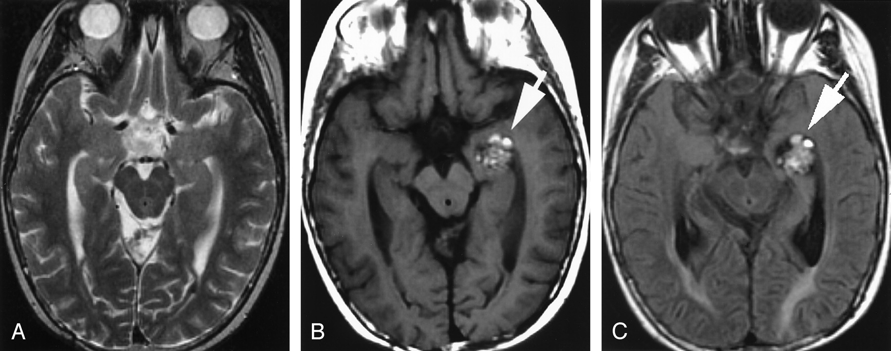
- Fig 1.
Case 1.
A, Axial T2-weighted MR image obtained 12 years after craniospinal radiation therapy at 3 years of age for a posterior fossa medulloblastoma showing no lesion in the left temporal lobe/temporal horn.
B, Axial T1-weighted and, C, fluid-attenuated inversion recovery (FLAIR) MR images obtained 15 years after the radiation therapy, showing a lobulated area of mixed signal intensity with peripheral hypointense rim (white arrow) in left medial temporal lobe/temporal horn, typical “popcorn” MR appearance of a cavernous angioma.
- Fig 2.
Case 2.
A, Axial T2- and, B, T1-weighted MR images obtained 3 years after involved field radiation therapy at 3 years of age for a posterior fossa ependymoma showing a large hemorrhagic, lobulated lesion of mixed signal intensity (white arrow) in left cerebellar hemisphere and vermis. Two more small cavernomas (black arrowheads) are seen in the right cerebellum. Postoperative changes (black arrow) are noted in the right cerebellum.
C, Axial T2‐weighted MR image shows an area of blooming due to another small cavernoma (black arrow) in the right temporo-occipital region.
- Fig 3.
Case 4.
A, Axial T2-weighted MR image obtained 6 years after craniospinal radiation therapy at age 13 years for a suprasellar germinoma showing no lesion in right frontoparietal region.
B, Axial T2-weighted and, C, FLAIR MR images obtained 8 years after the radiation therapy showing interval appearance a round lesion of mixed signal intensity with a fluid-fluid level and peripheral hypointense rim (black arrow) in right frontoparietal region.


Tables
Case Material
Case No. Age*/Sex Primary CNS Neoplasm Or Disease Age at RT (years) RT and Dose Latency Interval (years) Clinical Presentation Location of CM’s Number of CM’s Treatment of CM’s Associated Findings 1 18/F Posterior fossaMedulloblastoma 3 Craniospinal RT54 Gy (T)36 Gy (CS) 15 Incidental Lt temporal lobe/Intraventricular 1 Observation Meningioma Scalp DFS Panhypopitutarism Rt cerebellum 1 Observation 2 6/M Posterior fossaEpendymoma 3 Involved field RT50.4 Gy 3 Incidental (Subsequent asymptomatic Hemorrhage) Lt cerebellumRt cerebellumRt temporal lobe 121 SurgeryObservationObservation TH & GH Deficiency 3 34/M Posterior fossaMedulloblastoma 3 Craniospinal RT54 Gy (T) 36 Gy (CS) 31 Incidental Lt frontal lobe 1 Observation Meningioma 4 21/M Supra-sellarGerminoma 13 Craniospinal RT50.4 Gy (T)36 Gy (CS) 8 Incidental (Subsequent asymptomatic hemorrhage) Rt fronto-parietal lobe 1 Surgery - 5 57/F Cushing’s Disease 16 Cobalt RT Unknown dose 41 Incidental Rt frontal lobe 1 Observation Panhypopitutarism Note.—Age* indicates Age at the time of diagnosis of cavernomas; RT, Radiation therapy; T, Total Dose; CS, Craniospinal Dose; CM’s, cavernous malformations; DFS, Dermatofibrosarcoma; TH, Thyroid hormones; GH, Growth hormone.
In this issue


Jump to section
Related Articles
Cited By...
- Young stroke as a late complication of cranial irradiation
- Unusual delayed postradiation vasculopathy mimicking tumour in an early adolescent with a remote history of metastatic medulloblastoma
- Accumulation of Brain Hypointense Foci on Susceptibility-Weighted Imaging in Childhood Ataxia Telangiectasia
- Hyperintense Dentate Nuclei on T1-Weighted MRI: Relation to Repeat Gadolinium Administration
- A Drosophila model to investigate the neurotoxic side effects of radiation exposure
- Accelerated Aging, Decreased White Matter Integrity, and Associated Neuropsychological Dysfunction 25 Years After Pediatric Lymphoid Malignancies
- Cerebral cavernous malformations proteins inhibit Rho kinase to stabilize vascular integrity
- Focal Neuronal Gigantism: A Rare Complication of Therapeutic Radiation