Advertisement

Main menu

  • Home
  • Content
    • Current Issue
    • Accepted Manuscripts
    • Article Preview
    • Past Issue Archive
    • Video Articles
    • AJNR Case Collection
    • Case of the Week Archive
    • Case of the Month Archive
    • Classic Case Archive
  • Special Collections
    • AJNR Awards
    • Low-Field MRI
    • Alzheimer Disease
    • ASNR Foundation Special Collection
    • Photon-Counting CT
    • View All
  • Multimedia
    • AJNR Podcasts
    • AJNR SCANtastic
    • Trainee Corner
    • MRI Safety Corner
    • Imaging Protocols
  • For Authors
    • Submit a Manuscript
    • Submit a Video Article
    • Submit an eLetter to the Editor/Response
    • Manuscript Submission Guidelines
    • Statistical Tips
    • Fast Publishing of Accepted Manuscripts
    • Graphical Abstract Preparation
    • Imaging Protocol Submission
    • Author Policies
  • About Us
    • About AJNR
    • Editorial Board
    • Editorial Board Alumni
  • More
    • Become a Reviewer/Academy of Reviewers
    • Subscribers
    • Permissions
    • Alerts
    • Feedback
    • Advertisers
    • ASNR Home

User menu

  • Alerts
  • Log in

Search

  • Advanced search
American Journal of Neuroradiology

American Journal of Neuroradiology

ASHNR American Society of Functional Neuroradiology ASHNR American Society of Pediatric Neuroradiology ASSR
  • Alerts
  • Log in

Advanced Search

  • Home
  • Content
    • Current Issue
    • Accepted Manuscripts
    • Article Preview
    • Past Issue Archive
    • Video Articles
    • AJNR Case Collection
    • Case of the Week Archive
    • Case of the Month Archive
    • Classic Case Archive
  • Special Collections
    • AJNR Awards
    • Low-Field MRI
    • Alzheimer Disease
    • ASNR Foundation Special Collection
    • Photon-Counting CT
    • View All
  • Multimedia
    • AJNR Podcasts
    • AJNR SCANtastic
    • Trainee Corner
    • MRI Safety Corner
    • Imaging Protocols
  • For Authors
    • Submit a Manuscript
    • Submit a Video Article
    • Submit an eLetter to the Editor/Response
    • Manuscript Submission Guidelines
    • Statistical Tips
    • Fast Publishing of Accepted Manuscripts
    • Graphical Abstract Preparation
    • Imaging Protocol Submission
    • Author Policies
  • About Us
    • About AJNR
    • Editorial Board
    • Editorial Board Alumni
  • More
    • Become a Reviewer/Academy of Reviewers
    • Subscribers
    • Permissions
    • Alerts
    • Feedback
    • Advertisers
    • ASNR Home
  • Follow AJNR on Twitter
  • Visit AJNR on Facebook
  • Follow AJNR on Instagram
  • Join AJNR on LinkedIn
  • RSS Feeds

AJNR Awards, New Junior Editors, and more. Read the latest AJNR updates

Getting new auth cookie, if you see this message a lot, tell someone!
OtherBrain

Tumefactive Cysts: A Delayed Complication following Radiosurgery for Cerebral Arterial Venous Malformations

Whitney B. Edmister, John I. Lane, Julie R. Gilbertson, Robert D. Brown and Bruce E. Pollock
American Journal of Neuroradiology May 2005, 26 (5) 1152-1157;
Whitney B. Edmister
  • Find this author on Google Scholar
  • Find this author on PubMed
  • Search for this author on this site
John I. Lane
  • Find this author on Google Scholar
  • Find this author on PubMed
  • Search for this author on this site
Julie R. Gilbertson
  • Find this author on Google Scholar
  • Find this author on PubMed
  • Search for this author on this site
Robert D. Brown
  • Find this author on Google Scholar
  • Find this author on PubMed
  • Search for this author on this site
Bruce E. Pollock
  • Find this author on Google Scholar
  • Find this author on PubMed
  • Search for this author on this site
  • Article
  • Figures & Data
  • Info & Metrics
  • Responses
  • References
  • PDF
Loading

Article Figures & Data

Figures

  • Tables
  • Fig 1.
    • Download figure
    • Open in new tab
    • Download powerpoint
    Fig 1.

    Radiation therapy-induced cyst 89 months after AVM treatment.

    A, T2-weighted axial image shows a 3-cm cyst in the frontoparietal region with vasogenic edema (arrow).

    B, Contrast-enhanced T1-weighted axial image shows nodular contrast enhancement superficial to the cyst (arrow).

    C, Postoperative CT showed successful cyst decompression (arrow) after cystoperitoneal shunt placement.
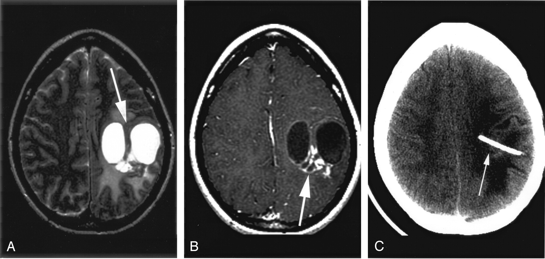
  • Fig 2.
    • Download figure
    • Open in new tab
    • Download powerpoint
    Fig 2.

    MR image obtained 40 months after AVM ablation.

    A, T2-weighted axial image showed a 5-cm complex, multiloculated cyst in the left frontoparietal region with vasogenic edema and local mass effect (arrow).

    B, Contrast-enhanced T1-weighted axial image shows nodular parenchymal enhancement (arrow).

    C, CT after successful cystoperitoneal shunt placement shows decompression of the cysts.
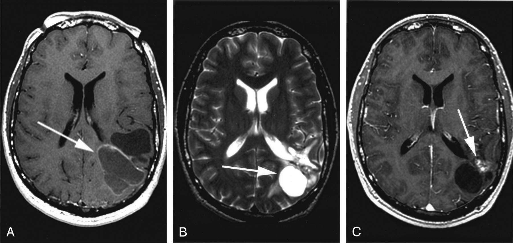
  • Fig 3.
    • Download figure
    • Open in new tab
    • Download powerpoint
    Fig 3.

    MR image obtained 34 months after AVM ablation.

    A, T2-weighted axial image shows multiple complex cysts in the posterior right temporal, occipital, and parietal region with vasogenic edema and mass effect.

    B, Contrast-enhanced T1-weighted axial image shows nodular parenchymal enhancement.

    C and D, MR images obtained after successful cystoperitoneal placement. Vasogenic edema and enhancement persist, but the patient remains asymptomatic after 4 years.

  • Fig 4.
    • Download figure
    • Open in new tab
    • Download powerpoint
    Fig 4.

    MR image obtained 59 months after AVM ablation.

    A, T2-weighted axial image demonstrates multilocular cyst with surrounding vasogenic edema (arrow).

    B, Contrast-enhanced T1-weighted axial image shows nodular parenchymal enhancement (arrow).

    C and D, MR after cystectomy show postoperative encephalomalacia, resolution of cysts and minimal residual parenchymal enhancement (arrow).

  • Fig 5.
    • Download figure
    • Open in new tab
    • Download powerpoint
    Fig 5.

    MR image obtained 36 months after AVM ablation.

    A, Contrast-enhanced axial T1-weighted image shows a peripherally enhancing, multiloculated cyst in the left parietal lobe.

    B and C, T2-weighted and post-contrast T1-weighted images obtained after cystoperitoneal shunting show residual cyst and enhancement (arrows).

Tables

  • Figures
  • Summary of patient demographics and clinical history

    CaseAge at Gamm a TxSexRx isodose vol (cc), margin/max dose (Gy)Tx to cyst Dx (months)Size at Dx (cc)SxTxAVM location
    133F28.7, 16/208914.0HA, L arm weakness.Aspiration; CP shunt.R parietal
    223F13.4, 16/32661.0 (40 at 106 months)None. HA, mild aphasia, transient R arm paresthesia at 106 months.Aspiration; CP shunt.L parietal
    320F13.5, 28/22.53411.9HA.Aspiration; CP shunt.R temporal
    456F8.0, 16/32 4.9, 18/36594.0Repeat Tx at 44 months. HA, ataxia.Aspiration; CP shunt; cystectomy.R temporal
    524M11.1, 18/36 16.5, 16/32373.5 (63 at 50 months)Repeat Tx at 38 months. None. HA at 50 months.CP shunt; cystectomy.L parietal
PreviousNext
Back to top

In this issue

American Journal of Neuroradiology: 26 (5)
American Journal of Neuroradiology
Vol. 26, Issue 5
1 May 2005
  • Table of Contents
  • Index by author
Advertisement
Print
Download PDF
Email Article

Thank you for your interest in spreading the word on American Journal of Neuroradiology.

NOTE: We only request your email address so that the person you are recommending the page to knows that you wanted them to see it, and that it is not junk mail. We do not capture any email address.

Enter multiple addresses on separate lines or separate them with commas.
Tumefactive Cysts: A Delayed Complication following Radiosurgery for Cerebral Arterial Venous Malformations
(Your Name) has sent you a message from American Journal of Neuroradiology
(Your Name) thought you would like to see the American Journal of Neuroradiology web site.
CAPTCHA
This question is for testing whether or not you are a human visitor and to prevent automated spam submissions.
Cite this article
Whitney B. Edmister, John I. Lane, Julie R. Gilbertson, Robert D. Brown, Bruce E. Pollock
Tumefactive Cysts: A Delayed Complication following Radiosurgery for Cerebral Arterial Venous Malformations
American Journal of Neuroradiology May 2005, 26 (5) 1152-1157;

Citation Manager Formats

  • BibTeX
  • Bookends
  • EasyBib
  • EndNote (tagged)
  • EndNote 8 (xml)
  • Medlars
  • Mendeley
  • Papers
  • RefWorks Tagged
  • Ref Manager
  • RIS
  • Zotero
0 Responses
Respond to this article
Share
Bookmark this article
Tumefactive Cysts: A Delayed Complication following Radiosurgery for Cerebral Arterial Venous Malformations
Whitney B. Edmister, John I. Lane, Julie R. Gilbertson, Robert D. Brown, Bruce E. Pollock
American Journal of Neuroradiology May 2005, 26 (5) 1152-1157;
del.icio.us logo Twitter logo Facebook logo Mendeley logo
  • Tweet Widget
  • Facebook Like
  • Google Plus One

Jump to section

  • Article
    • Abstract
    • Case Reports
    • Case 1
    • Case 2
    • Case 3
    • Case 4
    • Case 5
    • Discussion
    • Conclusion
    • Footnotes
    • References
  • Figures & Data
  • Info & Metrics
  • Responses
  • References
  • PDF

Related Articles

  • No related articles found.
  • PubMed
  • Google Scholar

Cited By...

  • Long-term efficacy of gamma knife radiosurgery in mesial temporal lobe epilepsy
  • Crossref
  • Google Scholar

This article has not yet been cited by articles in journals that are participating in Crossref Cited-by Linking.

More in this TOC Section

  • Predictors of Reperfusion in Patients with Acute Ischemic Stroke
  • Enhanced Axonal Metabolism during Early Natalizumab Treatment in Relapsing-Remitting Multiple Sclerosis
  • Progression of Microstructural Damage in Spinocerebellar Ataxia Type 2: A Longitudinal DTI Study
Show more Brain

Similar Articles

Advertisement

Indexed Content

  • Current Issue
  • Accepted Manuscripts
  • Article Preview
  • Past Issues
  • Editorials
  • Editor's Choice
  • Fellows' Journal Club
  • Letters to the Editor
  • Video Articles

Cases

  • Case Collection
  • Archive - Case of the Week
  • Archive - Case of the Month
  • Archive - Classic Case

More from AJNR

  • Trainee Corner
  • Imaging Protocols
  • MRI Safety Corner
  • Book Reviews

Multimedia

  • AJNR Podcasts
  • AJNR Scantastics

Resources

  • Turnaround Time
  • Submit a Manuscript
  • Submit a Video Article
  • Submit an eLetter to the Editor/Response
  • Manuscript Submission Guidelines
  • Statistical Tips
  • Fast Publishing of Accepted Manuscripts
  • Graphical Abstract Preparation
  • Imaging Protocol Submission
  • Evidence-Based Medicine Level Guide
  • Publishing Checklists
  • Author Policies
  • Become a Reviewer/Academy of Reviewers
  • News and Updates

About Us

  • About AJNR
  • Editorial Board
  • Editorial Board Alumni
  • Alerts
  • Permissions
  • Not an AJNR Subscriber? Join Now
  • Advertise with Us
  • Librarian Resources
  • Feedback
  • Terms and Conditions
  • AJNR Editorial Board Alumni

American Society of Neuroradiology

  • Not an ASNR Member? Join Now

© 2025 by the American Society of Neuroradiology All rights, including for text and data mining, AI training, and similar technologies, are reserved.
Print ISSN: 0195-6108 Online ISSN: 1936-959X

Powered by HighWire