
Article Figures & Data
Figures
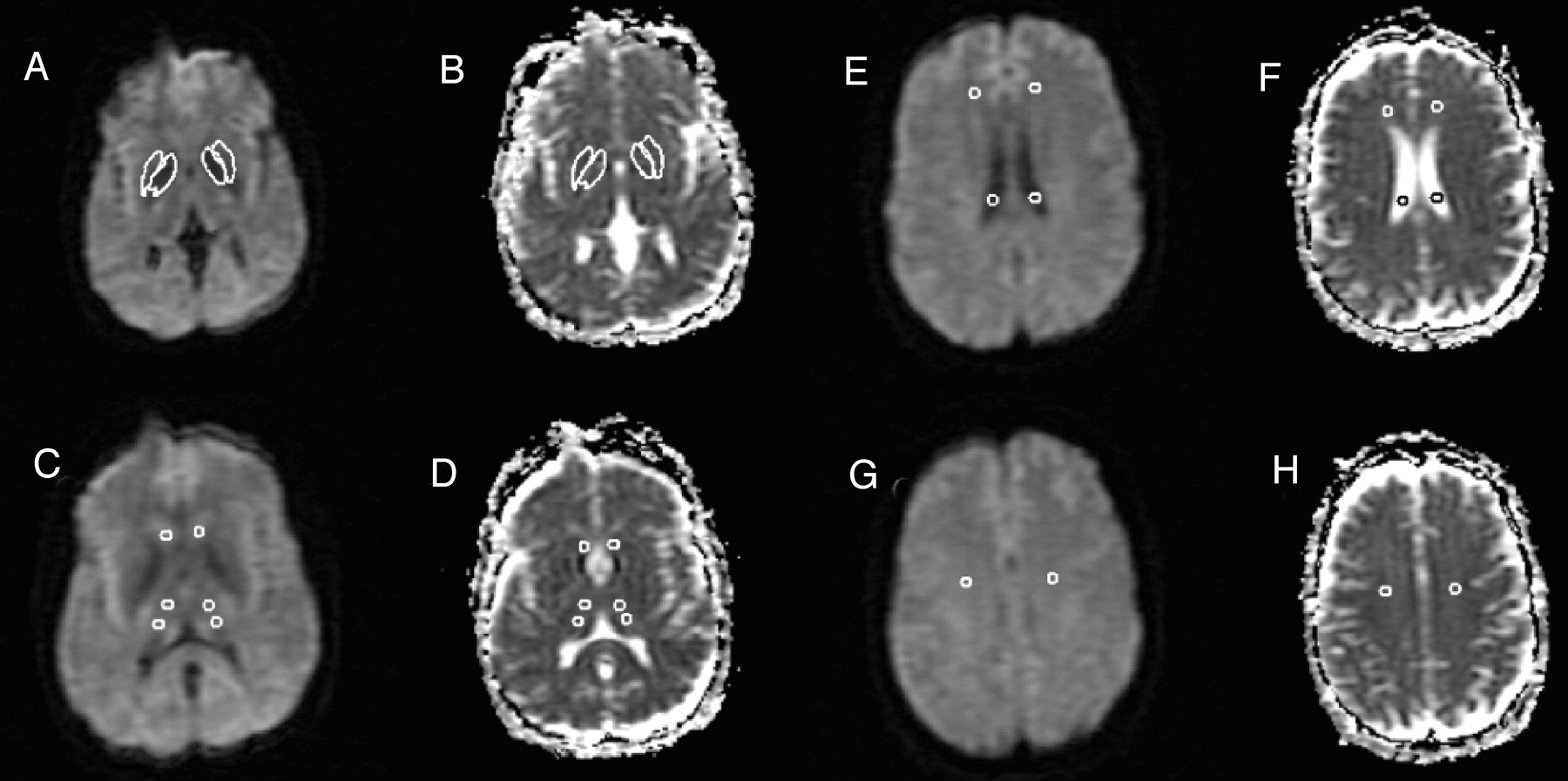
- Fig 1.
DW images for each section were collected with diffusion weighting in three orthogonal directions and a b factor of 1000 s/mm2. The DW images, together with a T2-weighted image (b = 0), were used to calculate three ADC images (one for each weighting direction), which were then averaged to form the mean diffusivity image used in analysis (B, D, F, H). In addition, the three DW images were averaged to form an average DW imaging (A, C, E, G). Region of interest analysis was then used to determine quantitatively the mean diffusivity in the selected gray matter structures. Five-millimeter-diameter circular regions of interest were drawn on the average DW images (C, E, G), because of the higher anatomic detail visualized on these images, for the anterior and posterior thalamus (C), caudate (C), lateral ventricle (E), anterior frontal white matter (E), and centrum semiovale (G). Freehand structure tracing was performed by a trained observer on the average DW image to create the region of interest for the putamen (A). The globus pallidus region of interest was created on the average DW image by a semiautomated edge-finding technique (A). The regions of interest were then copied to the mean diffusivity maps (B, D, F, H). The data from the anterior and posterior thalamus regions of interest were averaged to estimate the mean diffusivity for each thalamus (D).
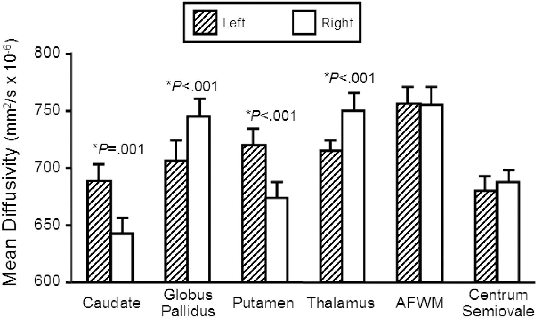
- Fig 2.
Comparison of regional mean diffusivity values (mm2/s × 10−6) between the left and right hemisphere in 23 normal individuals. Those structures in which there was significant difference (P < .005) between hemispheres are marked with an asterisk and the P value is given. Bar heights indicate the mean, and error bars are standard error of the mean. AF WM, anterior frontal white matter.


Tables
Mean Apparent Diffusion Coefficient (ADC) Values of Gray Matter Structures in Normal Individuals
Region Left hemisphere mean (SD) ADC mm2/s × 10−3 Right hemisphere mean (SD) ADC mm2/s × 10−3 Greater side, Percent Difference Left vs. Right P Value Caudate 0.689 (.069) 0.642 (.071) left, 7.0% P = .001 Globus pallidus 0.706 (.050) 0.745 (.053) right, 5.2% P < .001 Putamen 0.720 (.059) 0.674 (.052) left, 6.4% P < .001 Thalamus 0.716 (.031) 0.750 (.040) right, 4.5% P < .001 CSF 3.357 (.507) 3.312 (.524) left, 2.1% P = .291 Centrum semiovale 0.680 (.054) 0.688 (.049) right, 1.1% P = .320 Anterior white matter 0.757 (.042) 0.755 (.052) left, 0.2% P = .834


