Advertisement

Main menu

  • Home
  • Content
    • Current Issue
    • Accepted Manuscripts
    • Article Preview
    • Past Issue Archive
    • Video Articles
    • AJNR Case Collection
    • Case of the Week Archive
    • Case of the Month Archive
    • Classic Case Archive
  • Special Collections
    • AJNR Awards
    • Low-Field MRI
    • Alzheimer Disease
    • ASNR Foundation Special Collection
    • Photon-Counting CT
    • View All
  • Multimedia
    • AJNR Podcasts
    • AJNR SCANtastic
    • Trainee Corner
    • MRI Safety Corner
    • Imaging Protocols
  • For Authors
    • Submit a Manuscript
    • Submit a Video Article
    • Submit an eLetter to the Editor/Response
    • Manuscript Submission Guidelines
    • Statistical Tips
    • Fast Publishing of Accepted Manuscripts
    • Graphical Abstract Preparation
    • Imaging Protocol Submission
    • Author Policies
  • About Us
    • About AJNR
    • Editorial Board
    • Editorial Board Alumni
  • More
    • Become a Reviewer/Academy of Reviewers
    • Subscribers
    • Permissions
    • Alerts
    • Feedback
    • Advertisers
    • ASNR Home

User menu

  • Alerts
  • Log in

Search

  • Advanced search
American Journal of Neuroradiology

American Journal of Neuroradiology

ASHNR American Society of Functional Neuroradiology ASHNR American Society of Pediatric Neuroradiology ASSR
  • Alerts
  • Log in

Advanced Search

  • Home
  • Content
    • Current Issue
    • Accepted Manuscripts
    • Article Preview
    • Past Issue Archive
    • Video Articles
    • AJNR Case Collection
    • Case of the Week Archive
    • Case of the Month Archive
    • Classic Case Archive
  • Special Collections
    • AJNR Awards
    • Low-Field MRI
    • Alzheimer Disease
    • ASNR Foundation Special Collection
    • Photon-Counting CT
    • View All
  • Multimedia
    • AJNR Podcasts
    • AJNR SCANtastic
    • Trainee Corner
    • MRI Safety Corner
    • Imaging Protocols
  • For Authors
    • Submit a Manuscript
    • Submit a Video Article
    • Submit an eLetter to the Editor/Response
    • Manuscript Submission Guidelines
    • Statistical Tips
    • Fast Publishing of Accepted Manuscripts
    • Graphical Abstract Preparation
    • Imaging Protocol Submission
    • Author Policies
  • About Us
    • About AJNR
    • Editorial Board
    • Editorial Board Alumni
  • More
    • Become a Reviewer/Academy of Reviewers
    • Subscribers
    • Permissions
    • Alerts
    • Feedback
    • Advertisers
    • ASNR Home
  • Follow AJNR on Twitter
  • Visit AJNR on Facebook
  • Follow AJNR on Instagram
  • Join AJNR on LinkedIn
  • RSS Feeds

AJNR Awards, New Junior Editors, and more. Read the latest AJNR updates

Research ArticleBrain

Metabolite Phantom Correction of the Nonuniform Volume-Selection Profiles in MR Spectroscopic Imaging: Application to Temporal Lobe Epilepsy

J. O. Jesper Lundbom, Kim E. Vuori, Eija K. Gaily, R. I. Marja-Liisa Granström, Göran C. Blomstedt, Anna-Maija Häkkinen, Sami M. Heikkinen and Nina M. I. Lundbom
American Journal of Neuroradiology May 2005, 26 (5) 1072-1077;
J. O. Jesper Lundbom
  • Find this author on Google Scholar
  • Find this author on PubMed
  • Search for this author on this site
Kim E. Vuori
  • Find this author on Google Scholar
  • Find this author on PubMed
  • Search for this author on this site
Eija K. Gaily
  • Find this author on Google Scholar
  • Find this author on PubMed
  • Search for this author on this site
R. I. Marja-Liisa Granström
  • Find this author on Google Scholar
  • Find this author on PubMed
  • Search for this author on this site
Göran C. Blomstedt
  • Find this author on Google Scholar
  • Find this author on PubMed
  • Search for this author on this site
Anna-Maija Häkkinen
  • Find this author on Google Scholar
  • Find this author on PubMed
  • Search for this author on this site
Sami M. Heikkinen
  • Find this author on Google Scholar
  • Find this author on PubMed
  • Search for this author on this site
Nina M. I. Lundbom
  • Find this author on Google Scholar
  • Find this author on PubMed
  • Search for this author on this site
  • Article
  • Figures & Data
  • Info & Metrics
  • Responses
  • References
  • PDF
Loading

Article Figures & Data

Figures

  • Tables
  • Fig 1.
    • Download figure
    • Open in new tab
    • Download powerpoint
    Fig 1.

    Axial/coronal T1-weighted image at the level of the hippocampi with the MRSI voxels 1, 2, and 3 chosen to represent the hippocampus. The 100- and 80-mm-wide VOIs are shown (white rectangles).
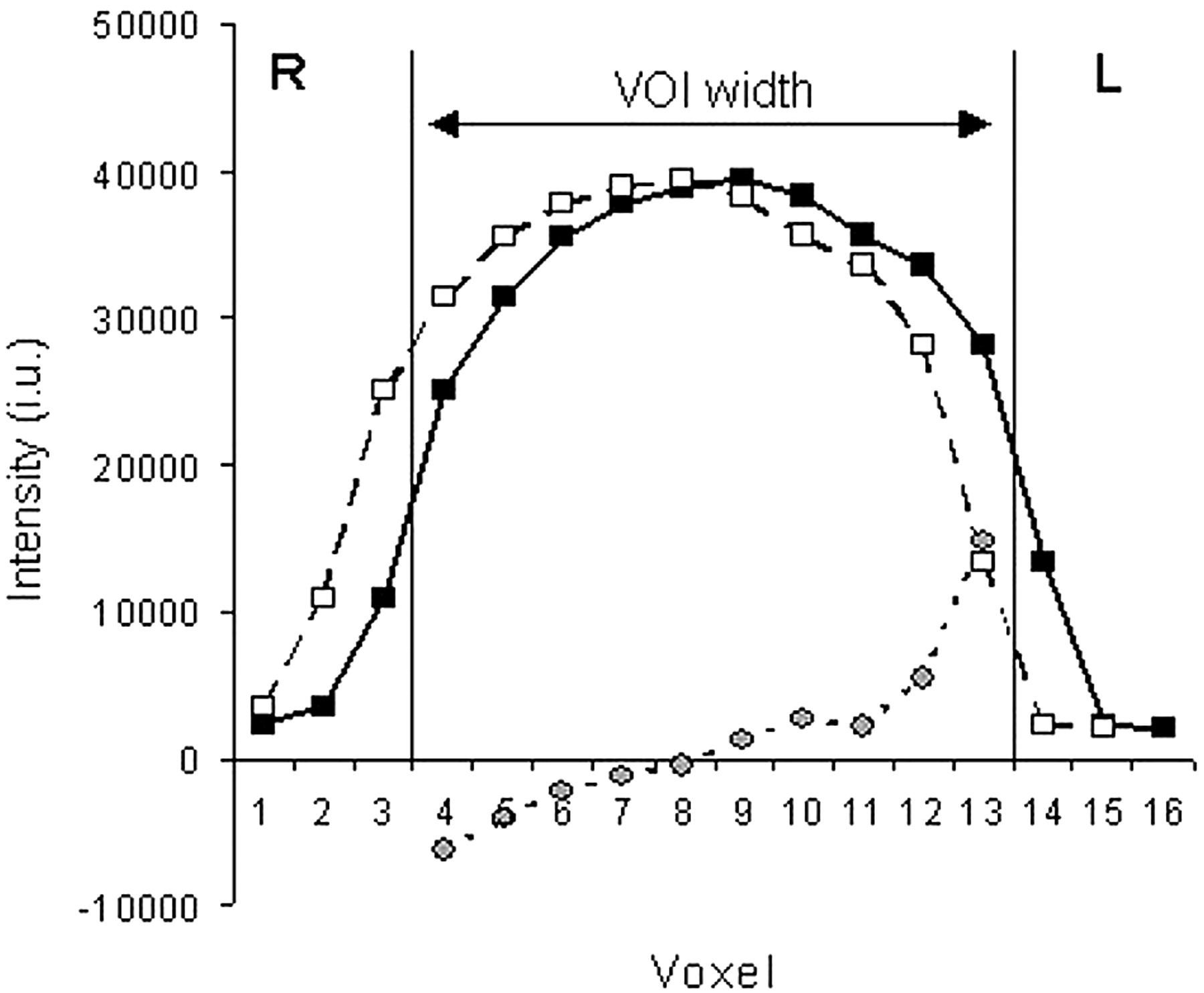
  • Fig 2.
    • Download figure
    • Open in new tab
    • Download powerpoint
    Fig 2.

    Phantom profile for water signal intensity with the 180° Hamming-weighted sinc pulse of the PRESS CSI sequence (solid line with black boxes) obtained by selecting the right-left dimension of the 100-mm VOI (vertical lines). Profile shifted by 10 mm (dashed line with white boxes). This displacement approximately corresponds to the chemical-shift difference between water and NAA with the typical excitation bandwidth of 1500 Hz for the section-selective 180° pulse. Images shows the difference between the original profile and the shifted profile spanning the VOI (dotted line with gray circles). The water intensity on the y axis is expressed in institutional units (i.u.), and the x axis corresponds to the 10-mm voxels in the 160-mm FOV.

Tables

  • Figures
    • View popup
    TABLE 1:

    Clinical, MR imaging, and MRSI findings in patients with TLE

    Patient/Age (y)/SexEpilepsy Clinical Lateralization and TypeMR ImagingMRSI LateralizationComment*
    No CorrectionPulse Correction
    1/9/ML TLEL T dysmyelinationNoneRSeizure-free without operation
    2/10/FL TLEL hippocampal dysgenesis (possible)NoneNoneSeizure-free without operation
    3/16/FL TLE, possible R FLENormalLLNo resection, VNS
    4/16/FL TLEL MTSLLL TL resection, seizure-free
    5/13/FR/L TLENormalLLNo operation, monthly seizures
    6/17/FR/L TLER/L hamartomas (tuberous sclerosis)NoneRDaily seizures from R TL, rare from L TL, VNS
    7/9/MR TLER hippocampal edemaNoneRNo operation, rare seizures
    8/12/FR TLER MTSLLR TL resection, seizure-free
    9/15/FR TLER hippocampus, smallLLR TL resection, seizure-free
    10/15/MR TLESturge-Weber syndrome R TORRSeizure-free without operation
    11/34/FR TLENormalNoneRR TL resection, seizure-free for 6 mo, postoperative seizures from L TL
    12/9/FMultifocalR hippocampus, smallNoneNoneSeizures not from TL
    13/14/ML TOEDysgenesis L TONoneNoneNone
    14/15/MUndeterminedL hippocampus, abnormalNoneNoneNot operation, rare seizures
    15/50/FR TLE or R FLENormalNoneNoneNot operation, monthly seizures
    • Note.—FLE = frontal lobe epilepsy, TOE = temporo-occipital epilepsy, MTS = mesial temporal sclerosis, TL = temporal lobe, VNS = vagal nerve stimulation.

    • * Rare seizures were fewer than one per 6 months.

    • View popup
    TABLE 2:

    Intensity asymmetry percentage in the phantom voxels corresponding to the hippocampi

    VOI Width (mm)ChoCrNAANAA/(Cho + Cr)
    80 mm7.9 ± 3.29.3 ± 3.418.1 ± 1.49.4 ± 1.5
    100 mm4.0 ± 4.47.5 ± 1.99.3 ± 1.33.8 ± 1.5
    • Note.—Intensity asymmetry percentage = 2 × 100[(R − L)/(R + L)]. Data are the mean percentage ± standard deviation.

    • View popup
    TABLE 3:

    Means and standard deviations for the right-left ratios of hippocampal metabolites in control subjects before and after correction

    Metabolite or IndexMeanStandard Deviation
    BeforeAfterBeforeAfter
    NAA1.150.980.070.05
    Cho + Cr1.101.020.090.08
    NAA/Cho + Cr1.050.970.060.06
PreviousNext
Back to top

In this issue

American Journal of Neuroradiology: 26 (5)
American Journal of Neuroradiology
Vol. 26, Issue 5
1 May 2005
  • Table of Contents
  • Index by author
Advertisement
Print
Download PDF
Email Article

Thank you for your interest in spreading the word on American Journal of Neuroradiology.

NOTE: We only request your email address so that the person you are recommending the page to knows that you wanted them to see it, and that it is not junk mail. We do not capture any email address.

Enter multiple addresses on separate lines or separate them with commas.
Metabolite Phantom Correction of the Nonuniform Volume-Selection Profiles in MR Spectroscopic Imaging: Application to Temporal Lobe Epilepsy
(Your Name) has sent you a message from American Journal of Neuroradiology
(Your Name) thought you would like to see the American Journal of Neuroradiology web site.
CAPTCHA
This question is for testing whether or not you are a human visitor and to prevent automated spam submissions.
Cite this article
J. O. Jesper Lundbom, Kim E. Vuori, Eija K. Gaily, R. I. Marja-Liisa Granström, Göran C. Blomstedt, Anna-Maija Häkkinen, Sami M. Heikkinen, Nina M. I. Lundbom
Metabolite Phantom Correction of the Nonuniform Volume-Selection Profiles in MR Spectroscopic Imaging: Application to Temporal Lobe Epilepsy
American Journal of Neuroradiology May 2005, 26 (5) 1072-1077;

Citation Manager Formats

  • BibTeX
  • Bookends
  • EasyBib
  • EndNote (tagged)
  • EndNote 8 (xml)
  • Medlars
  • Mendeley
  • Papers
  • RefWorks Tagged
  • Ref Manager
  • RIS
  • Zotero
0 Responses
Respond to this article
Share
Bookmark this article
Metabolite Phantom Correction of the Nonuniform Volume-Selection Profiles in MR Spectroscopic Imaging: Application to Temporal Lobe Epilepsy
J. O. Jesper Lundbom, Kim E. Vuori, Eija K. Gaily, R. I. Marja-Liisa Granström, Göran C. Blomstedt, Anna-Maija Häkkinen, Sami M. Heikkinen, Nina M. I. Lundbom
American Journal of Neuroradiology May 2005, 26 (5) 1072-1077;
del.icio.us logo Twitter logo Facebook logo Mendeley logo
  • Tweet Widget
  • Facebook Like
  • Google Plus One

Jump to section

  • Article
    • Abstract
    • Methods
    • Results
    • Discussion
    • Conclusion
    • Footnotes
    • References
  • Figures & Data
  • Info & Metrics
  • Responses
  • References
  • PDF

Related Articles

  • No related articles found.
  • PubMed
  • Google Scholar

Cited By...

  • No citing articles found.
  • Crossref
  • Google Scholar

This article has not yet been cited by articles in journals that are participating in Crossref Cited-by Linking.

More in this TOC Section

  • Enhanced Axonal Metabolism during Early Natalizumab Treatment in Relapsing-Remitting Multiple Sclerosis
  • Progression of Microstructural Damage in Spinocerebellar Ataxia Type 2: A Longitudinal DTI Study
  • SWI or T2*: Which MRI Sequence to Use in the Detection of Cerebral Microbleeds? The Karolinska Imaging Dementia Study
Show more Brain

Similar Articles

Advertisement

Indexed Content

  • Current Issue
  • Accepted Manuscripts
  • Article Preview
  • Past Issues
  • Editorials
  • Editor's Choice
  • Fellows' Journal Club
  • Letters to the Editor
  • Video Articles

Cases

  • Case Collection
  • Archive - Case of the Week
  • Archive - Case of the Month
  • Archive - Classic Case

More from AJNR

  • Trainee Corner
  • Imaging Protocols
  • MRI Safety Corner
  • Book Reviews

Multimedia

  • AJNR Podcasts
  • AJNR Scantastics

Resources

  • Turnaround Time
  • Submit a Manuscript
  • Submit a Video Article
  • Submit an eLetter to the Editor/Response
  • Manuscript Submission Guidelines
  • Statistical Tips
  • Fast Publishing of Accepted Manuscripts
  • Graphical Abstract Preparation
  • Imaging Protocol Submission
  • Evidence-Based Medicine Level Guide
  • Publishing Checklists
  • Author Policies
  • Become a Reviewer/Academy of Reviewers
  • News and Updates

About Us

  • About AJNR
  • Editorial Board
  • Editorial Board Alumni
  • Alerts
  • Permissions
  • Not an AJNR Subscriber? Join Now
  • Advertise with Us
  • Librarian Resources
  • Feedback
  • Terms and Conditions
  • AJNR Editorial Board Alumni

American Society of Neuroradiology

  • Not an ASNR Member? Join Now

© 2025 by the American Society of Neuroradiology All rights, including for text and data mining, AI training, and similar technologies, are reserved.
Print ISSN: 0195-6108 Online ISSN: 1936-959X

Powered by HighWire