
Article Figures & Data
Figures
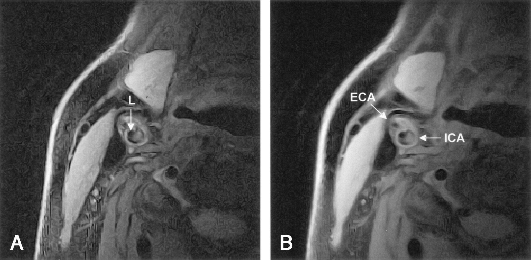
- Fig 1.
Axial lack-blood fast spin-echo images of the internal carotid artery (ICA) and external carotid artery (ECA). Comparison of images reveals no changes in relative signal intensity of the plaque. Hyperintense region is fibrous tissue, and hypointense spots are calcifications.
A, Proton density-weighted image. L = lumen.
B, Corresponding T2-weighted image. Plaque in the ICA is mainly hyperintense, with hypointense spots.
- Fig 2.
Axial lack-blood fast spin-echo images of the ICA (arrow). Plaque in the ICA is hyperintense. L = lumen.
A and B, Proton density-weighted (A) and corresponding T2-weighted (B) images. Large region of the plaque has intermediate signal intensity, with hyperintense edges.
C and D, Another patient. Proton density-weighted (C) and corresponding T2-weighted (D) images. Plaque in the ICA has a large region of intermediate signal intensity, which is relatively decreased in D, while the rest of the plaque remains hyperintense.
- Fig 3.
t-FLAIR images of the brain.
A, Cortical infarct in the territory of the left middle cerebral artery.
B, Centrum semiovale infarct (arrow) in the left hemisphere.
C, White matter lesion (arrow) in the right hemisphere.


Tables
Parameters Black-Blood Fast Spin-Echo Proton Density Weighted T2 Weighted t-FLAIR* TR (msec) 2 heartbeats 2 heartbeats 8000 TE (msec) 14 50 100 Band width (kHz) 20 20 Field of view (mm) 60 60 240 Section thickness (mm) 3 3 5 MATRIX 256 × 256 256 × 256 256 × 192 NEX 2 2 1 Echo train length 12 12 TI (msec) NA NA 2000 Gating Cardiac Cardiac NA Sections 8 8 24 Resolution (μm) 234 × 234 234 × 234 937 × 1250 Imaging time (min) 8 8 2.5 Note.—NA = not applicable.
* Turbo fluid-attenuated inversion recovery.
Lesion Lipid Core (n = 25) No Lipid Core (n = 16) P Value* Basal ganglia infarct No. of patients 2 (8) 0 (0) .51 Median no. of lesions 1.5 (1–2) NA Centrum semiovale infarct No. of patients 14 (56) 4 (25) .06 Median no. of lesions 1 (0–6) 0 (0–4) .04 Cortical infarct No. of patients 6 (24) 3 (19) .99 Any infarct No. of patients 17 (68) 5 (31) .03 White matter lesions No. of patients 21 (84) 13 (81) .99 Median no. of lesions 3 (0–9) 2 (0–12) .16 Note.—Data in parentheses are the percentage or the range. NA = not applicable.
* Group with a lipid core versus and group without a lipid core.
In this issue


Jump to section
Related Articles
Cited By...
- Plaque Components in Symptomatic Moderately Stenosed Carotid Arteries Related to Cerebral Infarcts: The Plaque At RISK Study
- Contrast-Enhanced 3T High-Resolution MR Imaging in Symptomatic Atherosclerotic Basilar Artery Stenosis
- Carotid Magnetization-Prepared Rapid Acquisition With Gradient-Echo Signal Is Associated With Acute Territorial Cerebral Ischemic Events Detected by Diffusion-Weighted MRI
- Carotid Artery Wall Thickness and Leukoaraiosis: Preliminary Results Using Multidetector Row CT Angiography
- 2010 ACCF/AHA Guideline for Assessment of Cardiovascular Risk in Asymptomatic Adults: A Report of the American College of Cardiology Foundation/American Heart Association Task Force on Practice Guidelines
- 2010 ACCF/AHA Guideline for Assessment of Cardiovascular Risk in Asymptomatic Adults: A Report of the American College of Cardiology Foundation/American Heart Association Task Force on Practice Guidelines Developed in Collaboration With the American Society of Echocardiography, American Society of Nuclear Cardiology, Society of Atherosclerosis Imaging and Prevention, Society for Cardiovascular Angiography and Interventions, Society of Cardiovascular Computed Tomography, and Society for Cardiovascular Magnetic Resonance
- Window Settings for the Study of Calcified Carotid Plaques with Multidetector CT Angiography