Advertisement

Main menu

  • Home
  • Content
    • Current Issue
    • Accepted Manuscripts
    • Article Preview
    • Past Issue Archive
    • Video Articles
    • AJNR Case Collection
    • Case of the Week Archive
    • Case of the Month Archive
    • Classic Case Archive
  • Special Collections
    • AJNR Awards
    • Low-Field MRI
    • Alzheimer Disease
    • ASNR Foundation Special Collection
    • Photon-Counting CT
    • View All
  • Multimedia
    • AJNR Podcasts
    • AJNR SCANtastic
    • Trainee Corner
    • MRI Safety Corner
    • Imaging Protocols
  • For Authors
    • Submit a Manuscript
    • Submit a Video Article
    • Submit an eLetter to the Editor/Response
    • Manuscript Submission Guidelines
    • Statistical Tips
    • Fast Publishing of Accepted Manuscripts
    • Graphical Abstract Preparation
    • Imaging Protocol Submission
    • Author Policies
  • About Us
    • About AJNR
    • Editorial Board
    • Editorial Board Alumni
  • More
    • Become a Reviewer/Academy of Reviewers
    • Subscribers
    • Permissions
    • Alerts
    • Feedback
    • Advertisers
    • ASNR Home

User menu

  • Alerts
  • Log in

Search

  • Advanced search
American Journal of Neuroradiology

American Journal of Neuroradiology

ASHNR American Society of Functional Neuroradiology ASHNR American Society of Pediatric Neuroradiology ASSR
  • Alerts
  • Log in

Advanced Search

  • Home
  • Content
    • Current Issue
    • Accepted Manuscripts
    • Article Preview
    • Past Issue Archive
    • Video Articles
    • AJNR Case Collection
    • Case of the Week Archive
    • Case of the Month Archive
    • Classic Case Archive
  • Special Collections
    • AJNR Awards
    • Low-Field MRI
    • Alzheimer Disease
    • ASNR Foundation Special Collection
    • Photon-Counting CT
    • View All
  • Multimedia
    • AJNR Podcasts
    • AJNR SCANtastic
    • Trainee Corner
    • MRI Safety Corner
    • Imaging Protocols
  • For Authors
    • Submit a Manuscript
    • Submit a Video Article
    • Submit an eLetter to the Editor/Response
    • Manuscript Submission Guidelines
    • Statistical Tips
    • Fast Publishing of Accepted Manuscripts
    • Graphical Abstract Preparation
    • Imaging Protocol Submission
    • Author Policies
  • About Us
    • About AJNR
    • Editorial Board
    • Editorial Board Alumni
  • More
    • Become a Reviewer/Academy of Reviewers
    • Subscribers
    • Permissions
    • Alerts
    • Feedback
    • Advertisers
    • ASNR Home
  • Follow AJNR on Twitter
  • Visit AJNR on Facebook
  • Follow AJNR on Instagram
  • Join AJNR on LinkedIn
  • RSS Feeds

AJNR Awards, New Junior Editors, and more. Read the latest AJNR updates

Research ArticleBRAIN

Dynamic Spin Labeling Angiography in Extracranial Carotid Artery Stenosis

Carsten Warmuth, Maria Rüping, Annette Förschler, Hans-Christian Koennecke, Jose Manuel Valdueza, Andreas Kauert, Stephan J. Schreiber, Ralf Siekmann and Claus Zimmer
American Journal of Neuroradiology May 2005, 26 (5) 1035-1043;
Carsten Warmuth
  • Find this author on Google Scholar
  • Find this author on PubMed
  • Search for this author on this site
Maria Rüping
  • Find this author on Google Scholar
  • Find this author on PubMed
  • Search for this author on this site
Annette Förschler
  • Find this author on Google Scholar
  • Find this author on PubMed
  • Search for this author on this site
Hans-Christian Koennecke
  • Find this author on Google Scholar
  • Find this author on PubMed
  • Search for this author on this site
Jose Manuel Valdueza
  • Find this author on Google Scholar
  • Find this author on PubMed
  • Search for this author on this site
Andreas Kauert
  • Find this author on Google Scholar
  • Find this author on PubMed
  • Search for this author on this site
Stephan J. Schreiber
  • Find this author on Google Scholar
  • Find this author on PubMed
  • Search for this author on this site
Ralf Siekmann
  • Find this author on Google Scholar
  • Find this author on PubMed
  • Search for this author on this site
Claus Zimmer
  • Find this author on Google Scholar
  • Find this author on PubMed
  • Search for this author on this site
  • Article
  • Figures & Data
  • Info & Metrics
  • Responses
  • References
  • PDF
Loading

Article Figures & Data

Figures

  • Tables
  • Fig 1.
    • Download figure
    • Open in new tab
    • Download powerpoint
    Fig 1.

    DSLA sequence scheme. After the section selective/nonselective labeling pulse, multiphase data acquisition started in a cine-like fashion. Any segmentable gradient-echo readout may be used. Figure depicts FLASH readout with threefold segmentation and flow compensation in the section and read directions. ADC indicates analog-to-digital converter; RF, radio frequency; S, section direction gradient.
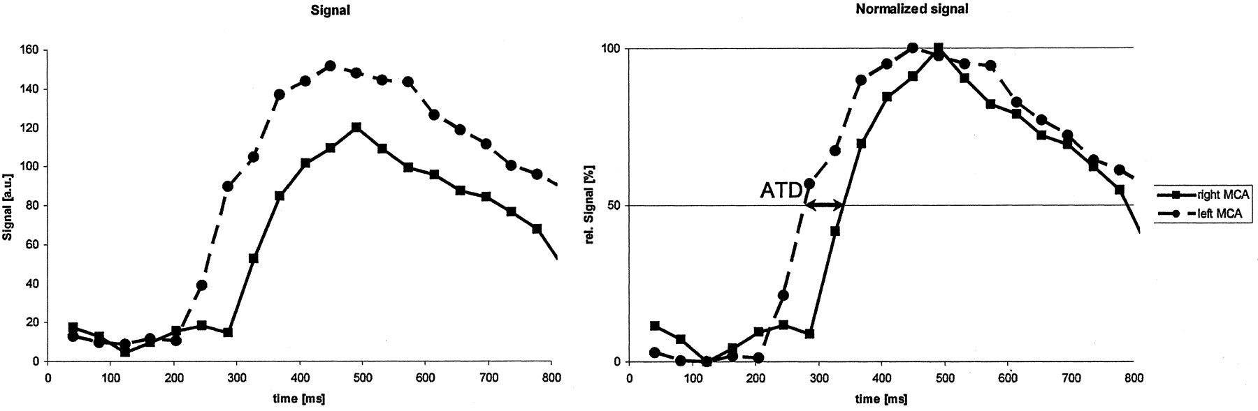
  • Fig 2.
    • Download figure
    • Open in new tab
    • Download powerpoint
    Fig 2.

    Determination of ATDs. Left, Original signal intensity-time courses measured in the left and right MCAs. Right, Normalized curves (divided by the respective maximum value). By using linear interpolation between the measured points, the intersection at which the signals reached 50% was determined. Difference was referred to as the ATD (arrows).

  • Fig 3.
    • Download figure
    • Open in new tab
    • Download powerpoint
    Fig 3.

    Patient 30, with an 85% stenosis of the left ICA. Dynamic angiograms of the circle of Willis in foot-to-head projections at 60, 100, 140, 220, 300, and 580 ms after labeling in a-f. In a, the right ICA (upper arrow) and basilar artery (lower arrow) fill first. In b, Collateral flow into the left MCA via the left posterior communicating artery and the anterior communicating artery is shown. In d, Left ICA fills. In f, because of the finite length of the labeled bolus, all vessels but the left ICA and MCA contain unlabeled blood at this late phase.

  • Fig 4.
    • Download figure
    • Open in new tab
    • Download powerpoint
    Fig 4.

    ATDs calculated for the CS (left) and MCA (right) increase significantly with the degree of stenosis (P < .01). Values at 60% and 75% stenosis are those of individual patients.
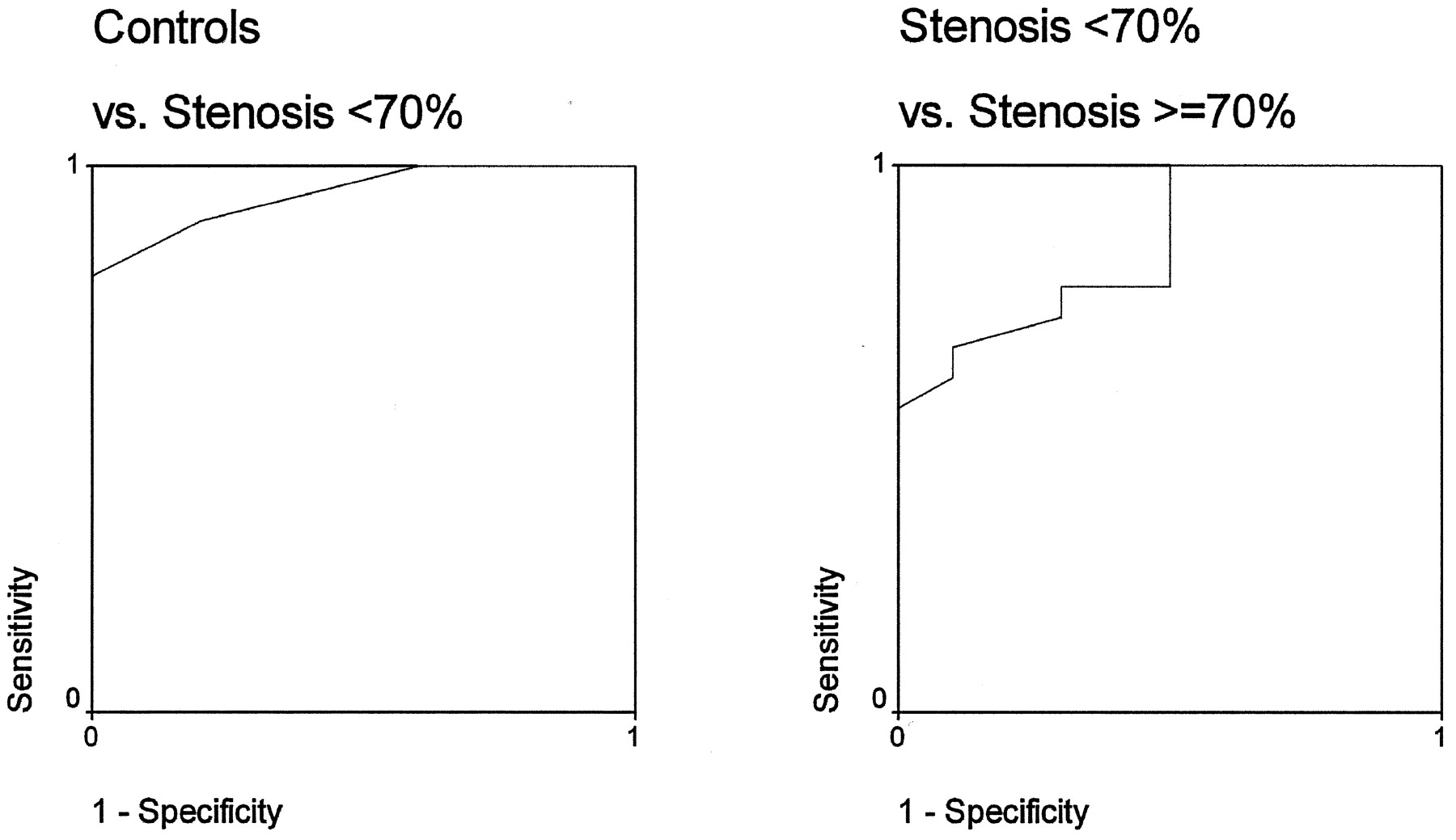
  • Fig 5.
    • Download figure
    • Open in new tab
    • Download powerpoint
    Fig 5.

    ROC curves for ATDs in the CS. Left, Distinction of control subjects and patients with 30–60% stenosis. Right, Distinction of control subjects and patients with <70% stenoses from those with 70–95% stenosis. ATD of >150 ms proved a high-grade stenosis (specificity, 100%; sensitivity, 56%).

  • Fig 6.
    • Download figure
    • Open in new tab
    • Download powerpoint
    Fig 6.

    Scatterplot of the ATDs calculated for the MCA versus those for the CS. Patients were subdivided by the degree of collateral flow visible on DSLA. Degree of collateralization was determined by subjectively assessing flow in the anterior and posterior communicating arteries. Patients with pronounced collateral flow are in the group in whom MCA ATD was less than CS ATD.

  • Fig 7.
    • Download figure
    • Open in new tab
    • Download powerpoint
    Fig 7.

    In a, DSLA difference images of the circle of Willis in patient 40, with a 95% stenosis before and after endarterectomy. In b, Signal intensity-time courses in the CS. After intervention, arrival time on the affected side was the same as on that the unaffected side (about 125 ms). Amplitudes were not calibrated; hence, higher signal intensity was not associated with higher blood flow or blood volume in a vessel.

Tables

  • Figures
    • View popup
    TABLE 1:

    Quantitative results in the control group

    SubjectATD (msec)
    CSMCA
    13010
    21020
    32020
    41020
    530−20
    6040
    72030
    82030
    92030
    1000
    All*16 ± 1119 ± 17
    • * Data are the mean ± standard deviation.

    • View popup
    TABLE 2:

    Quantitative results in patients

    PatientDegree of Stenosis (%)ATD (msec)
    CSCS, After SurgeryMCAMCA, After Surgery
    11304010
    123020−100
    1330100110
    14304010
    153030−30
    16504040
    175080103020
    185014070
    195010080200
    20607050
    11–20*66 ± 39†21 ± 57‡
    217010090
    22706060
    237016050
    2475500
    2580300160
    2680120018020
    27803507016030
    288050202040
    2985140−4050−60
    30856006032010
    318550110
    3285380§32010
    3390200100
    349090180
    3590170280
    3690510800
    3790350180
    21–37*216 ± 170†180 ± 187‡
    3895§70
    3995§01304
    4095220408020
    38–40*22093 ± 32
    • * Data are the mean or the mean ± standard deviation.

    • † P < .05.

    • ‡ P < .05.

    • § No signal.

PreviousNext
Back to top

In this issue

American Journal of Neuroradiology: 26 (5)
American Journal of Neuroradiology
Vol. 26, Issue 5
1 May 2005
  • Table of Contents
  • Index by author
Advertisement
Print
Download PDF
Email Article

Thank you for your interest in spreading the word on American Journal of Neuroradiology.

NOTE: We only request your email address so that the person you are recommending the page to knows that you wanted them to see it, and that it is not junk mail. We do not capture any email address.

Enter multiple addresses on separate lines or separate them with commas.
Dynamic Spin Labeling Angiography in Extracranial Carotid Artery Stenosis
(Your Name) has sent you a message from American Journal of Neuroradiology
(Your Name) thought you would like to see the American Journal of Neuroradiology web site.
CAPTCHA
This question is for testing whether or not you are a human visitor and to prevent automated spam submissions.
Cite this article
Carsten Warmuth, Maria Rüping, Annette Förschler, Hans-Christian Koennecke, Jose Manuel Valdueza, Andreas Kauert, Stephan J. Schreiber, Ralf Siekmann, Claus Zimmer
Dynamic Spin Labeling Angiography in Extracranial Carotid Artery Stenosis
American Journal of Neuroradiology May 2005, 26 (5) 1035-1043;

Citation Manager Formats

  • BibTeX
  • Bookends
  • EasyBib
  • EndNote (tagged)
  • EndNote 8 (xml)
  • Medlars
  • Mendeley
  • Papers
  • RefWorks Tagged
  • Ref Manager
  • RIS
  • Zotero
0 Responses
Respond to this article
Share
Bookmark this article
Dynamic Spin Labeling Angiography in Extracranial Carotid Artery Stenosis
Carsten Warmuth, Maria Rüping, Annette Förschler, Hans-Christian Koennecke, Jose Manuel Valdueza, Andreas Kauert, Stephan J. Schreiber, Ralf Siekmann, Claus Zimmer
American Journal of Neuroradiology May 2005, 26 (5) 1035-1043;
del.icio.us logo Twitter logo Facebook logo Mendeley logo
  • Tweet Widget
  • Facebook Like
  • Google Plus One

Jump to section

  • Article
    • Abstract
    • Methods
    • Results
    • Discussion
    • Conclusion
    • Footnotes
    • References
  • Figures & Data
  • Info & Metrics
  • Responses
  • References
  • PDF

Related Articles

  • No related articles found.
  • PubMed
  • Google Scholar

Cited By...

  • Highly Accelerated Vessel-Selective Arterial Spin Labelling Angiography using Sparsity and Smoothness Constraints
  • MR Imaging of Individual Perfusion Reorganization Using Superselective Pseudocontinuous Arterial Spin-Labeling in Patients with Complex Extracranial Steno-Occlusive Disease
  • Assessment of Intracranial Collateral Flow by Using Dynamic Arterial Spin Labeling MRA and Transcranial Color-Coded Duplex Ultrasound
  • Crossref
  • Google Scholar

This article has not yet been cited by articles in journals that are participating in Crossref Cited-by Linking.

More in this TOC Section

  • Predictors of Reperfusion in Patients with Acute Ischemic Stroke
  • Enhanced Axonal Metabolism during Early Natalizumab Treatment in Relapsing-Remitting Multiple Sclerosis
  • Progression of Microstructural Damage in Spinocerebellar Ataxia Type 2: A Longitudinal DTI Study
Show more Brain

Similar Articles

Advertisement

Indexed Content

  • Current Issue
  • Accepted Manuscripts
  • Article Preview
  • Past Issues
  • Editorials
  • Editor's Choice
  • Fellows' Journal Club
  • Letters to the Editor
  • Video Articles

Cases

  • Case Collection
  • Archive - Case of the Week
  • Archive - Case of the Month
  • Archive - Classic Case

More from AJNR

  • Trainee Corner
  • Imaging Protocols
  • MRI Safety Corner
  • Book Reviews

Multimedia

  • AJNR Podcasts
  • AJNR Scantastics

Resources

  • Turnaround Time
  • Submit a Manuscript
  • Submit a Video Article
  • Submit an eLetter to the Editor/Response
  • Manuscript Submission Guidelines
  • Statistical Tips
  • Fast Publishing of Accepted Manuscripts
  • Graphical Abstract Preparation
  • Imaging Protocol Submission
  • Evidence-Based Medicine Level Guide
  • Publishing Checklists
  • Author Policies
  • Become a Reviewer/Academy of Reviewers
  • News and Updates

About Us

  • About AJNR
  • Editorial Board
  • Editorial Board Alumni
  • Alerts
  • Permissions
  • Not an AJNR Subscriber? Join Now
  • Advertise with Us
  • Librarian Resources
  • Feedback
  • Terms and Conditions
  • AJNR Editorial Board Alumni

American Society of Neuroradiology

  • Not an ASNR Member? Join Now

© 2025 by the American Society of Neuroradiology All rights, including for text and data mining, AI training, and similar technologies, are reserved.
Print ISSN: 0195-6108 Online ISSN: 1936-959X

Powered by HighWire