
Article Figures & Data
Figures
- Fig 1.
Spin-echo T2-weighted MR images obtained at 1.5T show marked hypointensity in the dentate nuclei (A and D), substantia nigra and red nuclei (B), neostriatum and thalamic nuclei (C and D), and superior and inferior colliculi (B and D). Note the relative hyperintensity of the internal medullary lamina of the thalamus (arrow in C) and the hyperintensity of the pyramidal tract in the posterior limb of the internal capsule. The white matter of the parietal and occipital lobes and of the cerebellar hemispheres is diffusely hyperintense. The cerebral cortex is questionably hypointense (D).
A–C, Axial images.
D, Coronal image.
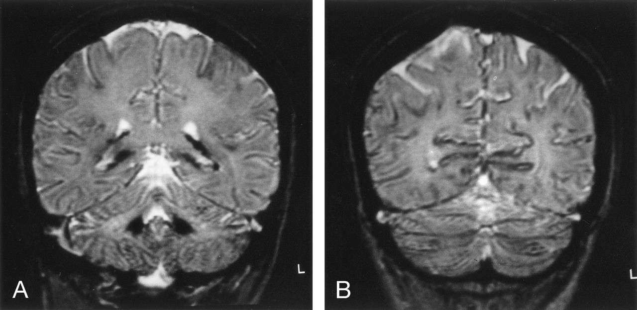
- Fig 2.
Coronal T2*-weighted gradient recalled-echo images at the level of the fourth ventricle (A) and 20 mm dorsally (B) show definite hypointensity in the superficial cortical layers of the cerebral and cerebellar hemispheres.
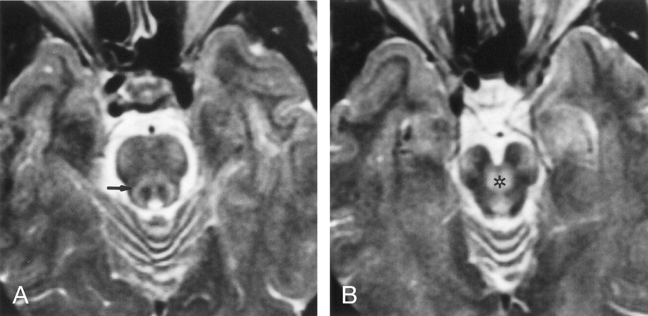
- Fig 3.
Axial SE T2-weighted images show mild hyperintensity of the corticospinal and corticopontine tracts in the cerebral peduncles and basis pontis.
A, Superior cerebellar peduncles are hyperintense (arrow).
B, Their decussation (asterisk) are also hyperintense.


