
Article Figures & Data
Figures
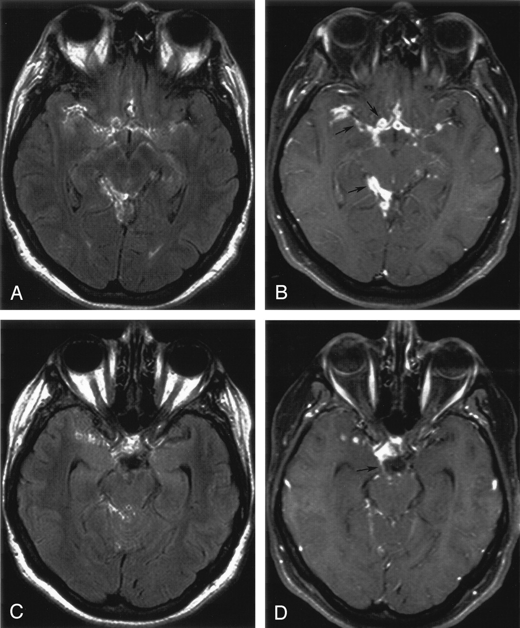
- Fig 1.
Cryptococcal meningitis.
A, Contrast-enhanced FLAIR image shows slight leptomeningeal enhancement in the right frontoparietal region (arrows). It is difficult to separate meningeal enhancement from other high signal intensity within the sulci and from adjacent parenchymal disease.
B, Contrast-enhanced T1-weighted image with FS shows greater contrast between the enhancing tissue and the adjacent brain and is better for depicting enhancement within the sulci and the interhemispheric fissure (arrows).
C, Contrast-enhanced FLAIR image of the same patient obtained at the level of the sylvian fissures shows subtle enhancing lesions in the basal ganglia bilaterally, but without a noncontrast FLAIR image for comparison it is not possible to distinguish enhancement from parenchymal edema.
D, Contrast-enhanced T1-weighted image with FS shows multiple punctuate enhancing areas within the basal ganglia bilaterally, due to cryptococcomas within dilated Virchow-Robin spaces (arrows). The enhancement is much more conspicuous on the T1-weighted image. Additional enhancement is present in several inferior sulci in both frontal lobes.
- Fig 2.
Tuberculous meningitis.
A, Contrast-enhanced FLAIR image shows mildly enhancing subarachnoid space lesions in the basilar cisterns, with extension into the sylvian fissures bilaterally, right ambient cistern, and quadrigeminal cistern.
B, Contrast-enhanced T1-weighted image with FS shows the enhancing lesions in the subarachnoid space more definitively (arrows). The contrast difference between the enhancing meninges and the adjacent brain is visually greater in the T1-weighted image.
C, Contrast-enhanced FLAIR image of the same patient shows enhancement in the region of the right third cranial nerve, but the nerve is not clearly seen.
D, The enhancement of the right third cranial nerve (arrow) is more distinct on the contrast-enhanced T1-weighted image with FS.
- Fig 3.
Transitional cell carcinoma of the urinary bladder with calvarial metastases and meningeal extension.
A, Contrast-enhanced FLAIR image shows localized mild pachymeningeal enhancement (arrows) adjacent to the focal destructive lesion of the right parietooccipital bone. The hyperintensity of adjacent white matter edema is clearer on contrast-enhanced FLAIR image. Note that the internal cerebral veins and cortical veins do not enhance on the FLAIR image. Similarly, the hypervascular pachymeninges (arrows) with increased blood pool does not enhance nearly as much as on the T1-weighted image in panel B.
B, The pachymeningeal enhancement is more apparent and appears thicker on a contrast-enhanced T1-weighted image with FS (arrow). The enhancing cortex is distinct from the underlying hypointense edema.
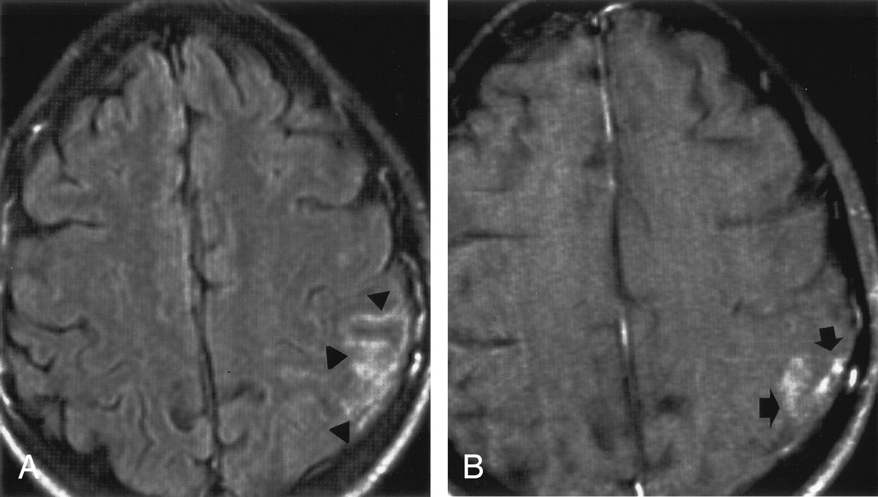
- Fig 4.
Tuberculous meningitis.
A, Contrast-enhanced FLAIR image shows hyperintensity along the meninges and within several sulci of the left parietal lobe (arrowheads). A precontrast FLAIR image was not available to assess how much of the hyperintensity reflected T2 signal intensity and how much was true enhancement.
B, Contrast-enhanced T1-weighted image with FS reveals enhancement in the same area (arrows), but the enhancement is less intense and less extensive.


Tables
- TABLE 1:
Results of readings of contrast-enhanced T1-weighted imaging with FS and contrast-enhanced FLAIR imaging for meningeal disease
Contrast-Enhanced T1-Weighted with FS (n = 35) Contrast-Enhanced FLAIR (n = 35) Positive 35 33 Equivocal 0 0 Negative 0 2 Note.—Data are the number of studies (n = 35).
- TABLE 2:
Comparison between contrast-enhanced T1-weighted MR imaging with FS and contrast-enhanced FLAIR imaging for depicting meningeal disease
T1-Weighted Images with FS Acquired First FLAIR Images Acquired First T1W Better 11 7 Equal 4 3 T1-Weighted Worse 6 2 Note.—Data are the number of studies (n = 33).
Patient No. Meningeal Pathology Initial Meningeal Findings and Changes on Follow-Up Studies MR 1 MR 2 (time interval) MR 3 (time interval) 1 Tuberculosis Meningeal enhancement along right sylvian fissure and adjacent sulci of temporal, high frontal lobes Slightly more meningeal enhancement and thickening (20 days) Unchanged (9 days) 2 Tuberculosis Meningeal enhancement along left sylvian fissure and adjacent sulci of temporoparietal region Markedly increased meningeal enhancement with adjacent brain parenchymal abnormality (21 days) ——— 3 Bacterial Abscess Diffuse pachymeningeal enhancement Unchanged (7 days) Decreased meningeal enhancement in both frontal regions (7 days) 4 Bacterial Abscess Focal pachymeningeal enhancement in both frontal regions Decreased meningeal thickening and enhancement (6 days) ——— 5 Bacterial Abscess Pachymeningeal enhancement in parasagittal, left frontal region and left occipital region Decreased meningeal thickening and enhancement (1 month) ——— 6 Lymphoma Pachymeningeal enhancement in right frontotemporal region with adjacent subdural fluid collection Unchanged (5 months) Slightly increased meningeal enhancement and decreased size of fluid collection (2 months) 7 Breast Carcinoma Marked focal nodular meningeal thickening at right temporal region Increased thickening and enhancement of lesion (1 month) ——— 8 Aseptic Meningitis Diffuse leptomeningeal enhancement Near disappearance of enhancing abnormality (2 months) ———
In this issue


Jump to section
Related Articles
Cited By...
- Diagnostic Accuracy of MRI for Detection of Meningitis in Infants
- Potentially Reversible and Recognizable Acute Encephalopathic Syndromes: Disease Categorization and MRI Appearances
- Comparison of the Added Value of Contrast-Enhanced 3D Fluid-Attenuated Inversion Recovery and Magnetization-Prepared Rapid Acquisition of Gradient Echo Sequences in Relation to Conventional Postcontrast T1-Weighted Images for the Evaluation of Leptomeningeal Diseases at 3T