
Article Figures & Data
Figures
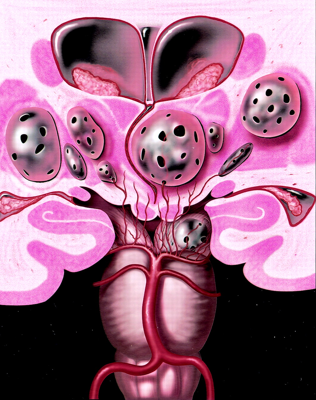
- Fig 1.
Coronal anatomic diagram depicts multiple bilateral giant perivascular spaces (PVSs) in the mesencephalothalamic region. There are fenestrations of the giant PVSs, which may allow accumulation of interstitial fluid between the vessel and pia or within the interpial space causing enlargement of the PVSs. Note the mass effect upon the third ventricle with associated obstructive hydrocephalus. Graphic courtesy of James Cooper, MD and AMIRSYS, Inc (35).
- Fig 2.
Axial T2-weighted (A), postcontrast axial T1-weighted (B), and postcontrast coronal T1-weighted (C) images obtained in a 46-year-old man with headaches show a nonenhancing multiloculated cystic mass in the right midbrain, thalamus, and right medial temporal lobe. The cysts follow CSF signal intensity on all pulse sequences and do not enhance. Follow-up imaging 13 years later showed no change. Biopsy proved pial-lined giant perivascular spaces.
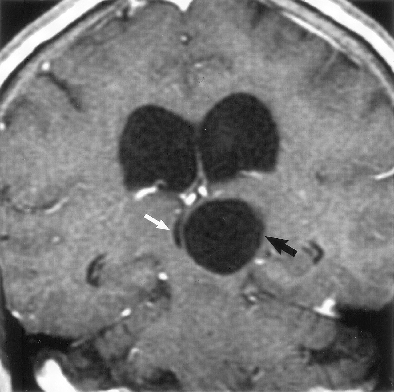
- Fig 3.
Coronal postcontrast T1-weighted image obtained in a 56-year-old woman with headaches shows a nonenhancing unilocular giant perivascular space (black arrow) in the left thalamus with compression and displacement of the third ventricle (white arrow). Note associated hydrocephalus. Surgery disclosed a smooth walled cyst with no abnormality in the adjacent brain. The patient initially underwent a cyst fenestration with a decrease in the size of the PVS. Four months later, there was reaccumulation of fluid and the PVS enlarged to its original size. A ventriculoperitoneal shunt was then placed, which relieved the hydrocephalus. Follow-up studies showed no change in cyst size over 4 years.
- Fig 4.
Sagittal T1-weighted (A), axial FLAIR (B), and coronal T2-weighted (C) images obtained in a 71-year-old man with dementia show extensive involvement of the hemispheric and subcortical white matter with multilocular giant perivascular spaces. The coronal image shows the marked asymmetry of the lesions. Note the scattered focal white matter changes surrounding some of the lesions (black arrows) seen best on the FLAIR image. Case courtesy of Anthony Doyle, MD.
- Fig 5.
Sagittal T1-weighted (A), axial FLAIR (B), and axial T2-weighted (C) images obtained in a 46-year-old woman with a visual field defect show extensive involvement of the corpus callosum and cingulate gyrus (A, white arrow) with extension to the subcortical white matter of the parietal and occipital lobes. There is slight increased signal intensity surrounding the lesions, best seen on FLAIR image (B, white arrows). The gray matter is stretched and displaced over the multiloculated giant perivascular spaces. Case courtesy of Leena Valanne, MD.
- Fig 6.
Images obtained in a 37-year-old man with headaches. Axial T2-weighted (A) and axial FLAIR (B) images show diffuse, confluent white matter hyperintensity surrounding the giant perivascular spaces. There is mild gyral expansion over the perivascular spaces. The diffusion-weighted image (C) shows no diffusion restriction.
- Fig 7.
Images obtained in a 6-year-old boy with a history of minor trauma. Axial T1-weighted (A), coronal T2-weighted (B), axial FLAIR (C) and postcontrast coronal T1-weighted (D) images show multiloculated giant perivascular spaces in the left dentate gyrus. There is mild focal mass effect upon the fourth ventricle (white arrow). The clustered cysts follow CSF on all pulse sequences.
- Fig 8.
Images obtained in a 35-year-old man with headache who underwent a biopsy and a third ventriculostomy procedure. Axial T1-weighted MR image (A) shows the giant perivascular space in the left midbrain. A section through the cyst wall (B) shows red-stained collagen in the leptomeninges on the outer aspect of the cyst wall coating the underlying brain tissue that forms the bulk of the cyst wall. No lining of pia matter is present on the inner aspect of the cyst. (Hematoxylin van Gieson stain, original magnification ×20) There is extensive gliosis in the cyst wall as demonstrated by the numerous brown stained fibrillary processes within the brain tissue (C). (Immunocytochemistry for GFAP, original magnification ×20) A high-power view (D) of the cyst wall near the pia arachnoid outer coating (top of picture). Reactive astrocytes are seen toward the bottom of the illustration. No neurons were identified. (Hematoxylin and eosin stain, original magnification ×40)


Tables
Headache 15 Dizziness 3 Dementia 3 Visual changes 3 Post-traumatic evaluation 2 Cranial neuropathy 2 Seizure 1 Syncope 1 Stroke 1 Memory problems 1 Poor balance and concentration 1 N = 33 Age Sex Giant PVS Location Biopsy Follow-up Imaging 39 M Thalamus Y 2 years 76 M Thalamus N N 56 F Thalamus N 4 years 86 M Thalamus N 12 years 35 M Thalamus N 2 years 42 F Thalamus N N 49 F Thalamus N 7 years 35 M Midbrain Y 1 year 57 M Midbrain Y N 44 F Midbrain N N 75 M Midbrain N N 40 M Midbrain Y N 28 M Midbrain N N 35 M Midbrain/Pons Y 1 year 47 F Thalamus/Midbrain N 9 years 62 M Thalamus/Midbrain N N 46 M Thalamus/Midbrain Y N 75 M Thalamus/Midbrain N 1 year 72 F Thalamus/Basal Ganglia N 10 years 41 M Basal Ganglia N 1 year 47 M Basal Ganglia N N 15 M White Matter N 7 years 70 M White Matter N N 62 F White Matter N N 37 M White Matter N 1 year 68 M White Matter N N 56 F White Matter N N 44 F White Matter N 1 year 21 M White Matter Y 6 years 36 M White Matter N 2 years 68 F White Matter N N 46 F White Matter N N 20 F White Matter N N 40 F White Matter N N 10 M White Matter N 3 years 17 M Cerebellum N N 6 M Cerebellum N N
In this issue


Jump to section
Related Articles
Cited By...
- Giant Tumefactive Perivascular Spaces in a Patient Presenting With a First Seizure
- Teaching NeuroImage: Traumatic Dissection of Lenticulostriate Arteries Within an Enlarged Perivascular Space
- Quantitative MRI of Perivascular Spaces at 3T for Early Diagnosis of Mild Cognitive Impairment
- Lacunar Infarcts, but Not Perivascular Spaces, Are Predictors of Cognitive Decline in Cerebral Small-Vessel Disease
- Perivascular Spaces in Old Age: Assessment, Distribution, and Correlation with White Matter Hyperintensities
- The structure of the perivascular compartment in the old canine brain: a case study
- Tumefactive perivascular spaces: a rare incidental finding
- Brain Perivascular Spaces as Biomarkers of Vascular Risk: Results from the Northern Manhattan Study
- Hydrocephalus due to extreme dilation of Virchow-Robin spaces
- Subcortical Cystic Lesions within the Anterior Superior Temporal Gyrus: A Newly Recognized Characteristic Location for Dilated Perivascular Spaces
- Obstructive hydrocephalus due to cavernous dilation of Virchow-Robin spaces
- Neuropathological Correlates of Temporal Pole White Matter Hyperintensities in CADASIL
- Acute mesencephalic stroke associated with dilated cystic perivascular spaces